2541
Verifying the concordance between motion corrected and conventional MPRAGE for pediatric morphometric analysis using minimal motion data1Centre for the Developing Brain, School of Biomedical Engineering and Imaging Sciences, King's College London, London, United Kingdom, 2Biomedical Engineering Department, School of Biomedical Engineering and Imaging Sciences, King's College London, London, United Kingdom, 3Department for Forensic and Neurodevelopmental Sciences, Institute of Psychiatry, Psychology and Neuroscience, King's College London, London, United Kingdom, 4Guy’s and St Thomas’ NHS Foundation Trust, London, United Kingdom, 5MR Research Collaborations, Siemens Healthcare Limited, Camberley, United Kingdom, 6Biomedical Image Technologies, ETSI Telecomunicación, Universidad Politécnica de Madrid and CIBER-BNN, ISCIII, Madrid, Spain
Synopsis
Keywords: Neuro, Motion Correction, Segmentation
Motivation: Head motion is a common cause of image degradation in pediatric neuroimaging. Multiple strategies are available for correcting intrascan motion, including DISORDER - a retrospective motion correction approach.
Goal(s): We aimed to validate the use of DISORDER for brain morphometric analyses in a pediatric population.
Approach: We compared a wide range of morphometry measures obtained from high quality linear phase-encoding MPRAGE and DISORDER MPRAGE acquisitions in 21 children aged 7-8 years.
Results: DISORDER reduced data loss due to motion and brain morphometric analyses obtained using both MPRAGE acquisitions were highly consistent for most brain regions.
Impact: DISORDER, a retrospective motion correction technique, reduces data loss due to head motion in pediatric populations and produces quantitative brain morphometric measures that are largely consistent with measures derived from a standard acquisition.
Introduction
Obtaining high quality neuroimaging data suitable for morphometric analyses in children is challenging. Multiple strategies are available to correct for head motion, including a retrospective motion correction technique: Distributed and Incoherent Sample Orders for Reconstruction Deblurring by using Encoding Redundancy (DISORDER)1.In addition to the sequence used to acquire the images, the choice of software for automated segmentation of brain structures needs to be considered. Previous work has suggested that FreeSurfer performs best in cortical analyses2, and FSL is closer to manual segmentation in subcortical analyses3. HippUnfold is a recently developed automated approach to segment hippocampal subfields4.
Our aim was to evaluate the use of DISORDER for morphometric analysis by comparing brain cortical and subcortical measures in children, aged 7-8 years, who had both motion-free linear phase-encoding MPRAGE data and MPRAGE data acquired with the DISORDER scheme. In addition, we compared subcortical volumes segmented using FreeSurfer and FSL-FIRST and used HippUnfold to compare hippocampal subfield volumes derived from both MPRAGE acquisitions.
Methods
Subjects and data acquisition: 60 children were recruited between August 2022 - August 2023. (Ethical Committee approval 22/WA/0014). Of these, 20 had no linear phase-encoding MPRAGE data and 3 had no MPRAGE with DISORDER data, so only 37 subjects were included; Scanning was undertaken on a 3T scanner (MAGNETOM Vida, Siemens Healthcare, Erlangen, Germany) with acquisition parameters as follows for both sequence variants; TR = 2200 ms, TE = 2.46 ms, flip angle = 8°, voxel size = 1.1 x 1.071 x 1.071 mm3.Motion correction: Motion and reconstruction were jointly estimated using the DISORDER scheme1,5. The DISORDER acquisition and reconstruction was implemented based on its open source implementation (https://github.com/mriphysics/DISORDER).
Image preprocessing: MPRAGE data underwent intensity normalisation using ANTs6. Removal of Gibbs ringing artefacts was performed using a 3D subvoxel shift method7,8. Images were preprocessed using the Human Connectome Project (HCP) minimal preprocessing pipeline9.
Brain structural measures: Cortical measures were obtained using FreeSurfer. Segmentation of subcortical structures was performed using FreeSurfer10 and FSL-FIRST11 and hippocampal subfields were segmented using HippUnfold4.
Statistical analysis: Correlation of morphometric measures between the two MPRAGE acquisitions were analysed with Pearson’s correlation (R) for normally distributed variables and Spearman’s correlation (ρ) for data which were not normally distributed.
Results
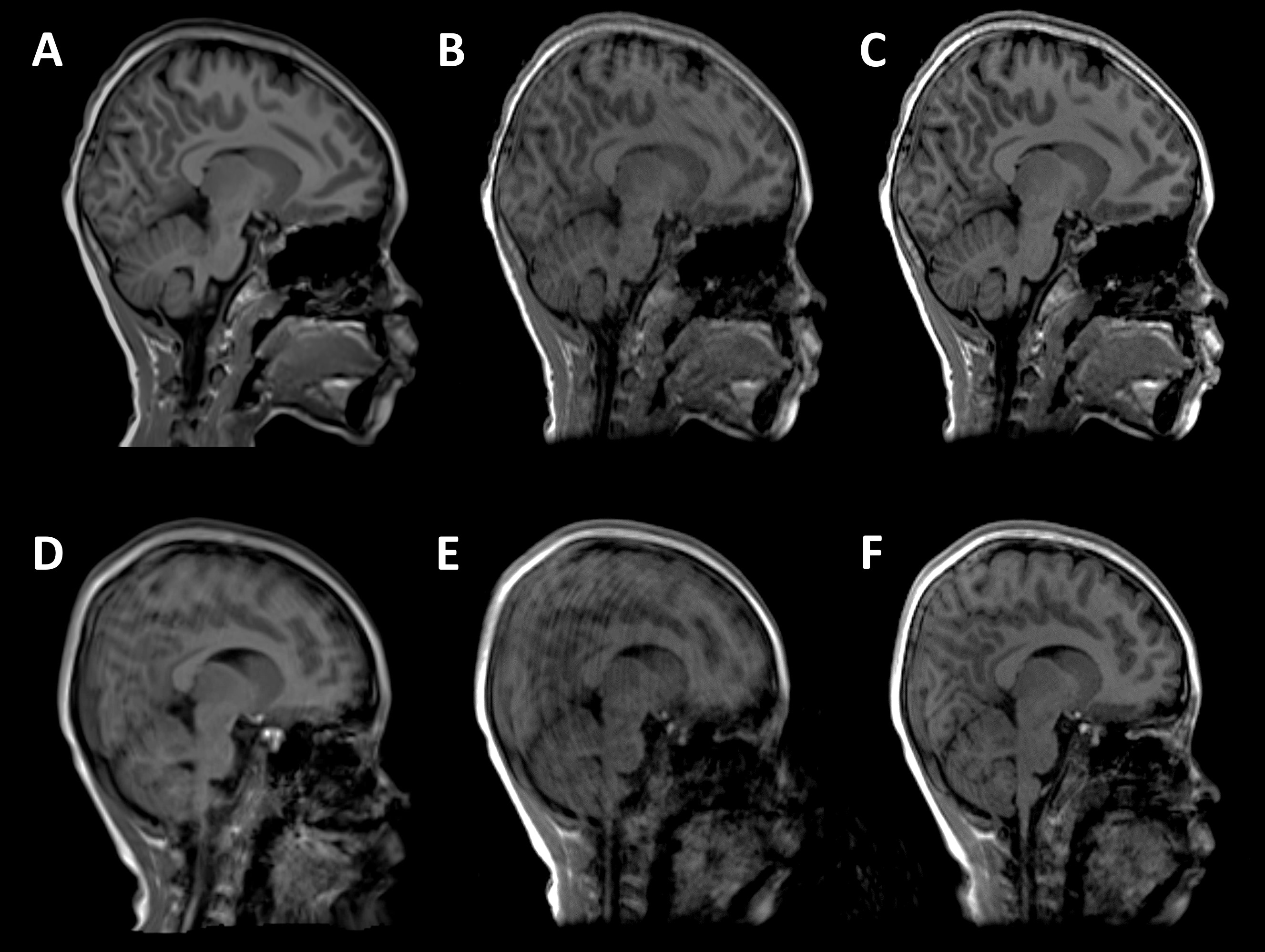
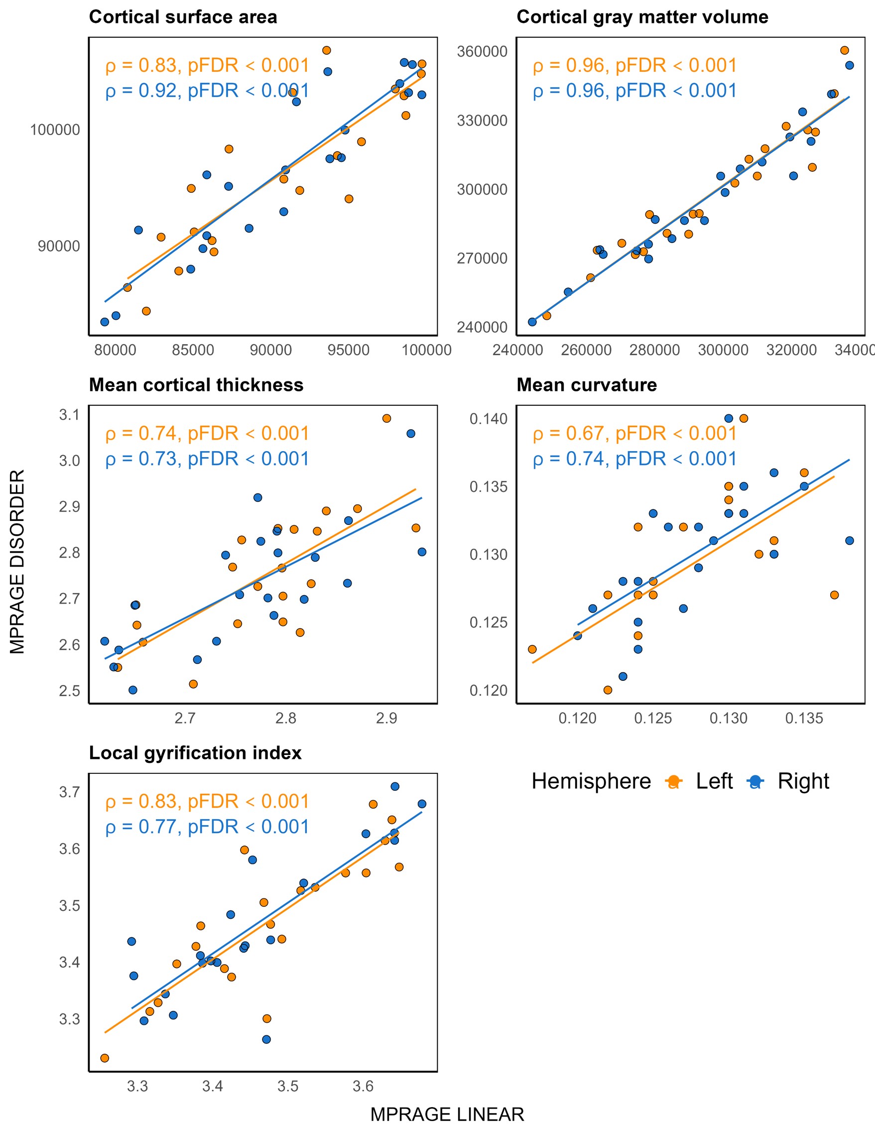
A total of 16 subjects were excluded because of motion artefact on the linear MPRAGE images, but all motion corrected DISORDER scans were suitable. Thus data from 21 subjects (10 male, imaged at a median [range] age 7.75 [7.5-8.17] years) were analysed. Figure 1 shows linear phase-encoding, and uncorrected and corrected MPRAGE DISORDER images for 2 subjects, one included and one excluded due to the quality of the linear MPRAGE.All cortical measures were highly correlated between the two MPRAGE acquisitions (PFDR < 0.05, ρ ≥ 0.67, Figure 2).
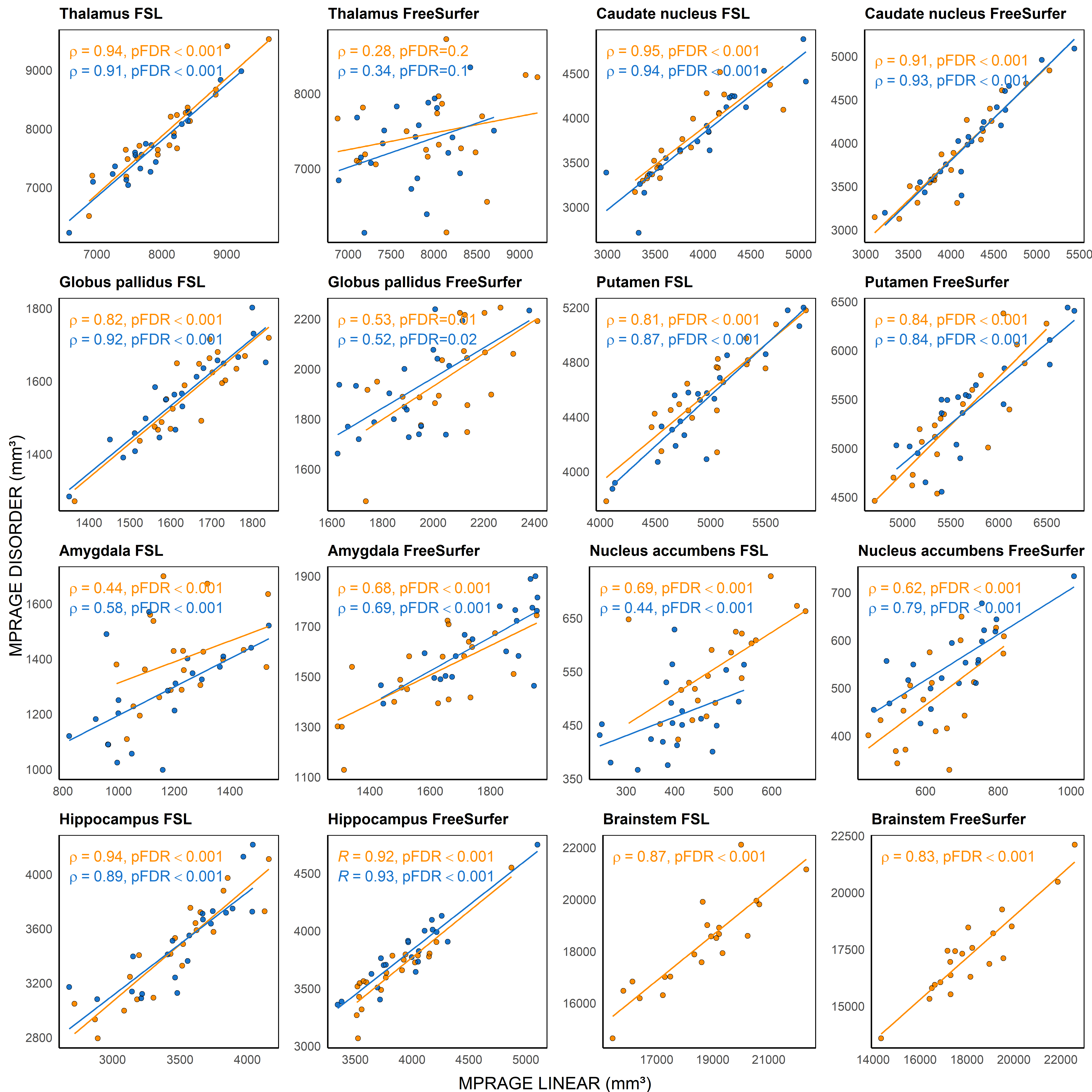
Caudate, putamen, hippocampus and brainstem volumes showed strong correlations between the two MPRAGE acquisitions using both FreeSurfer and FSL-FIRST segmentations (PFDR < 0.05, ρ ≥ 0.81). Correlations were strong for thalamus and globus pallidus volumes using FSL-FIRST (PFDR < 0.05, ρ ≥ 0.82) but were moderate-weak using FreeSurfer (thalamus: PFDR ≥ 0.1, ρ ≤ 0.34; global pallidus: PFDR < 0.05, ρ ≥ 0.52). Correlations for amygdala and nucleus accumbens volumes were moderate using both FreeSurfer and FSL-FIRST (PFDR < 0.05, ρ ≥ 0.44). Figure 3 shows results of correlation analyses for subcortical structures.
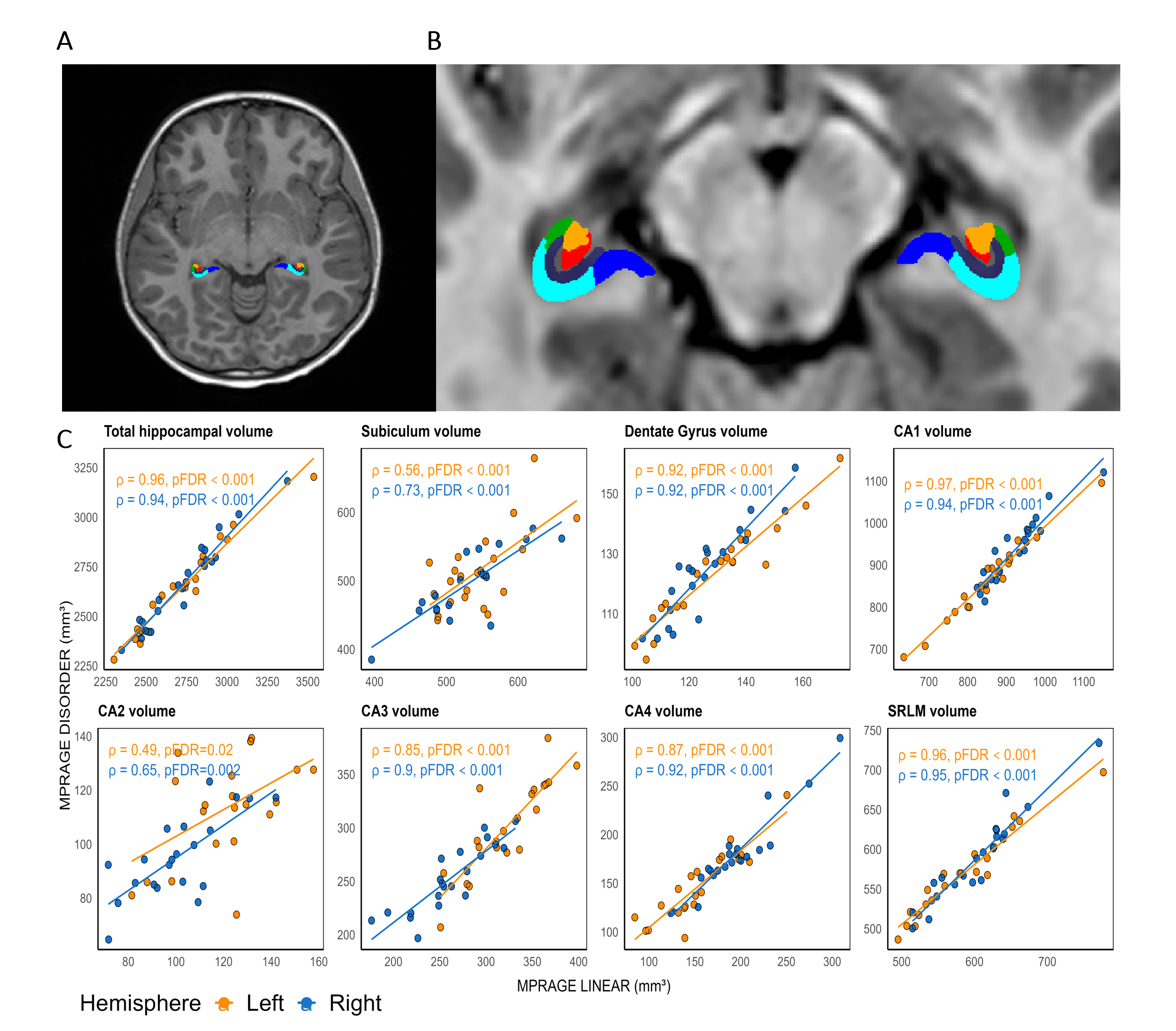
Volumes of hippocampal subfields were similar between both MPRAGE sequences (PFDR < 0.05, ρ ≥ 0.49). Figure 4 shows HippUnfold hippocampal segmentation from one subject and the results of correlation analyses for hippocampal subfield volumes and total hippocampus volumes obtained using both MPRAGE acquisitions.
Discussion
We evaluated the use of the DISORDER scheme for pediatric volumetric neuroimaging by comparing results obtained from high quality motion-free linear phase encoded MPRAGE data with those acquired using DISORDER. Of note, a high proportion of data acquired with the linear phase encoded acquisition had evidence of motion artefact, 16 out of 37 children (43%), and so were not included in the analysis, highlighting the need for motion correction approaches in pediatric populations.We undertook a comprehensive study of brain structures to include regions that would be of interest in a wide range of pediatric neuroimaging studies. Our results suggest that morphometric measures obtained from MPRAGE DISORDER are consistent with those obtained using a linear phase-encoding MPRAGE acquisition.
Conclusion
DISORDER enables quantitative structural MRI in difficult-to-image pediatric populations and will facilitate quantitative morphometry in clinical populations and research studies. This study validates the use of DISORDER for brain morphometric analyses in children.Acknowledgements
This research was funded by the Medical Research Council UK (MR/V002465/1).References
1. Cordero‐Grande, L. et al. Motion‐corrected MRI with DISORDER: Distributed and incoherent sample orders for reconstruction deblurring using encoding redundancy. Magn. Reson. Med. 84, 713–726 (2020).
2. Pulli, E. P. et al. Feasibility of FreeSurfer Processing for T1-Weighted Brain Images of 5-Year-Olds: Semiautomated Protocol of FinnBrain Neuroimaging Lab. Front. Neurosci. 16, (2022).
3. Lidauer, K. et al. Subcortical and hippocampal brain segmentation in 5‐year‐old children: Validation of FSL‐FIRST and FreeSurfer against manual segmentation. Eur. J. Neurosci. 56, 4619–4641 (2022).
4. DeKraker, J. et al. Automated hippocampal unfolding for morphometry and subfield segmentation with HippUnfold. eLife 11, (2022).
5. Cordero-Grande, L. et al. Sensitivity Encoding for Aligned Multishot Magnetic Resonance Reconstruction. IEEE Trans. Comput. Imaging 2, 266–280 (2016).
6. Tustison, N. J. et al. N4ITK: Improved N3 Bias Correction. IEEE Trans. Med. Imaging 29, 1310–1320 (2010).
7. Kellner, E., Dhital, B., Kiselev, V. G. & Reisert, M. Gibbs-Ringing Artifact Removal Based on Local Subvoxel-Shifts. Magn. Reson. Med. 76, 1574–1581 (2016).
8. Bautista, T., O’Muircheartaigh, J., Hajnal, J. & Tournier, J.-D. Removal of Gibbs ringing artefacts for 3D acquistions using subvoxel shifts. Proc. Intl. Soc. Mag. Reson. Med. 29, 3535 (2021).
9. Glasser, M. F. et al. The minimal preprocessing pipelines for the Human Connectome Project. NeuroImage 80, 105–124 (2013).
10. Fischl, B. et al. Whole Brain Segmentation. Neuron 33, 341–355 (2002).
11. Patenaude, B., Smith, S. M., Kennedy, D. N. & Jenkinson, M. A Bayesian model of shape and appearance for subcortical brain segmentation. NeuroImage 56, 907–922 (2011).
Figures