2540
Application of Synthetic MRI Technology in Preoperative Brain Development of Pediatric Congenital Heart Disease1Lanzhou University Second Hospital, Lanzhou, China, 2Gansu Provincial Maternity and Child-Care Hospital, Lanzhou, China, 3Siemens Healthineers, Shanghai, China
Synopsis
Keywords: Neuro, Quantitative Imaging
Motivation: Children with congenital heart disease (CHD) may exhibit brain development abnormalities, making it crucial to understand the risk and improve neurological function in these patients.
Goal(s): In this study, we aimed to quantitative assessment of preoperative brain development in CHD children using Synthetic Magnetic Resonance Imaging (SyMRI).
Approach: Through the quantitative evaluation and correlation analysis of T1, T2 and and PD values of SyMRI in the preoperative brain development of children with CHD.
Results: The results indicate that the presence of neurological development abnormalities existed in children with CHD before surgery. SyMRI can provid a basis for early clinical evaluation and timely intervention.
Impact: SyMRI quantitative assessment can serve as a reference indicator for brain development abnormalities in CHD children, aiding in guiding early clinical evaluation and timely intervention.
Introduction
Understanding the risks associated with Congenital heart disease (CHD) related brain development and improving their neurological function is one of the challenges in treating these children[1]. SyMRI allows the acquisition of multiple contrast images in a short time through a single scan, while simultaneously providing quantitative spectra for tissue analysis[2-4]. This study primarily investigates the value of SyMRI quantitative assessment in preoperative brain development of CHD children.Methods
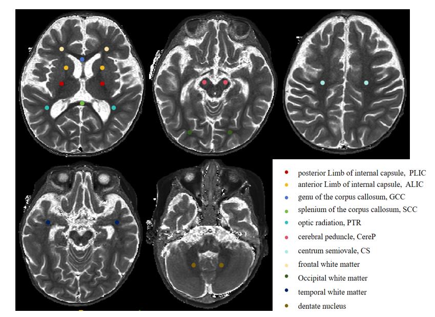
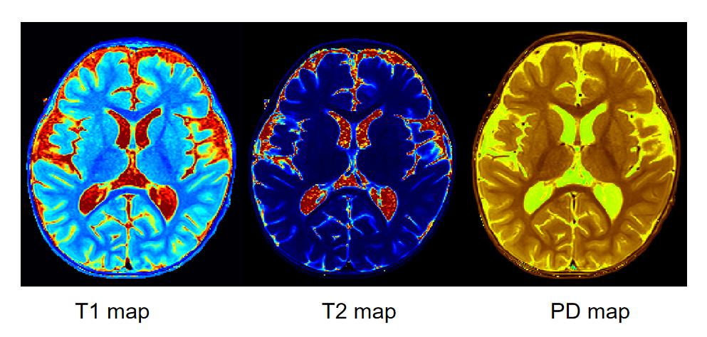
This study obtained approval from our institutional ethics committee and obtained informed consent from the parents or guardians. A total of 43 infants with CHD, aged 2-24 months, were included in the observation group. In addition, 43 healthy infants of matched age and gender were selected as the control group. Both groups of subjects underwent Gesell Development Schedules (GESSELL) developmental assessment. All subjects underwent routine MRI and SyMRI examinations on a 3.0T MRI scanner(MAGNETOM Lumina, Siemens Healthcare, Erlangen, Germany). SyMRI utilized a multi-dynamic multi-echo (MDME) sequence with the following scanning parameters: TR 4450 ms, TE 23 ms, receiver bandwidth 150 Hz/pixel, FOV = 180 mm × 180 mm, matrix size = 256x180, 30 slices, 3 mm slice thickness, 0.6 mm slice gap, and a total scan time of 4 min 50 s. The acquired scan data was processed using an in-house MDME Toolbox (Designed and provided by collaboration scientist from Siemens Healthineers) to automatically calculate T1, T2, and proton density (PD) maps. Manual delineation of two brain regions of interest (ROI) for both groups was performed on the parameter maps using ITK-SNAP 4.0.1 software, and T1, T2, and PD values were computed as shown in Figure 2.This study employed SPSS 27.0 software for data analysis. Comparative analyses were performed on T1, T2, and PD values in different brain ROI between the two groups of subjects, as well as on the five functional area scores from the Gesell Development Schedules. Pearson correlation analysis was utilized to examine the relationship between the regions with differing T1, T2, PD values and the scores in the five functional areas of the Gesell Development Schedules.Receiver operating characteristic (ROC) curves were employed to assess the diagnostic efficacy of T1 and T2 values in various brain regions with statistically significant differences for identifying preoperative brain developmental abnormalities in children with CHD. A significance level of P < 0.05 was used to indicate statistical significance.Results
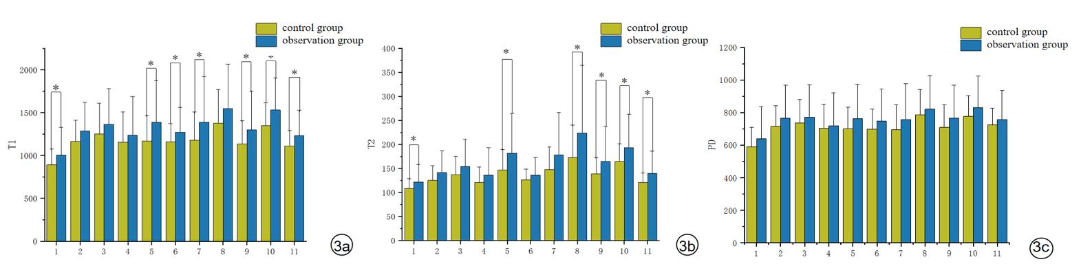
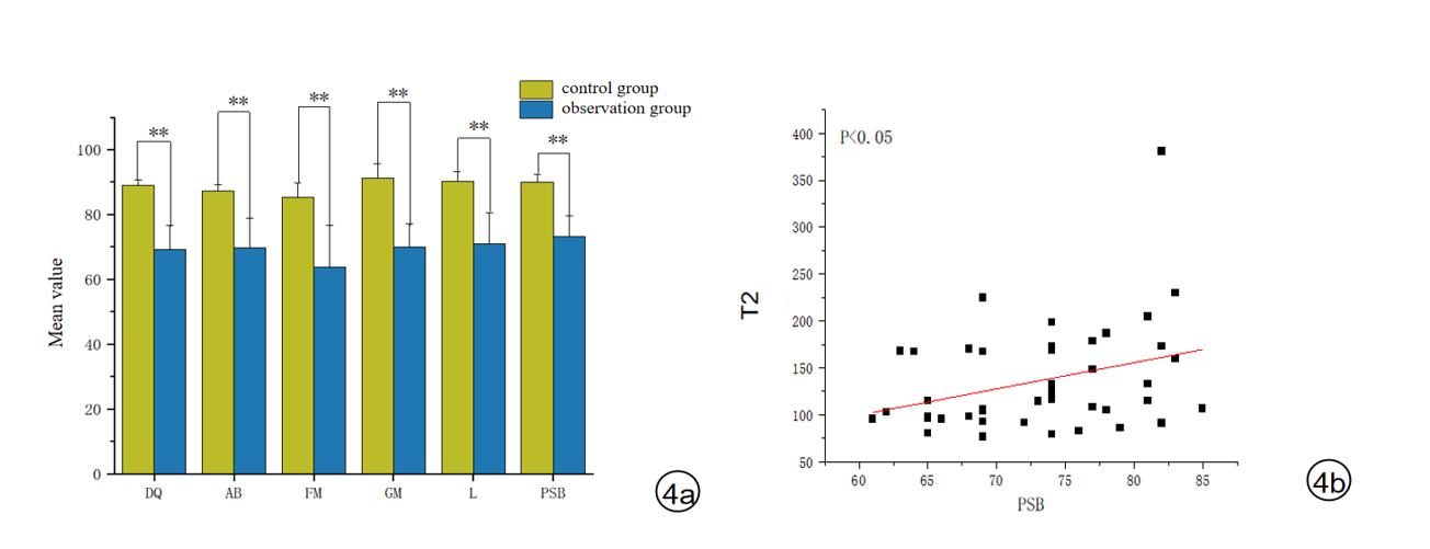
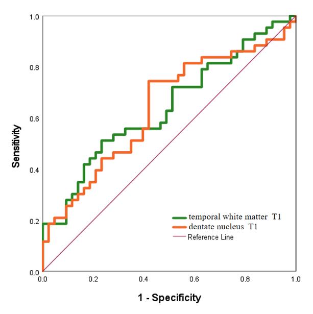
There were no statistically significant differences in age and gender between the two groups. In the observation group, the T1 values of the PLIC, PTR, CereP, CS, occipital white matter, temporal white matter, and dentate nucleus were all higher than those in the control group, and these differences were statistically significant (P < 0.05) (Figure 3a). In the observation group, the T2 values of PLIC, PTR, frontal white matter, occipital white matter, temporal white matter, and dentate nucleus were all greater than in the control group, and these differences were statistically significant (P < 0.05) (Figure 3b). Pearson correlation analysis revealed that the observation group had significantly lower scores in DQ, AB,FM, GM, L,PSB, with all of these differences being statistically significant (P < 0.05) when compared to the control group (Figure 4). In the observation group, the T2 value of the splenium of the corpus callosum (SCC) was significantly positively correlated with the PSB score (r=0.325, p=0.033) (Figure 4). The ROC curve results indicated that the area under the curve (AUC) for diagnosing preoperative brain developmental abnormalities in children with CHD using T1 values of the temporal white matter and dentate nucleus was both greater than 0.60 (Figure 5).Discussion
This study utilized SyMRI for quantitative assessment of preoperative brain development in children with CHD. The results suggest that there are differences in the relaxation values (T1 and T2) at various brain regions between the two groups, indicating the presence of preoperative brain developmental abnormalities in children with CHD. Pearson correlation analysis revealed that the observation group had lower scores in DQ, AB, FM, GM, L, and PSB in comparison to the control group. The extension of T2 relaxation values in the splenium of the corpus callosum was significantly positively correlated with the PSB score in the observation group, indicating developmental delay in these children.The ROC curve analysis demonstrated that T1 values of the temporal white matter and dentate nucleus had good diagnostic efficacy for identifying preoperative brain developmental abnormalities in children with CHD.Conclusion
Quantitative assessment using SyMRI is beneficial in early detection of preoperative brain developmental abnormalities in children with CHD. It provides a valuable reference for clinical early assessment and timely intervention.Acknowledgements
We would like to thank the whole study team at the Gansu provincial Maternity and Child-care Hospital and the NMR Room of Lanzhou University Second Hospital, for their continued support.References
[1] Bhattacharjee I, Mohamed MA, Nandakumar V, et al. Scoring of brain magnetic resonance imaging and neurodevelopmental outcomes in infants with congenital heart disease. Early Hum Dev. 2022 ;169:105574.
[2] Schmidbauer V, Geisl G, Cardoso Diogo M, et al. Validity of SyMRI for Assessment of the Neonatal Brain. Clin Neuroradiol. 2021 ;31(2):315-323.
[3] Kim JS, Cho HH, Shin JY, et al. Diagnostic performance of synthetic relaxometry for predicting neurodevelopmental outcomes in premature infants: a feasibility study. Eur Radiol. 2023 ;33(10):7340-7351.
[4] Schmidbauer VU, Yildirim MS, Dovjak GO, et al. Synthetic MR Imaging-Based WM Signal Suppression Identifies Neonatal Brainstem Pathways in Vivo. AJNR Am J Neuroradiol. 2022 ;43(12):1817-1823.
Figures