2538
Discriminating High-Grade and Low-Grade Pediatric Gliomas by time-dependent diffusion MRI1Xinhua Hospital, Shanghai Jiao Tong University School of Medicine, Shanghai, China, 2Philips Healthcare, Shanghai, China, 3Philips Healthcare (Shenzhen) Ltd., Shenzhen, China
Synopsis
Keywords: Neuro, Diffusion/other diffusion imaging techniques, OGSE; IMPULSED; Glioblastoma; cellularity;cell diameter
Motivation: Gliomas are the most common type of central nervous system tumors in children, and the histological classification plays a crucial role in determining the prognosis and the treatment. A non-invasive approach is needed for pre-surgery characterization of the histopathology of the tumor.
Goal(s): The aim of this study was to explore the feasibility of applying time-dependent diffusion magnetic resonance imaging (td-dMRI) parameters in discriminating between high-grade gliomas (HGG) and low-grade gliomas (LGG).
Approach: Nine pediatric glioma patients were involved and underwent td-dMRI scan.
Results: The td-dMRI parameters may serve as potential markers for the differentiation between high-grade gliomas (HGG) and low-grade gliomas (LGG).
Impact: Time-dependent diffusion MRI parameters can distinguish pediatric high-grade and low-grade gliomas noninvasively, aiding personalized treatment and enhancing prognosis, among other potential applications.
Background
Gliomas are the most common type of central nervous system tumors in children, and the histological classification plays a crucial role in determining the prognosis and and the treatment. However, that always requires an invasive procedure such as biopsy. Therefore, a non-invasive approach is needed for the pre-surgery characterization of the histopathology of the tumor. Time-dependent diffusion magnetic resonance imaging (td-dMRI) is an emerging technique, which can provide cellular-level information of the tissue. The application of td-dMRI in various kind of tumors has been explored. This study was conducted to explore the utility of td-dMRI parameters in discriminating between high-grade gliomas (HGG) and low-grade gliomas (LGG) in pediatric patients.Methods
This prospective study included a cohort of nine pediatric glioma patients, comprising six in the LGG group and three in the HGG group, with an average age of 7.25 ± 4.15 years. They underwent a td-dMRI scan, which consisted of pulsed and oscillating gradient diffusion sequences, performed on a 3T MR scanner (Ingenia CX, Philips, Best, the Netherlands). The acquired dMRI data was then fitted by the IMPULSED (imaging microstructural parameters using limited spectrally edited diffusion) model to derive parameters including cellularity, cell diameter (d), intracellular volume fraction (Vin), and extracellular diffusion coefficient (Dex)[1-3] (see Supporting Information Figure 1). All these postprocessing was executed in MATLAB 2022b(MathWorks). Histopathological examination with H&E staining was employed to validate the microstructural properties.Results
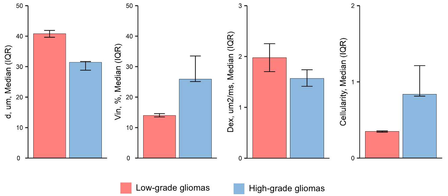
Representative images of td-dMRI and corresponding parametric maps were shown in Figure 1. In general, values of microstructural parameters were different between LGG and HGG patients (Figure 2). In HGG group, the mean cellularity was significantly higher compared to LGG group, and the former demonstrated a greater variance, indicating the heterogeneity of the tumor. Conversely, low-grade gliomas were characterized by a higher average cell diameter and extracellular diffusion coefficient when compared to high-grade gliomas. Furthermore, high-grade gliomas exhibited a higher intracellular volume fraction in comparison to low-grade gliomas.Conclusions
The time-dependent diffusion MRI parameters may serve as potential markers for the differentiation between high-grade gliomas HGG and low-grade gliomas LGG in pediatric patients.Acknowledgements
The authors wish to thank the staffs of Philips Healthcare in Shanghai for their support.References
1. Xu, J., et al., Magnetic resonance imaging of mean cell size in human breast tumors. Magn Reson Med, 2020. 83(6): p. 2002-2014.
2. Jiang, X., et al., In vivo imaging of cancer cell size and cellularity using temporal diffusion spectroscopy. Magn Reson Med, 2017. 78(1): p. 156-164.
3. Jiang, X., et al., Quantification of cell size using temporal diffusion spectroscopy. Magn Reson Med, 2016. 75(3): p. 1076-85.
Figures
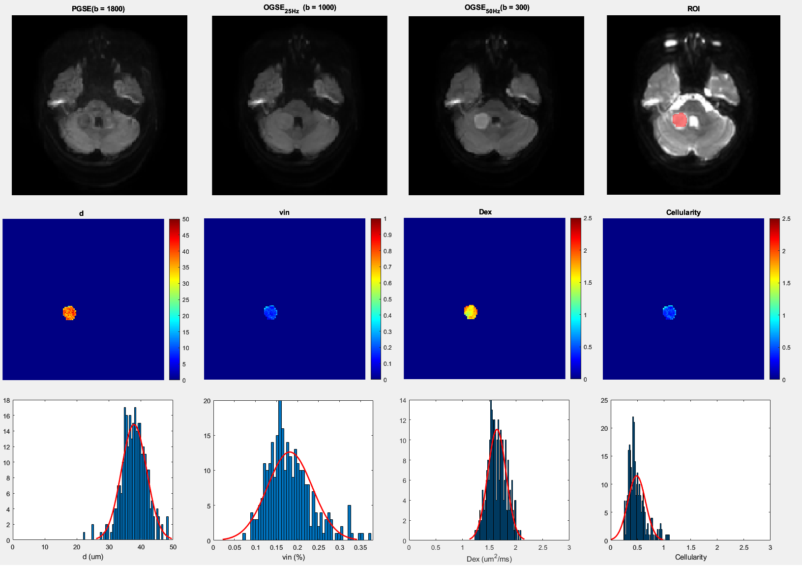
Figure 1. Representative images of raw td-MRI and responding reconstructed parametric maps.
Top: Diffusion weighted images of PGSE (b = 1800), OGSE25Hz(b = 1000), OGSE50Hz(b = 300), and the drawn ROI(red) fused on a b = 0 image.
Middle: Parametric maps derived from IMPULSED model (left to right): mean cell diameter(d), intracellular volume fraction (Vin), and extracellular diffusion coefficient (Dex), and cellularity.
Bottom: The responding histograms of fitted IMPULSED metrics.