2532
4D Flow MRI in Pediatric Brain Arteriovenous Malformation: The Role of Hemodynamic Markers in Monitoring Disease Progression1Radiology and Biomedical Imaging, University of California - San Francisco, San Francisco, CA, United States, 2Neurological Surgery, University of California - San Francisco, San Francisco, CA, United States, 3Neurology, University of California - San Francisco, San Francisco, CA, United States, 4Anesthesia, University of California - San Francisco, San Francisco, CA, United States
Synopsis
Keywords: Neuro, Velocity & Flow, Brain Arteriovenous Malformation
Motivation: Approximately 10-20% of pediatric patients with brain arteriovenous malformation (bAVM) present with reoccurring formation or growth even after initial successful treatment.
Goal(s): Evaluate the feasibility and efficacy of 4D Flow MRI to quantitatively assess the hemodynamic changes in pediatric bAVMs following treatment and establish its use for early detection of high-risk changes.
Approach: Prospective 4D Flow MRI study of pediatric bAVM patient cohort pre and post treatment
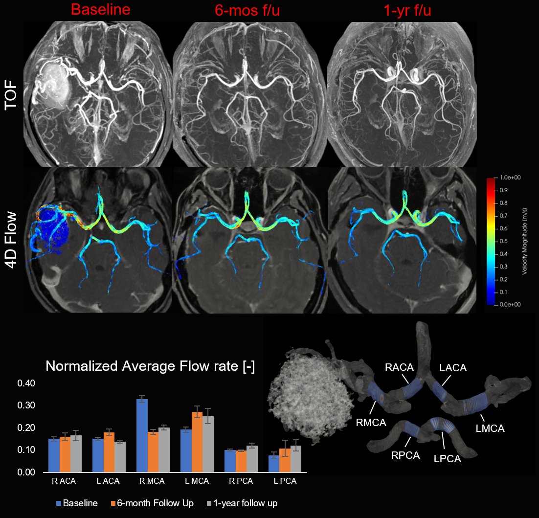
Results: Decreased flow rate and maximum velocity in main bAVM feeding arteries post treatment, resulting in reduced flow laterality mismatch between hemispheres.
Impact: 4D Flow MRI provides a quantitative tool to assess pediatric bAVM flow characteristics and the hemodynamic impact of intervention, which may help to identify patients at risk of bAVM reoccurrence.
Introduction
Brain arteriovenous malformations (bAVM) are tangles of blood vessels which create direct arterial-venous connections prone to rupture. In the pediatric patient population, bAVMs are the leading cause of hemorrhagic stroke. While some bAVM demonstrate long-term stability after intervention, approximately 10-20% of pediatric patients present with dynamic vascular lesions, which can result in a new feeding artery aneurysm, venous outflow stenosis, or reoccurrence of bAVM, even after initial successful intervention1. Previous work on hemodynamic assessment of bAVMs has so far been limited to the adult population2,3. The objective of this study is to evaluate the feasibility and efficacy of 4D Flow MRI to quantitatively assess the hemodynamic changes in pediatric bAVMs following treatment and its use for early detection of high-risk changes.Methods
We have prospectively enrolled 20 children (mean age 11.8 ± 3.8 years) with bAVM, who underwent either surgical resection or gamma knife as part of their treatment, into a single center cohort study. As part of a clinical MRI protocol, 4D Flow MRI was acquired in a transverse slab covering the circle of Willis using prospective gating, compressed sensing, and a resolution of 0.7 x 0.7 x 1.0 mm3 on a 3T GE Discovery MR750 scanner with a 32-channel head coil. MRI at baseline and post-intervention (6-month and 1-year follow ups) has been acquired for 5 patients thus far. 4D Flow data was post-processed using our in-house processing pipeline, which includes correction of background phase, segmentation of the main cerebral arteries, and calculation of cardiac-averaged flow rates in cross-sections orthogonal to the centerline of each vessel segment. Flow rates in each vessel segment were normalized by the total flow to the brain to facilitate the comparison across follow-ups.Results
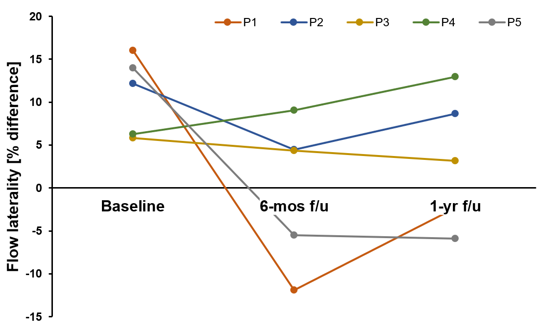
The flow rate precision was evaluated by calculating the flow conservation at the right/left internal carotid and basilar junctions, yielding average errors of 10.7% ± 12.7, 14.0% ± 9.5, and 19.3% ± 13.8 respectively. Across post-intervention studies, we observed a decrease in flow rates and maximum velocities, as well as an increase in pulsatility index, in the primary feeding arteries of the bAVM. One patient example is shown in Figure 1. The hemisphere containing the bAVM received an average of 57.2% ± 2.8 of total brain flow at baseline, which decreased to an average of 51.2% ± 2.9 at 1-year follow-up. In 2 out of 5 patients, the flow laterality between hemispheres flipped post-intervention resulting in increased flow to the contralateral hemisphere (Figure 2).Discussion
In this study, we demonstrated the feasibility of acquiring 4D flow MRI in the pediatric bAVM patient population with high quality and adequate flow conservation in the Circle of Willis. 4D Flow MRI is a very attractive option for evaluating the response to treatment in intracranial lesions given the inability of Doppler ultrasound to penetrate the skull. 4D Flow MRI also provides a powerful ability to compare flow in the same anatomy over multiple time points as opposed to 2D methods which are challenging to co-localize. In the 5 patients who have completed follow-up scans thus far, we have seen a reduction in flow in the primary bAVM supplying arteries and in general a reduction in the flow laterality mismatch between hemispheres, in line with clinical expectation. However, we did encounter several instances where flow laterality flipped post-intervention, resulting in increased flow to the patient’s contralateral hemisphere. Further investigation is needed to determine the physiological basis for these observations and to determine the impact of these flow changes on future risk.Conclusion
These preliminary results indicate that 4D Flow MRI may be a robust and useful tool for monitoring changes in intracranial flow patterns after bAVM treatment in children.Acknowledgements
This work is funded by the the American Heart Association-Bugher Foundation award for hemorrhagic Stroke Research.References
[1] Hak JF, Boulouis G, Kerleroux B, et al. Pediatric brain arteriovenous malformation recurrence: a cohort study, systematic review and meta-analysis. J Neurointerv Surg. 2022 Jun;14(6):611-617. doi: 10.1136/neurintsurg-2021-017777
[2] Aristova M, Vali A, Ansari SA, et al. Standardized Evaluation of Cerebral Arteriovenous Malformations Using Flow Distribution Network Graphs and Dual-venc 4D Flow MRI. J Magn Reson Imaging. 2019 Dec;50(6):1718-1730. doi: 10.1002/jmri.26784.
[3] Wu C, Ansari SA, Honarmand AR, et al. Evaluation of 4D vascular flow and tissue perfusion in cerebral arteriovenous malformations: influence of Spetzler-Martin grade, clinical presentation, and AVM risk factors. AJNR Am J Neuroradiol. 2015 Jun;36(6):1142-9. doi: 10.3174/ajnr.A4259
Figures