2530
An in-vitro deuterium NMR study to measure the efficacy of combinatorial therapy for diffuse intrinsic pontine glioma1St. Jude Children's Research Hospital, Memphis, TN, United States
Synopsis
Keywords: Cancer, Spectroscopy, DIPG, Pediatrics, NMR
Motivation: There is a need for developing next generation of clinical trials by targeting selective pathways in diffuse intrinsic pontine glioma (DIPG).
Goal(s): We explore the impact of Vehicle (DMSO), Paxalisib, BAY-876, and their combination in patient-derived SJ-DIPGX7 cell line to evaluate their potential as a therapeutic strategy for patients with DIPG.
Approach: 2H-NMR spectra were longitudinally obtained in a cell suspension. We calculated the lactate flux turnover, which was validated using 100-run Monte-Carlo simulation. This outcome was further confirmed by conducting a glycolysis stress test.
Results: The combined therapy (Paxalisib + BAY-876) holds potential for enhancing its therapeutic effectiveness against DIPGs.
Impact: This study paves the way for future in-vitro and in-vivo studies to be conducted for monitoring efficacy of targeted therapeutic combination for DIPG.
Introduction
Diffuse Intrinsic Pontine Glioma (DIPG) is a highly aggressive pediatric brain tumor targeting the central nervous system with very limited treatment options [1]. There is a clear need for developing next generation of clinical trials by targeting selective pathways. PI3K/AKT/mTOR pathway has been shown to be commonly upregulated in DIPG [2]. A recently developed blood-brain barrier penetrant PI3K inhibitor Paxalisib (GDC-0084) [3] has been shown to have excellent efficacy in in-vitro setting. However, Paxalisib did not extend survival of patient derived xenograft models of DIPG [4]. There was an enhanced efficacy of Paxalisib when combined with ERK pathway inhibitor [5]. Here, we explore the impact of Paxalisib, the selective GLUT1-inhibitor BAY-876 [6], and their combination in the SJ-DIPGX7 cell line to evaluate their potential as a therapeutic strategy for patients with DIPG.Materials and Methods
1x10⁶ SJ-DIPGX7 cells were washed with phosphate-buffered saline (PBS) and suspended in a colorless Dulbecco's Modified Eagle Medium (DMEM) devoid of glucose and glutamine, supplemented with 10 mM [6,6-2H2] D-glucose and 4 mM L-glutamine. The treatment groups included: (i) DMSO, (ii) Paxalisib (0.8 µM), (iii) BAY-876 (4.0 µM), and Paxalisib + BAY-876 (0.8 µM + 4.0 µM). High-resolution 2H NMR spectra were recorded at 310 K on a Bruker Avance III HD 600 MHz NMR spectrometer (Bruker Biospin, Germany) equipped with a 5 mm TCI cryoprobe. NMR experiments were performed at 1-hour intervals for 23 hours. For each experiment, 64 transients were collected using a relaxation delay of 2 seconds and an acquisition time of 1.5 seconds. Lactate concentration at each time point was measured for all the treatment groups. Lactate turnover flux was determined by multiplying the steady-state concentration with the rate constant of lactate synthesis after fitting an exponential curve. A 100-run Monte Carlo simulation was performed to estimate the uncertainty in the derived flux by generating 100 new data sets containing the same amount of noise as the experimental data [7]. To further test our findings, we performed a glycolysis stress test using a XF pro Seahorse Analyzer (Agilent Technologie-103020-100) [8]. 3x10⁴ cells were seeded in a 96-well plate following the manufacturer's recommendations in the four treatment groups.Results and Discussion
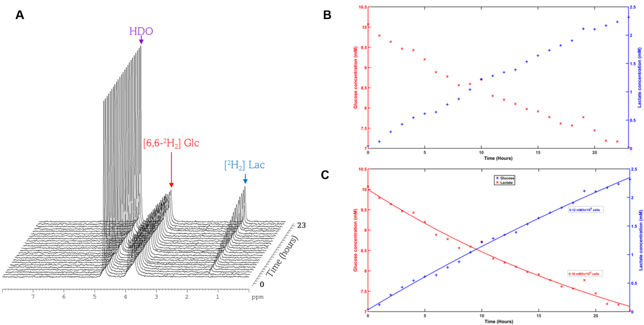
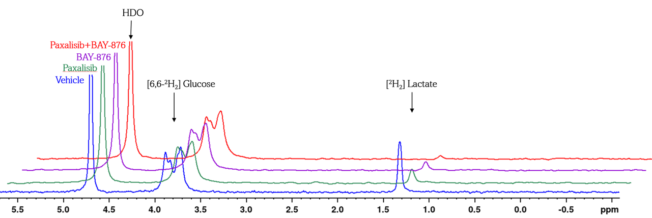
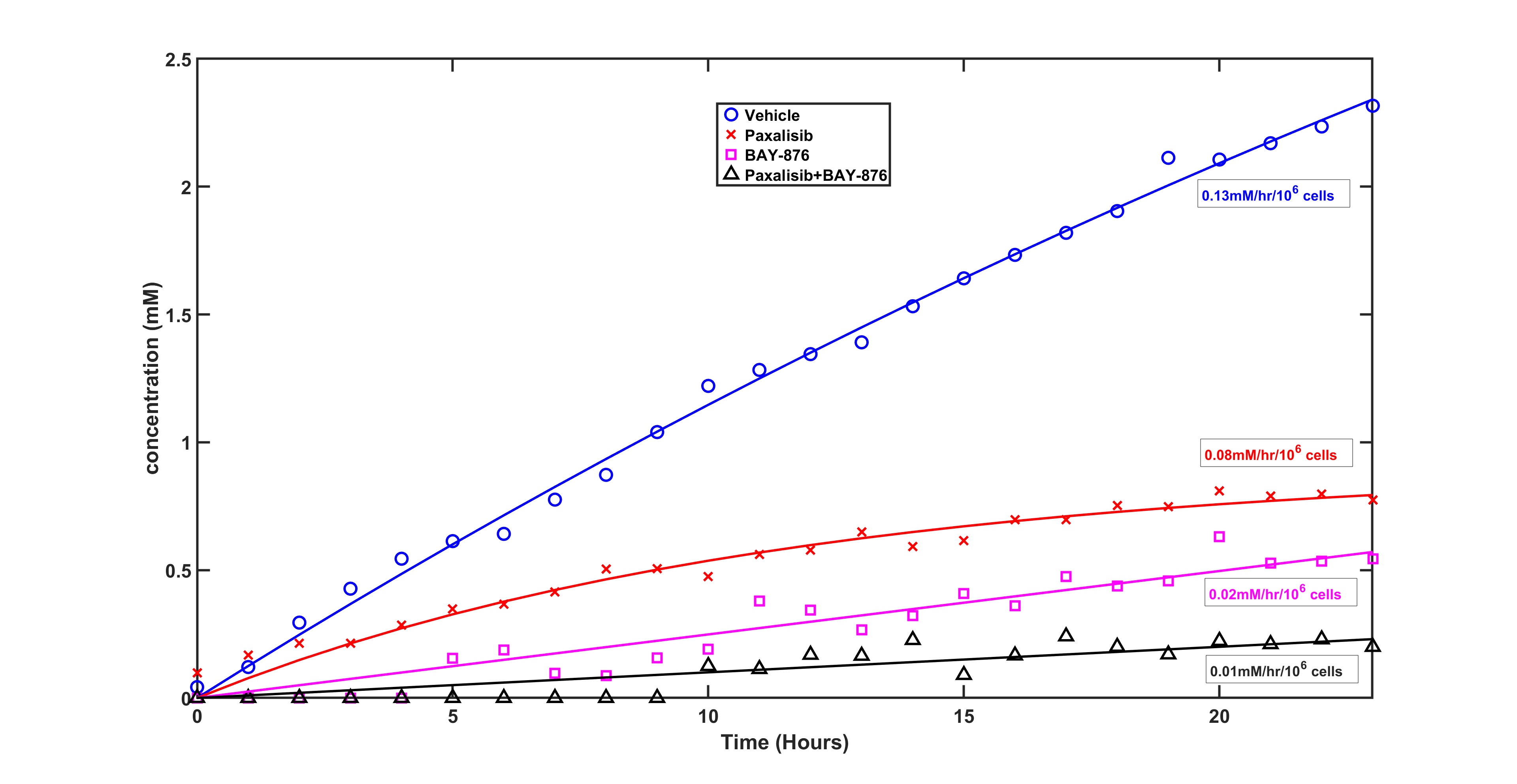
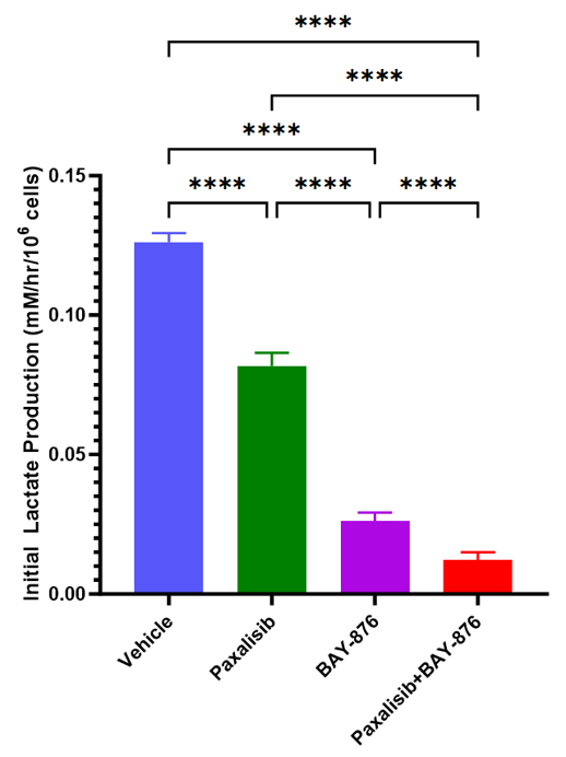
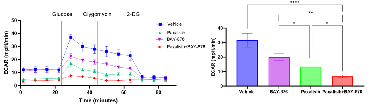
Fig. 1 shows a time-lapse of high-resolution 2H NMR spectra of an untreated SJ-DIPGX7 cell line for 23 hours depicting glucose reduction (3.8 ppm) and lactate production (1.3 ppm) with time, signifying elevated anaerobic glycolysis in the untreated DIPG cells [9]. Fig. 2 presents a comparative snapshot of the 2H NMR spectra acquired at the 23rd hour for vehicle, Paxalisib, BAY-876 and Paxalisib+BAY-876 treated cell lines. The lactate concentrations at the 23rd hour was 2.31 mM in vehicle, 0.77 mM in Paxalisib, 0.54 mM in BAY-876 and 0.21 mM in the combination treatment. The exponential evolution of lactate concentration with time, following the second order kinetics of Michaelis–Menten equation, are shown in Fig. 3 for the four treatment scenarios. The initial lactate flux was 0.13 mM/hr/106 cells in untreated, 0.08 mM/hr/106 cells in Paxalisib-treated, 0.02 mM/hr/106 cells in BAY-876-treated, and 0.01 mM/hr/106 cells in combination therapy-treated cells. The Monte-Carlo simulation of the lactate turnover flux in the four cell lines, as shown in Fig. 4, shows a significant reduction in lactate flux rate (by as much as 90±2.1% compared to untreated cells) using combination therapy. Fig. 5 shows the glycolytic stress data and confirms that the extracellular acidification rate (ECAR) was reduced in the combination therapy indicating lower glycolytic activity. Our findings suggest that combined treatment is effective in reducing glycolysis. The simultaneous targeting of the PI3K/AKT/mTOR pathway and glucose uptake, as evidenced in the combination therapy, offers the prospect of enhanced therapeutic efficacy for DIPGs.Acknowledgements
No acknowledgement found.References
1. Warren KE. Diffuse intrinsic pontine glioma: poised for progress. Front Oncol. 2012;2:205.
2. Aziz-Bose R, Monje M. Diffuse intrinsic pontine glioma: molecular landscape and emerging therapeutic targets. Curr Opin Oncol. 2019;31(6):522-530.
3. Wen PY, Cloughesy TF, Olivero AG, et al. First-in-Human Phase I Study to Evaluate the Brain-Penetrant PI3K/mTOR Inhibitor GDC-0084 in Patients with Progressive or Recurrent High-Grade Glioma. Clin Cancer Res. 2020;26(8):1820-1828.
4. He C, Xu K, Zhu X, et al. Patient-Derived Orthotopic Xenografts and Cell Lines from Pediatric High-Grade Glioma Recapitulate the Heterogeneity of Histopathology, Molecular Signatures, and Drug Response. bioRxiv. 2020:2020.2012.2006.407973.
5. Wu YL, Maachani UB, Schweitzer M, et al. Dual Inhibition of PI3K/AKT and MEK/ERK Pathways Induces Synergistic Antitumor Effects in Diffuse Intrinsic Pontine Glioma Cells. Transl Oncol. 2017;10(2):221-228.
6. Chen Z, Vaeth M, Eckstein M, et al. Characterization of the effect of the GLUT-1 inhibitor BAY-876 on T cells and macrophages. Eur J Pharmacol. 2023;945:175552.
7. Lanz B, Gruetter R, Duarte JM. Metabolic Flux and Compartmentation Analysis in the Brain In vivo. Front Endocrinol (Lausanne). 2013;4:156.
8. Ludikhuize MC, Meerlo M, Burgering BMT, Rodriguez Colman MJ. Protocol to profile the bioenergetics of organoids using Seahorse. STAR Protoc. 2021;2(1):100386.
9. Autry AW, Hashizume R, James CD, Larson PEZ, Vigneron DB, Park I. Measuring Tumor Metabolism in Pediatric Diffuse Intrinsic Pontine Glioma Using Hyperpolarized Carbon-13 MR Metabolic Imaging. Contrast Media Mol Imaging. 2018;2018:3215658.
Figures