2525
Use of the Allen Human Brain spatial transcriptome for the validation of myelin content imaging using BMC-mcDESPOT.1National Institute on Aging, National Institute of Health, Baltimore, MD, United States
Synopsis
Keywords: Aging, Relaxometry, Transcriptomics, Myelin water fraction
Motivation: Myelin water imaging has demonstrated its ability to successfully detect changes of myelin content in different neuropathology. However, the validation of these measures remains challenging.
Goal(s): In this study, we aim to validate our MWF measurements with the gene expression that are relevant to myelin.
Approach: We utilized the Allen Human Brain Atlas (AHBA) transcriptomics dataset to validate our Myelin Water Fraction (MWF) measurements. We correlated the aggregated gene expression from AHBA with our derived MWF for different brain regions.
Results: Our results demonstrate strong correlations of gene expression related to myelin and the transcription of myelin with derived MWF measurements.
Impact: We utilized transcriptomics to validate derived Myelin Water Fraction measures, which strongly correlated with the gene expression specific to myelin. The use of transcriptomics further supports on the molecular basis of myelin synthesis and transcriptional changes with aging.
Introduction
Myelin degeneration has been associated with various neurological diseases such as multiple sclerosis1, schizophrenia2 and Alzheimer’s disease3. Therefore, myelin water imaging methods have been developed to probe the integrity of myelin. Those developments led to the investigation of myelination patterns during cerebral development4, 5, the role of myelin in cognitive and motor functions6, 7, and risk factors affecting myelination8. Several different relaxometry-based techniques5, 9, 10 have been proposed to measure myelin water fraction (MWF), however the validation of these measures remains challenging. A previous study11 attempted to correlate derived MWF measurements in multiple sclerosis (MS) patients with histopathology. While this study found a strong correlation between MWF and myelin stain, the histopathology has several limitations including its destruction and the denaturation of the sample during manipulation and preparation. Further, histology does not provide a molecular basis for MS pathology and its association with MWF measures. Therefore, innovative approaches to validate MR imaging metrics including MWF are necessary. Some of these approaches involve use of omics including proteomics and transcriptomics12-14. In our recent work15, we used proteomics to validate axonal density measures derived from our recently introduced multi-shell diffusion method15. In this study, we aim to use transcriptomics to investigate the correlation between gene expression related to myelin and MWF measurements derived from our myelin imaging technique, BMC-mcDESPOT5, 16-18Methods
MWF Imaging: We recruited 70 subjects from the Baltimore Longitudinal Study of Aging (age = 22-94, M/F=36/34) and 62 subjects from the Genetic and Epigenetic Signatures of Translational Aging Laboratory Testing (age=24-88, M/F=38/24). Each subject underwent our BMC-mcDESPOT protocols on a 3T Philips MRI system. MWF maps for each subject were calculated similarly to the previous study5, 9 and registered to the MNI template using FSL19. Mean values of MWF fraction from different white matter regions and deep gray matter structures were extracted for each subject using the JHU atlas20.Gene expressions: Microarray data were obtained from the Allen Human Brain Atlas21, which contains the gene expression data from 6 healthy adult donors. These data were processed with abagen toolbox22, which aggregates the gene expression across the 6 donors and registers expressions spatially according to a given atlas. We used the JHU atlas for obtaining gene expression for the white matter, and Desikan-Killiany23 atlas for the deep gray matter structure.
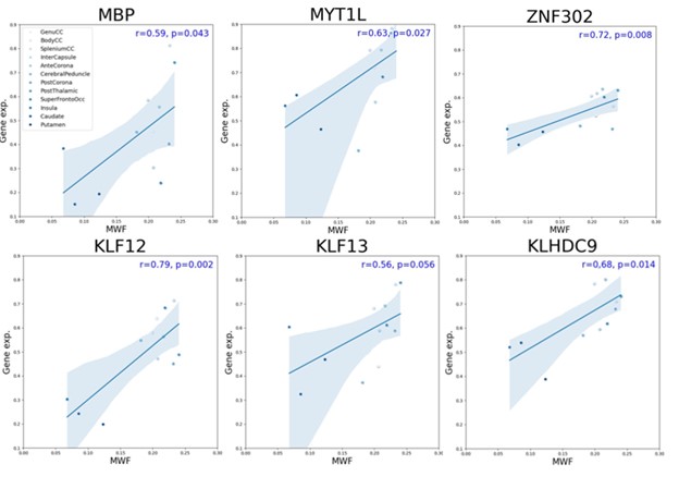
Correlation analysis: We correlated our regional mean MWF measurement with the aggregated regional gene expression for each brain region. Among the genes exhibiting significant correlations, we examined the genes that are related to myelin, such as Myelin Basic Protein (MBP), Myelin Transcription Factor 1 Like (MYT1L) and other transcription factors that are known to be related to myelin synthesis24.
Results & Discussion
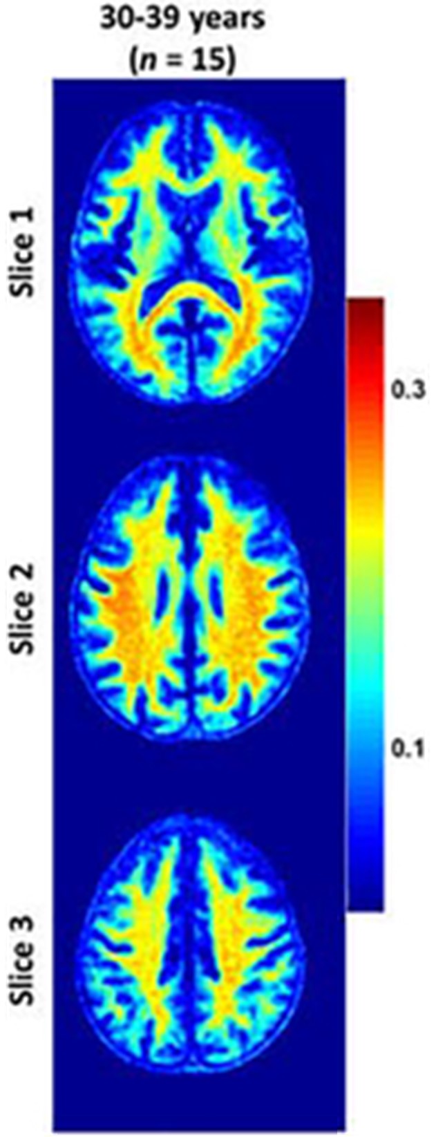
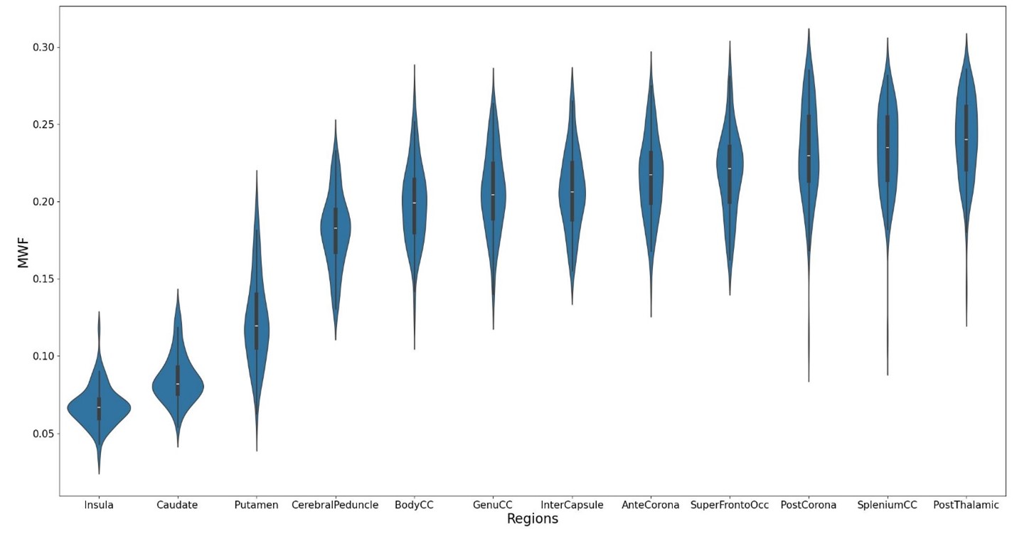
Figure 1 shows the MWF maps in one example subject, where different white matter structures exhibit different MWF values. Figure 2 shows the distributions of MWF from our cohort in different white matter and in the deep gray matter structure. As expected, higher MWF values are associated with the white matter as compared to those in the deep gray matter. Figure 3 shows the correlation between different gene expression in selected regions and respective MWF measurements. We found strong correlation between MWF and genes that are relevant to the myelin structures such as MBP, and those related to the transcription of myelin. Our results suggest that MWF measures derived from BMC-mcDESPOT correlate strongly with the genes associated with myelin transcriptions. Based on our current findings, future studies are also warranted to investigate age-stratifications correlations and infer transcriptional changes with aging.Acknowledgements
This work was supported by the Intramural Research Program of the National Institute on Aging of the National Institutes of Health.References
1. Lemus, H.N., A.E. Warrington, and M. Rodriguez, Multiple sclerosis: mechanisms of disease and strategies for myelin and axonal repair. Neurologic clinics, 2018. 36(1): p. 1-11.
2. Valdés-Tovar, M., et al., Insights into myelin dysfunction in schizophrenia and bipolar disorder. World Journal of Psychiatry, 2022. 12(2): p. 264.
3. Ettle, B., J.C. Schlachetzki, and J. Winkler, Oligodendroglia and myelin in neurodegenerative diseases: more than just bystanders? Molecular neurobiology, 2016. 53: p. 3046-3062.
4. Bouhrara, M., et al., Adult brain aging investigated using BMC-mcDESPOT–based myelin water fraction imaging. Neurobiology of aging, 2020. 85: p. 131-139.
5. Bouhrara, M. and R.G. Spencer, Rapid simultaneous high-resolution mapping of myelin water fraction and relaxation times in human brain using BMC-mcDESPOT. NeuroImage, 2017. 147: p. 800-811.
6. Akhonda, M.A., et al., The effect of the human brainstem myelination on gait speed in normative aging. The Journals of Gerontology: Series A, 2023: p. glad193.
7. Faulkner, M.E., et al., Lower myelin content is associated with lower gait speed in cognitively unimpaired adults. The Journals of Gerontology: Series A, 2023. 78(8): p. 1339-1347.
8. Park, M., et al., Brain myelin water fraction is associated with APOE4 allele status in patients with cognitive impairment. Journal of Neuroimaging, 2022. 32(3): p. 521-529.
9. Bouhrara, M. and R.G. Spencer, Improved determination of the myelin water fraction in human brain using magnetic resonance imaging through Bayesian analysis of mcDESPOT. Neuroimage, 2016. 127: p. 456-471.
10. Choi, J.Y., et al., Evaluation of normal‐appearing white matter in multiple sclerosis using direct visualization of short transverse relaxation time component (ViSTa) myelin water imaging and gradient echo and spin echo (GRASE) myelin water imaging. Journal of Magnetic Resonance Imaging, 2019. 49(4): p. 1091-1098.
11. Laule, C., et al., Myelin water imaging in multiple sclerosis: quantitative correlations with histopathology. Multiple Sclerosis Journal, 2006. 12(6): p. 747-753.
12. Martins, D., et al., Imaging transcriptomics: Convergent cellular, transcriptomic, and molecular neuroimaging signatures in the healthy adult human brain. Cell reports, 2021. 37(13).
13. Fornito, A., A. Arnatkevičiūtė, and B.D. Fulcher, Bridging the gap between connectome and transcriptome. Trends in cognitive sciences, 2019. 23(1): p. 34-50.
14. Rittman, T., et al., Regional expression of the MAPT gene is associated with loss of hubs in brain networks and cognitive impairment in Parkinson disease and progressive supranuclear palsy. Neurobiology of Aging, 2016. 48: p. 153-160.
15. Alsameen, M.H., et al., C-NODDI: a constrained NODDI model for axonal density and orientation determinations in cerebral white matter. Front Neurol, 2023. 14: p. 1205426.
16. Bouhrara, M., et al., Analysis of mcDESPOT- and CPMG-derived parameter estimates for two-component nonexchanging systems. Magn Reson Med, 2015.
17. Bouhrara, M. and R.G. Spencer, Incorporation of nonzero echo times in the SPGR and bSSFP signal models used in mcDESPOT. Magn Reson Med, 2015. 74(5): p. 1227-35.
18. Bouhrara, M. and R.G. Spencer, Improved determination of the myelin water fraction in human brain using magnetic resonance imaging through Bayesian analysis of mcDESPOT. Neuroimage, 2016. 127: p. 456-71.
19. Jenkinson, M., et al., Fsl. Neuroimage, 2012. 62(2): p. 782-790.
20. Eickhoff, S.B., et al., A new SPM toolbox for combining probabilistic cytoarchitectonic maps and functional imaging data. Neuroimage, 2005. 25(4): p. 1325-1335.
21. Sunkin, S.M., et al., Allen Brain Atlas: an integrated spatio-temporal portal for exploring the central nervous system. Nucleic acids research, 2012. 41(D1): p. D996-D1008.
22. Markello, R.D., et al., Standardizing workflows in imaging transcriptomics with the abagen toolbox. elife, 2021. 10: p. e72129.
23. Desikan, R.S., et al., An automated labeling system for subdividing the human cerebral cortex on MRI scans into gyral based regions of interest. Neuroimage, 2006. 31(3): p. 968-980.
24. Bernhardt, C., et al., KLF9 and KLF13 transcription factors boost myelin gene expression in oligodendrocytes as partners of SOX10 and MYRF. Nucleic Acids Research, 2022. 50(20): p. 11509-11528.
Figures