2519
Myelin Water Imaging on MAGNUS (high-performance Mesoscale Anatomy Gradient for Neuroimaging with Ultrafast Scanning) system1GE Healthcare, Mississauga, ON, Canada, 2Technology & Innovation Center, GE HealthCare, Niskayuna, NY, United States, 3Global MR Applications & Workflow, GE HealthCare, Menlo Park, CA, United States, 4Department of Radiology, University of British Columbia, Vancouver, BC, Canada
Synopsis
Keywords: White Matter, Brain, myelin water imaging, Gradient Echo Spin Echo (GRASE))
Motivation: Myelin water fraction (MWF) is conventionally measured using the T2 decay curve. Due to hardware limitations, for spin-echo based sequences, TR and TE times were restricted by SAR and gradient slew rate.
Goal(s): Our objective is to reduce the total scan time by shortening the TR time. A shorter TE time should also capture more signal from the short T2 components of myelin water.
Approach: We implemented myelin water imaging (MWI) on the MAGNUS system with reduced TR and TE times.
Results: When implemented on the MAGNUS system, MWI could be carried out in a shorter time and the MWF were slightly larger.
Impact: The work aims to optimize MWI by shortening scan times. It successfully achieves this, leading to quicker, high-quality MWI acquisition, benefiting medical diagnostics and research efficiency.
INTRODUCTION:
Myelin water imaging (MWI), a magnetic resonance imaging technique capable of resolving the fraction of water molecules located between the layers of myelin, is a valuable tool for investigating both normal and pathological brain structure in vivo both in developing brain and in neurodegenerative conditions. MWI using a spin-echo based MR sequence was considered as the reference method1. MWI based on 3D Gradient and Spin echo (GRASE) has been introduced to allow whole cerebrum imaging in less scan time2. This method has been used to visualize myelination in the brain and spinal cord in vivo3.The head-only MAGNUS MRI system is designed to help researchers image subtle neuro microstructures and changes that conventional MRI systems cannot achieve. It can deliver an unprecedented combination of gradient strength and slew rate for brain imaging4.
In this research, we implemented MWI GRASE sequence on the MAGNUS system. By enabling reduction of TR and TE times, it improves efficiency and image quality. The aim of this work is to explore the benefit of using this advanced MR system to study myelin. This advancement is valuable for medical diagnostics, as it enables quicker, high-quality myelin water imaging.
METHODS:
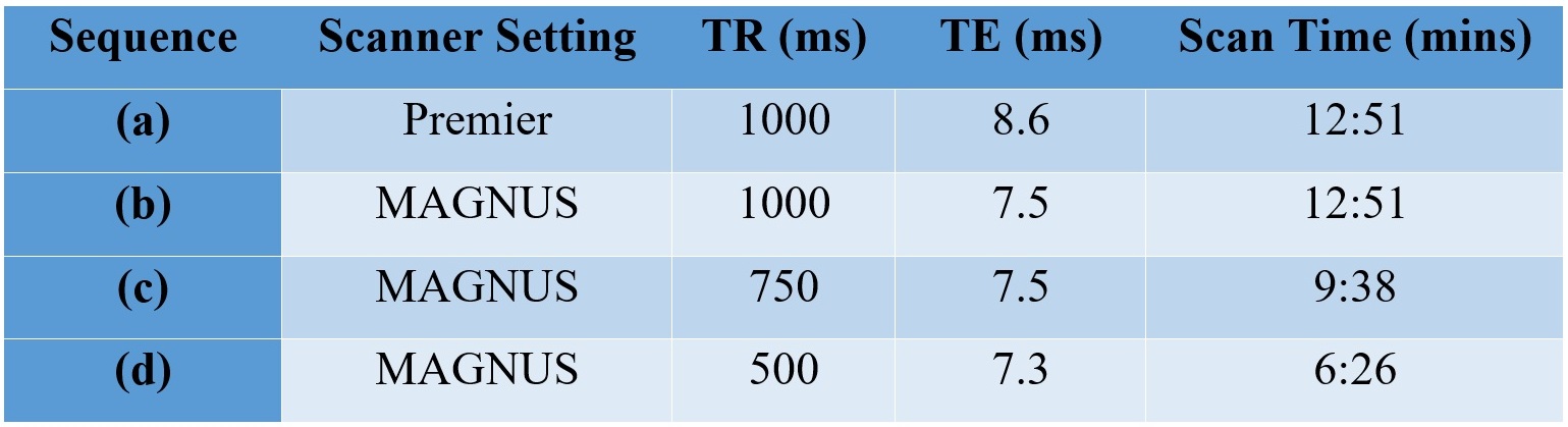
A normal volunteer underwent MRI scanning as per IRB-approved protocols using a 3.0 T MRI system equipped with both SIGNA Premier (Gmax=80 mT/m and SRmax=200 T/m/s) and MAGNUS gradient capabilities (Gmax=300 mT/m and SRmax=750 T/m/s) and standard clinical 2 MVA gradient drivers (GE HealthCare, Niskayuna, NY, USA). A 32-channel phased array head coil (NOVA Medical, Wilmington, MA, USA) was used in all experiments. The GRASE data acquisition featured a field of view (FOV) of 230 x 230 x 100 cm³, 32 echoes, and reconstructed images with a voxel size of 1 x 1 x 2.5 mm³. The readout bandwidth was 100 kHz, and the GRASE factor was set to 3. GRASE data were acquired with 4 different scanner settings, detailed in Table 1. The myelin water imaging from these scans was analyzed using DECAES software5.RESULTS:
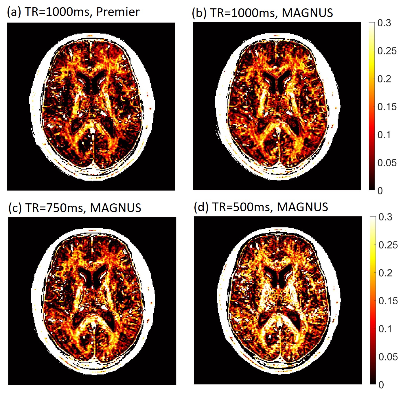
We conducted a comparison of MWF maps from Premier and MAGNUS MRs at TR=1000ms, as illustrated in Figure 1 (a) and (b). For sequence (a), both TR and TE were the minimum allowable due to slew rate and SAR (specific absorption rate) limitations on the Premier system. The MAGNUS system, benefiting from superior gradient performance and lower SAR, achieved a slightly shorter TE. A TR of 1000ms yielded MWF values similar to those reported in the literature for 3T2.Figure 1(c) and (d) show MWF images from MAGNUS with TR of 750ms and 500ms, respectively. Generally, lower TR values resulted in higher MWFs values compared to those obtained with a TR of 1000ms.
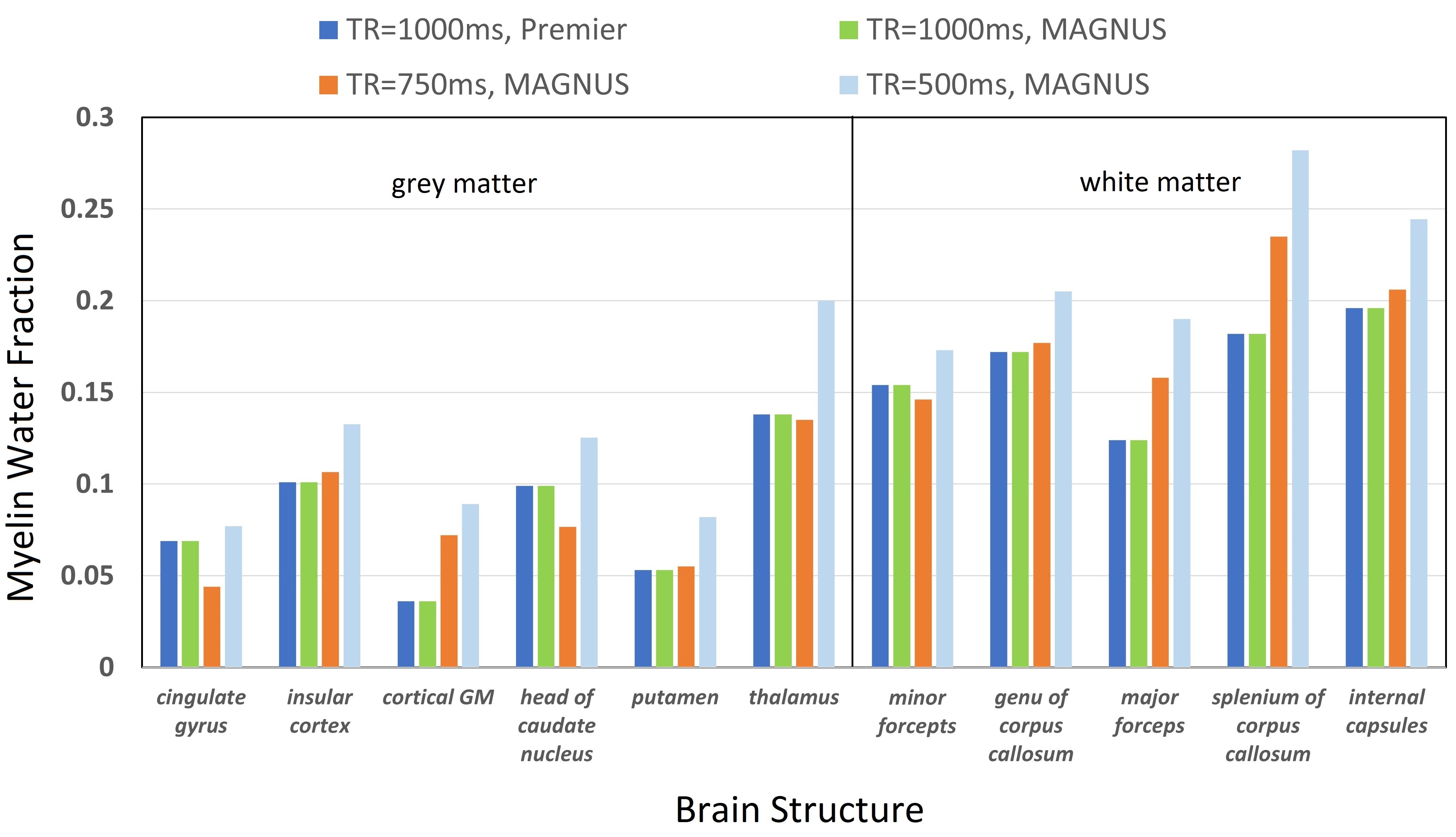
Figure 2 illustrates the mean ROI MWFs from various brain structures, highlighting increased MWFs with shorter TR in both white matter and grey matter regions.
DISCUSSION:
Qualitatively, when TR is the same, a shorter TE will capture more signal from the short T2 component, resulting in enhanced MWF maps. The MWFs from sequence (a) and (b) were similar as shown in Figure 1(a) and (b); likely because the TEs were not too different (8.6ms and 7.5ms).The SAR of MAGNUS is about 20% of the whole body Premier system's SAR level. This allows for substantial TR reduction, leading to significantly shorter scan times. For instance, a TR of 500ms reduces scan time by half while maintaining the same resolution. It is important to note that shorter TR times yield higher MWF values compared to those reported in the literature2. This can be better explained by the four-pool model, where shorter TR saturates intra-extracellular water signals relative to myelin water signals, resulting in a relative increase in the myelin water signal and a consequent increase in apparent myelin water fraction6,7.
With the advanced gradient performance of MAGNUS, future research will explore the potential of under-sampling and reconstruction techniques to enhance SNR efficiency and further reduce scan times in GRASE MWF acquisition8,9. Very recently, full brain MWI has been acquired with 1.7 mm3 resolution with under-sampling pattern in 7:26 minutes10. With the short TR time on MAGNUS, it may be possible to do MWI in about 3 minutes. These advancements are poised to accelerate myelin water imaging, making it more suitable for clinical applications.
CONCLUSION:
In this study, we introduced myelin water imaging on MAGNUS system. The reduced TR enables rapid MWF map acquisition, while shorter TE times capture early myelin signals. These advancements expand the potential applications of myelin water imaging in clinical and neuroscience research.Acknowledgements
We express our gratitude to all the healthy volunteers for their valuable assistance in sequence development and data collection.References
1. Alonso-Ortiz, E., Levesque, I. R. & Pike, G. B. MRI-based myelin water imaging: A technical review. Magnetic Resonance in Medicine 73, 70–81 (2015).
2. Prasloski, T. et al. Rapid whole cerebrum myelin water imaging using a 3D GRASE sequence. Neuroimage 63, 533–539 (2012).
3. MacKay, A. L. & Laule, C. Magnetic Resonance of Myelin Water: An in vivo Marker for Myelin. Brain Plast 2, 71–91 (2016).
4. Foo, T. et al. (ISMRM 2018) MAGNUS: An ultra-high efficiency head-only gradient coil for imaging the brain microstructure. https://archive.ismrm.org/2018/0839.html.
5. Doucette, J., Kames, C. & Rauscher, A. DECAES - DEcomposition and Component Analysis of Exponential Signals. Z Med Phys 30, 271–278 (2020).
6. Manning, A. P., MacKay, A. L. & Michal, C. A. Understanding aqueous and non-aqueous proton T1 relaxation in brain. Journal of Magnetic Resonance 323, 106909 (2021).
7. Zhang, J., Banerjee, S. & MacKay, A. L. TR effect on Myelin Water Imaging. ISMRM https://cds.ismrm.org/protected/23MPresentations/abstracts/2629.html (2023).
8. Lee, J. et al. Artificial neural network for myelin water imaging. Magnetic Resonance in Medicine 83, 1875–1883 (2020).
9. Liu, H. et al. Myelin water imaging data analysis in less than one minute. NeuroImage 210, 116551 (2020).
10. Dvorak, A. V. et al. The CALIPR framework for highly accelerated myelin water imaging with improved precision and sensitivity. Science Advances 9, (2023).
Figures