2516
Cross Site Reproducibility for standard and ultra-high b-value diffusion imaging in high-performance gradient (MAGNUS 3T) MRI systems.1Technology & Innovation Center, GE HealthCare, Niskayuna, NY, United States, 2University of Iowa, Iowa City, IA, United States, 3University of Wisconsin - Madison, Madison, WI, United States, 4GE HealthCare, Waukesha, WI, United States
Synopsis
Keywords: White Matter, Diffusion/other diffusion imaging techniques, High performance gradient inserts, cross-site, repeatability, reproducibility
Motivation: Cross-site reproducibility of ultra-high b-value diffusion MRI across multiple MAGNUS MRI systems is important for multi-site studies
Goal(s): To disentangle the contributions of physiological fluctuations vs. manufacturing tolerances to inform future cross-site studies for advanced and novel brain microstructural modelling and quantification
Approach: A traveling volunteer was recruited and imaged at three different MAGNUS (2nd generation) systems. An expanded multi-shell parameter space ranging from b=500-30,000 s/mm2 was analysed quantitatively to assess cross-site reproducibility.
Results: Statistical comparisons across sites for global white matter and white matter parcels highlight good agreement without harmonization efforts.
Impact: This dataset is expected to lay the ground work for multicenter collaboration for novel and advanced brain microstructural modelling and quantification. It can further be used to evaluate differences across scanners and to show the consistency of pipeline outputs.
Introduction
The 2nd generation MAGNUS[1] platform delivers 300mT/m and 750T/m/s using standard clinical 3.0T system power electronics (Signa Premier/Architect, GE HealthCare, Waukesha, USA), allowing for substantially shorter diffusion encoding pulse-widths, echo-times, with reduced distortion and blurring from shorter EPI echo spacing. This is partly due to higher PNS thresholds achievable compared to whole-body gradient systems [2, 3]. High-performance head-only gradient inserts allows for a broader dMRI parameter space and simplified biophysical models to be explored. These in-turn allow for inferences on the estimation of elusive contrasts such as effective intra-axonal radii, and time-dependent diffusivities.Multicenter trials for dMRI have drawn considerable interest due to the expanding need for statistical power in large-scale brain imaging studies, while a growing number of multi-shell diffusion models bolstered by advances in MR gradient design present an exciting avenue for non-invasive quantification of brain cyto- and myelo-architecture with the potential to generate individually specific estimates of physio-pathological information. Yet cross-site scanner variability including data acquisition tends to confound reliable individual-based analysis of diffusion measures.
In this study we present preliminary findings on cross-site reproducibility with minimal differences between hardware and software versions.
Methods
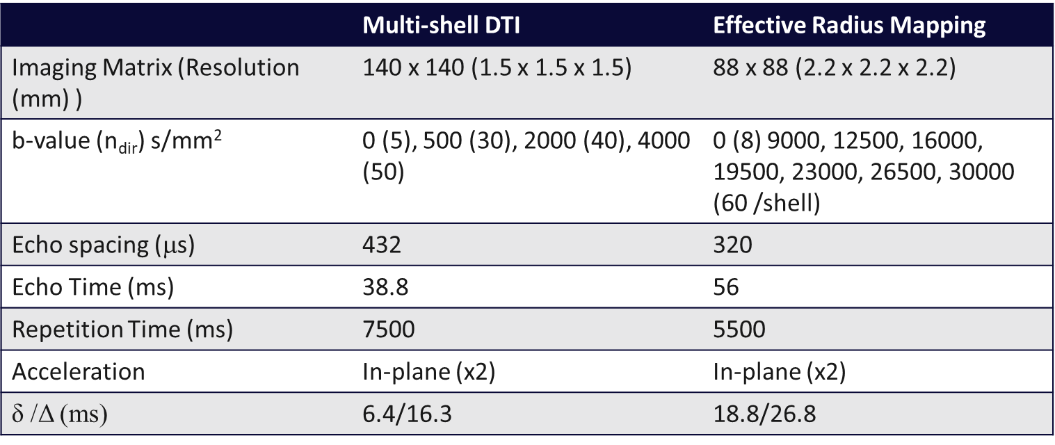
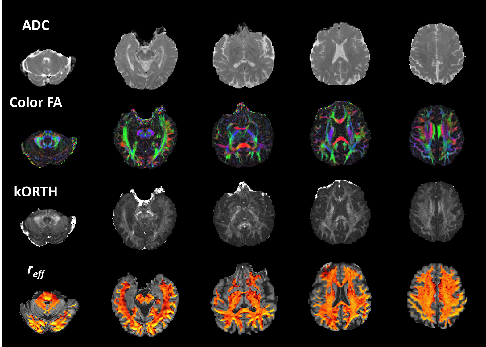
Acquisition: A healthy volunteer was recruited as a cross-site, traveling control subject, and scanned with MAGNUS (Gmax,SRmax=300mT/m,750T/m/s) across three locations, under IRB-approved protocols at TIC (test-retest), U of Iowa, and UW—Madison. A 32-channel phased array head coil (NOVA Medical, Wilmington, MA, USA) was used for all scanning. The subject was scanned at the three sites using the same protocol, with a focus on diffusion tensor-based acquisitions(Table 1). The average time between acquisitions on the three scanners was 1-month. Signal processing for DTI/DKI metrics: Diffusion-weighted images were corrected for eddy current distortion, bulk motion and susceptibility and gradient non-linearity for diffusion encoding[4] followed by generalized spherical deconvolution for denoising [3], using a custom image processing pipeline. Diffusion and kurtosis tensors were fitted using a non-negativity constrained least-squares approach. Signal processing for reff: To mitigate the influence of Rician bias, we adopt a decorrelated phase filtering technique which utilizes filter kernels optimized via spatial noise correlation patterns [5], to output real-valued data (RVD), maintaining a Gaussian noise distribution. RVD was further corrected for distortion, eddy currents, bulk motion and non-linearity of diffusion-encoding gradients in our reconstruction pipeline. The spherical mean signal was modeled to generate a projection of the tail-weighted reff (mm)[6] distribution in the in-vivo brain. Statistical analysis: Whole-brain white matter segmentation along with registration of the JHU-ICBM-DTI-81 White-Matter Labeled Atlas were used to define the white matter segments in subject space, and for statistical assessment of cross-site repeatability. A global white-matter posterior-probability mask with the top 55% of the voxels retained (to minimize partial volume effects) was used to generate correlation analysis. Estimated metrics for tensor, kurtosis, and reff were extracted from the datasets to evaluate reproducibility, by means of Bland-Altman parcel-based analysis. Further cross-site precision was estimated using the repeatability coefficient (CoR) with an expectation value of 95%.Results & Discussion
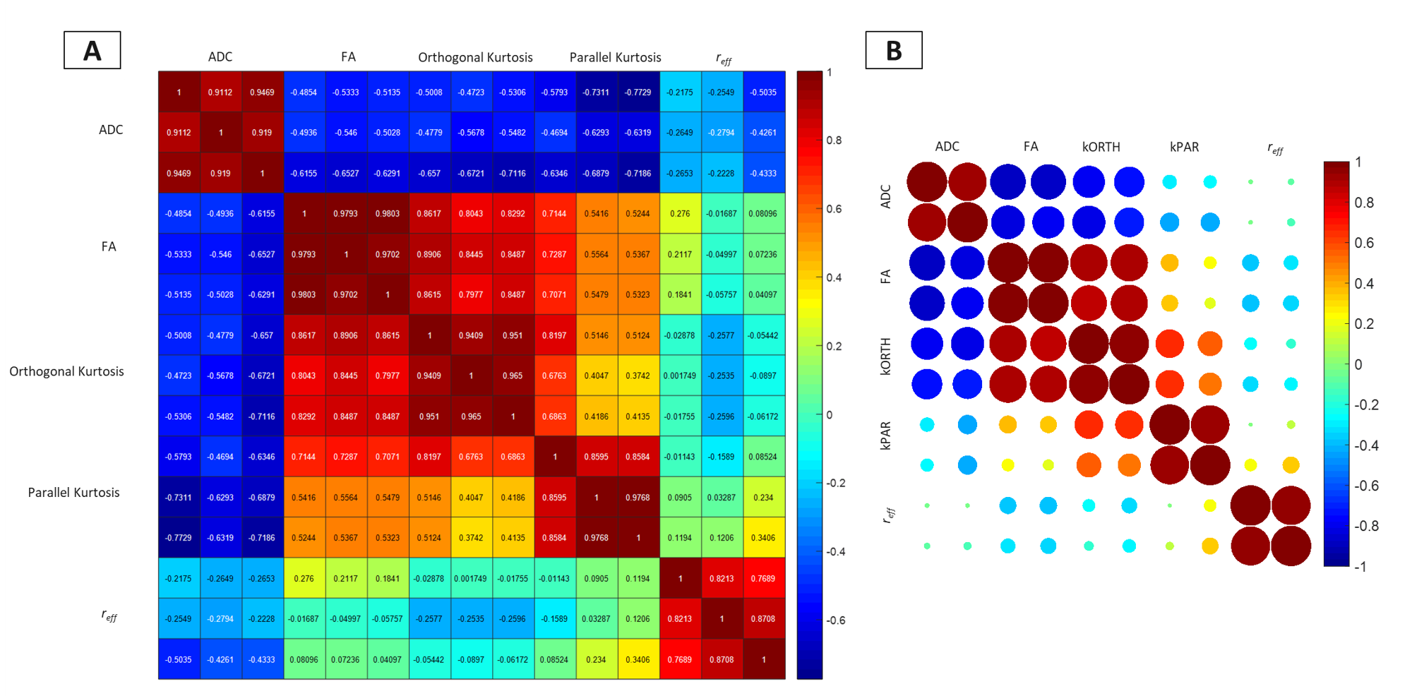
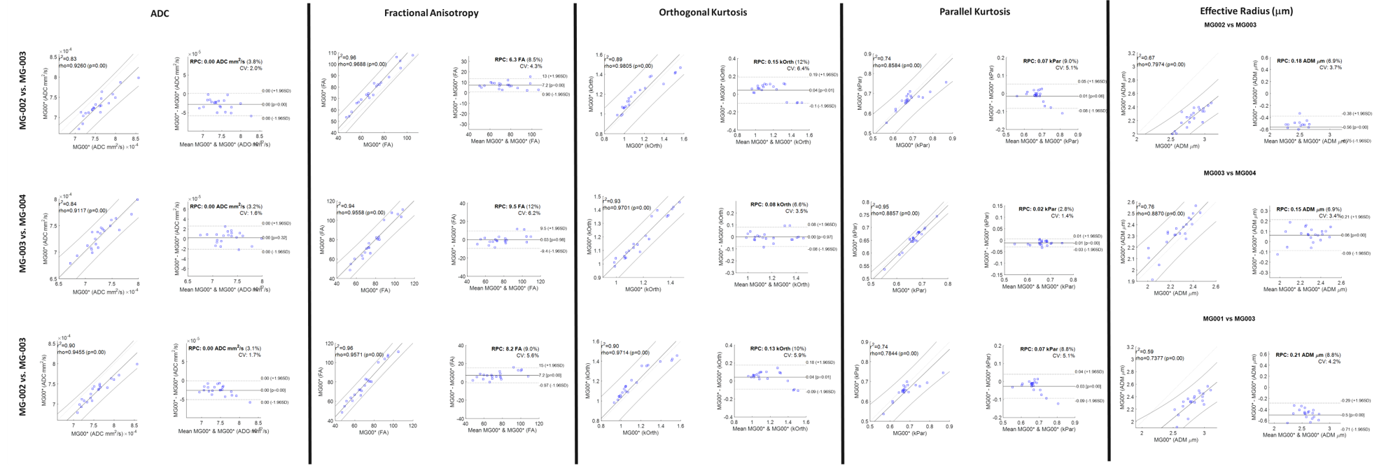
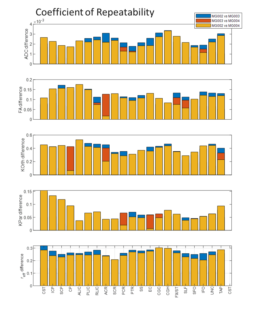
Pearson correlation coefficient was computed between all pairs of diffusion contrasts for whole brain white matter (Figure 3). High correlation coefficients(>0.75) are reported across the three sites – without treatment of the data for harmonization. Same site test-retest showed a high CoV(>0.8). Bland-Altman plots highlight cross-site reproducibility in parcel-wise estimation between the measurements(Figure 4). The absolute mean difference and the 95% confidence intervals are shown. Mean coefficient of variation (COV) of 1.7% (ADC), 5.2 % (FA), 5.2% (kOrth), 3.8% (kPar), and 3.8% (reff) were noted across the sites, while test-retest at the same site showed mean CoV <3%. The tight variation metrics (test-retest and cross-site) highlight that differences are dominated by physiological changes with minimal instrumental variance. The CoR(Figure 5) across the metrics highlights the 95% limits of agreement for the analyzed contrasts. A CoR of ~0.35 was observed with orthogonal kurtosis and effective radii which could be attributed to measurement noise. However, more importantly, the probability of detecting a test-retest change in this volunteer is only 2.5% with the data available across the sites.Conclusion
In this limited evaluation, a lack of systematic differences between scanners was observed, indicating decreased risk of bias in comparing datasets from the three different sites and highlighting tighter CoV in these research systems than has been previously reported [6-10]. Further efforts directed towards harmonization will only serve to minimize operator and protocol bias as cross-site traveling subject recruitment is increased. The cross-site agreement bodes well for future cross-site, large cohort studies – where multi-site studies are needed to achieve the necessary statistical power to make robust inferences regarding pathology.Acknowledgements
Grant funding from NIH S10OD030220, NIH S10OD030415References
1. Foo, T.K.F., et al., Highly efficient head-only magnetic field insert gradient coil for achieving simultaneous high gradient amplitude and slew rate at 3.0T (MAGNUS) for brain microstructure imaging. Magnetic Resonance in Medicine, 2020. 83(6): p. 2356-2369.
2. Tan, E.T., et al., Peripheral nerve stimulation limits of a high amplitude and slew rate magnetic field gradient coil for neuroimaging. Magn Reson Med, 2020. 83(1): p. 352-366.
3. Sperl, J.I., et al., Model-based denoising in diffusion-weighted imaging using generalized spherical deconvolution. Magnetic Resonance in Medicine, 2017. 78(6): p. 2428-2438.
4. Newitt, D.C., et al., Gradient nonlinearity correction to improve apparent diffusion coefficient accuracy and standardization in the american college of radiology imaging network 6698 breast cancer trial. J Magn Reson Imaging, 2015. 42(4): p. 908-19.
5. Sprenger, T., et al., Real valued diffusion-weighted imaging using decorrelated phase filtering. Magn Reson Med, 2017. 77(2): p. 559-570.
6. Veraart, J., et al., The variability of MR axon radii estimates in the human white matter. Human Brain Mapping, 2021. 42(7): p. 2201-2213.
7. Grech-Sollars, M., et al., Multi-centre reproducibility of diffusion MRI parameters for clinical sequences in the brain. NMR Biomed, 2015. 28(4): p. 468-85.
8. Kurokawa, R., et al., Cross-scanner reproducibility and harmonization of a diffusion MRI structural brain network: A traveling subject study of multi-b acquisition. Neuroimage, 2021. 245: p. 118675.
9. Palacios, E.M., et al., Toward Precision and Reproducibility of Diffusion Tensor Imaging: A Multicenter Diffusion Phantom and Traveling Volunteer Study. American Journal of Neuroradiology, 2017. 38(3): p. 537-545.
10. Prohl, A.K., et al., Reproducibility of Structural and Diffusion Tensor Imaging in the TACERN Multi-Center Study. Front Integr Neurosci, 2019. 13: p. 24.
Figures