2514
Hyperplane-based tract parcellations for improved anatomical coherence in tractometry1Medical Image Computing, German Cancer Research Center (DKFZ), Heidelberg, Germany, 2Pattern Analysis and Learning Group, Department of Radiation Oncology, Heidelberg University Hospital, Heidelberg, Germany, 3German Cancer Consortium (DKTK), DKFZ, core center Heidelberg, Heidelberg, Germany, 4Medical Faculty Heidelberg, Heidelberg University, Heidelberg, Germany, 5National Center for Tumor Diseases (NCT), NCT Heidelberg, a partnership between DKFZ and the university medical center Heidelberg, Heidelberg, Germany
Synopsis
Keywords: White Matter, Tractography & Fibre Modelling, Tractometry, Parcellation
Motivation: The subdivision of WM tracts into individual parcels required for tractometry analysis, suffers from blurred parcel borders and wrong assignments of complete tract regions, particularly in fanning tracts.
Goal(s): The goal of this work is to overcome these limitations with a new parcellation approach that yields clearly delineated tract parcels and avoids wrong parcel assignments even in challenging tracts.
Approach: We propose a self-supervised approach based on optimally separating hyperplanes, obtained using large-margin classifiers, to separate tracts into parcels.
Results: We compare our approach to two popular state-of-the-art approaches and clearly show markedly improved results in 24 tracts and 98 subjects.
Impact: A new method to parcellate tracts for fiber tractometry, avoiding frequent errors of state-of-the-art approaches, particularly in complex tracts with a fanning topology. This might lead to improved tractometry analysis and potentially insights that were not possible with previous approaches.
Introduction
Diffusion-MRI-based tractometry has become increasingly popular in recent years. It divides white matter (WM) tracts into smaller parcels and analyzes diffusion metrics in each parcel to provide a detailed picture of microstructural variations within the tract. Tractometry has been used in a variety of applications and is considered the state-of-the-art in tract-specific white matter analysis1-4.To analyze the image along the tract, it is evaluated at the points of each tract fiber and each value is assigned to a parcel depending on its position. There are two main approaches for parcel assignment: one statically resamples the streamlines to n points (static parcellation)5, and the other assigns each value at a streamline point to the closest point on a tract-centerline composed of n points (centerline-based parcellation)6.
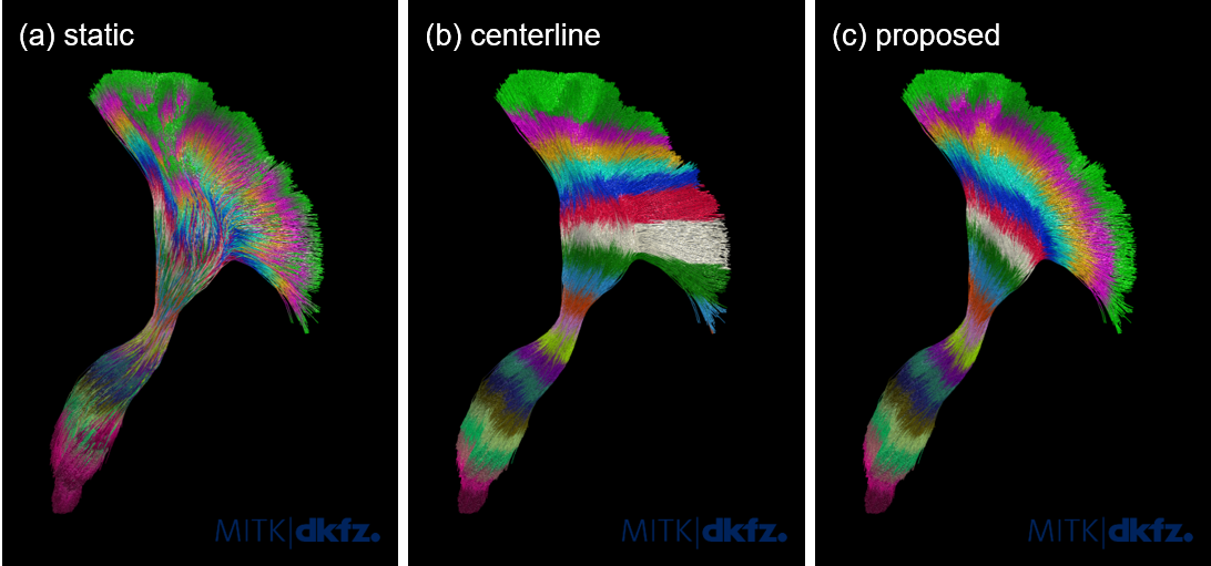
Both parcellation approaches suffer from assignment errors between tract positions and parcels. Static parcellations suffer from misalignment among the individual streamlines, causing blurred parcel borders and image values at the same spatial position to be assigned to different parcels (Figure 1a). Centerline-based parcellations on the other hand wrongly assign complete tract regions, particularly in tracts with a lot of fanning (Figure 1b).
We present a new approach for tract parcellation that yield clearly delineated tract parcels, in contrast to a static parcellation, and that avoids wrong parcel assignments that frequently occur in centerline-based parcellations (Figure 1c).
Methods
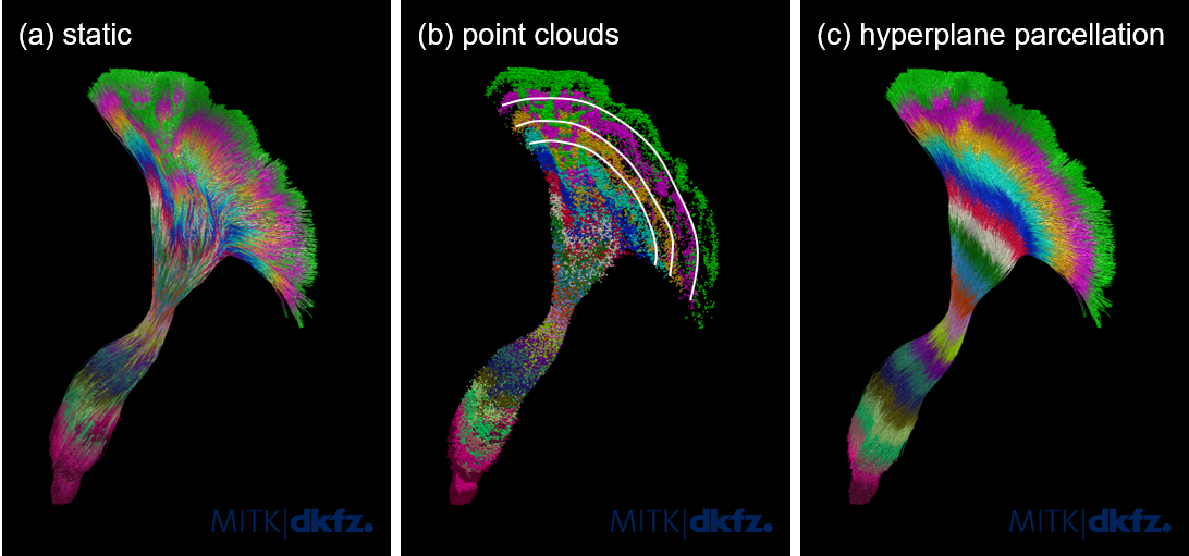
The points of a statically resampled tract can be viewed as partly overlapping point clouds of different classes. We propose to calculate the hyperplanes that optimally separate these point clouds with a minimal amount of wrong samples. These hyperplanes divide the tract into clearly delineated parcels while respecting splitting and fanning tract architectures. See Figures 2a and 2b.The task of finding such an optimal separation is solved by large margin classifiers. For each new tract, we train a support vector machine in a self-supervised way using the point coordinates along the statically resampled streamlines as features and their position index as class label l ∈[1 ...n]7. To ensure short training times, to avoid a bias towards dense tract regions and to avoid redundant data, the initial number of streamlines is reduced to about 500 using QuickBundles8. The trained classifier is then applied to all tract points, yielding the hyperplane-based parcellation (Figure 2c).
Experiments and Results
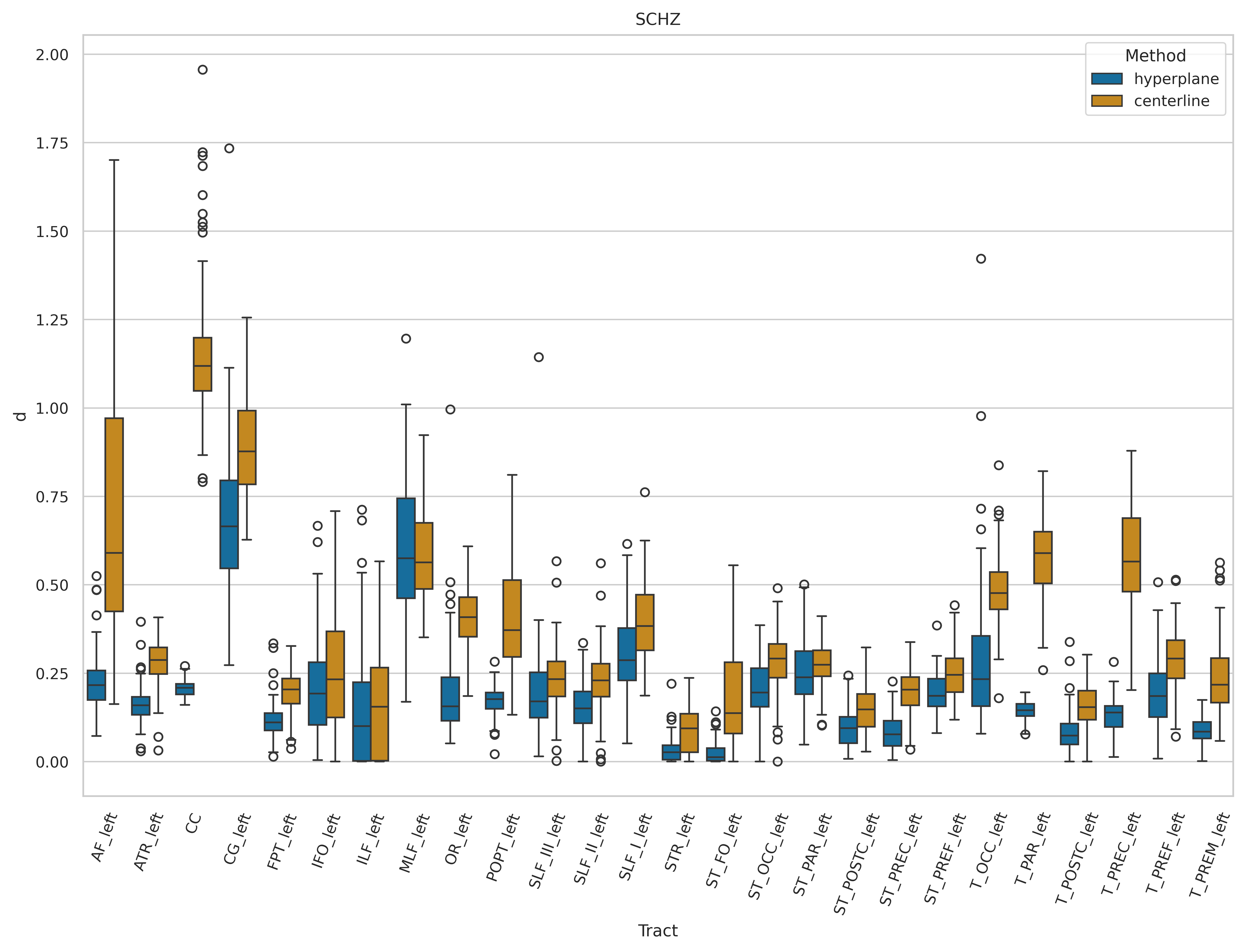
We performed parcellation experiments using the static, centerline-based and proposed hyperplane-based approach on 98 subjects from the OpenfMRI database (https://openfmri.org/dataset/ds000030/)9. Images were corrected for typical artifacts using MRtrix and FSL10,11. TractSeg (https://github.com/MIC-DKFZ/TractSeg/), was used for tract- and tract-endpoint-segmentation as well as for tractography 26 tracts12,13. The number of parcels per tract was estimated automatically to obtain parcels of about 5 voxels thickness.The goals of the presented approach are (1) to obtain clear parcel borders and (2) an improved anatomical coherence. Goal (1) is achieved by definition. Quantitative evaluation of (2) is challenging, since no ground truth is available. Nevertheless, an improved anatomical coherence should be reflected in a lower fraction of fiber endpoints being located outside the first or last parcel. We quantified this as the distance d between the true label and the actual label in the start/end parcels of each tract.
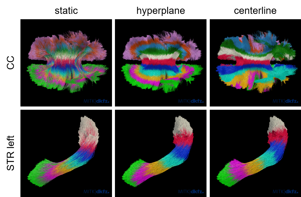
Figure 3 shows that d is indeed significantly lower (paired t-test, no correction for multiple comparisons) using the proposed hyperplane-based parcellation as compared to the centerline-based approach in 24 tracts with no significant difference in 2 tracts (ILF_left, MLF_left). The difference between the approaches is particularly large in tracts with a lot of fanning, e.g., the corpus callosum, and low in straight tracts without a lot of fanning, e.g., STR_left (see Figure 4).
Discussion
We presented a new tract parcellation approach for improved fiber tractometry. Our experiments show that the proposed approach avoids parcel assignment errors due to misaligned streamlines and shows improved anatomical coherence compared to two state of the art parcellation strategies.An aspect that renders the validation of a new tract parcellation approach challenging is the lack of a soundly defined reference for tract parcellations. On a whole-tract level, multiple expert-annotated datasets have been published recently, but no such annotations exist on a parcel level. While our results convincingly demonstrate that hyperplane-based parcellations avoid errors of state-of-the-art, a more quantitative evaluation of this aspect would be desirable. Beyond this aspect, further work will focus on the impact of the new hyperplane-based parcellation on downstream tasks such as tractometry-based group comparison or predictive modelling experiments.
The proposed hyperplane-based parcellation is implemented in RadTract and available on GitHub (https://github.com/MIC-DKFZ/radtract) or as a ready to use python package (https://pypi.org/project/radtract/).
Acknowledgements
This work was supported by the German Research Foundation (DFG) grant numbers MA6340/10-1 and MA6340/12-1References
- Colby, J. B. et al. Along-tract statistics allow for enhanced tractography analysis. NeuroImage 59, 3227–3242 (2012).
- Yeatman, J. D., Wandell, B. A. & Mezer, A. A. Lifespan maturation and degeneration of human brain white matter. Nat. Commun. 5, 4932 (2014).
- Wasserthal, J. et al. Multiparametric mapping of white matter microstructure in catatonia. Neuropsychopharmacology 45, 1750–1757 (2020).
- Yeh, P.-H. et al. Longitudinal changes of white matter microstructure following traumatic brain injury in U.S. military service members. Brain Commun. 4, fcac132 (2022).
- Yeatman, J. D., Dougherty, R. F., Myall, N. J., Wandell, B. A. & Feldman, H. M. Tract profiles of white matter properties: automating fiber-tract quantification. PloS One 7, e49790 (2012).
- Chandio, B. Q. et al. Bundle analytics, a computational framework for investigating the shapes and profiles of brain pathways across populations. Sci. Rep. 10, 17149 (2020).
- Pedregosa, F. et al. Scikit-learn: Machine Learning in Python. J. Mach. Learn. Res. 12, 2825–2830 (2011).
- Garyfallidis, E., Brett, M., Correia, M. M., Williams, G. B. & Nimmo-Smith, I. QuickBundles, a method for tractography simplification. Front. Neurosci. 6, (2012).
- Poldrack, R. A. et al. A phenome-wide examination of neural and cognitive function. Sci. Data 3, 160110 (2016).
- Tournier, J.-D. et al. MRtrix3: A fast, flexible and open software framework for medical image processing and visualisation. NeuroImage 202, 116137 (2019).
- Jenkinson, M., Beckmann, C. F., Behrens, T. E. J., Woolrich, M. W. & Smith, S. M. FSL. Neuroimage 62, 782–790 (2012).
- Wasserthal, J., Neher, P. & Maier-Hein, K. H. TractSeg - Fast and accurate white matter tract segmentation. NeuroImage 183, 239–253 (2018).
- Wasserthal,
J., Neher, P. F., Hirjak, D. & Maier-Hein, K. H. Combined tract
segmentation and orientation mapping for bundle-specific tractography. Med. Image Anal. 58, 101559 (2019).
Figures