2512
Abnormal brain perfusion detected in elderly Long COVID patients more than one year after initial mild infection1Medical College of Wisconsin, Milwaukee, WI, United States, 2The Ohio State University, Columbus, OH, United States
Synopsis
Keywords: Other Neurodegeneration, Arterial spin labelling
Motivation: Long COVID in aging has become a significant public health concern, but the neurological mechanisms of Long COVID symptoms remain elusive.
Goal(s): This study aimed to assess abnormal brain perfusion related to cognitive impairments in Long COVID, who had initial non-hospitalized mild COVID-19.
Approach: : 3D pCASL with Hadamard-encoded multiple PLDs was applied in 30 elderly Long COVID patients in comparison with 28 controls.
Results: Both prolonged arterial transit time (ATT) and reduced ATT-corrected CBF (cerebral blood flow) were detected in Long COVID patients compared with controls, which was linked to cognitive deficits.
Impact: This study demonstrates the usefulness of advanced MR perfusion techniques in evaluating Long COVID patients, where CBF and ATT showed distinctive patterns that correlated with cognitive decline, manifesting more than a year following the initial mild infection.
Introduction
While the majority of COVID-19 cases exhibit either no symptoms or mild symptoms followed by complete recovery, a significant number of individuals with COVID-19 continue to experience a range of symptoms long after overcoming the initial stages of the illness, known as "Long COVID" [1]. Among those affected by Long COVID, there is a noticeable prevalence of neurological symptoms, including "brain fog" [2]. However, the lack of understanding surrounding the specific mechanisms that contribute to the development of these neurological symptoms has impeded the identification of effective treatments for the neurological complications associated with Long COVID [3]. Moreover, there is a growing concern regarding the potential of Long COVID to provoke or expedite neurodegeneration in elderly survivors [4]. The objective of this study was to evaluate the abnormalities in brain perfusion that are linked to cognitive impairments in aging individuals with Long COVID, who had a mild initial non-hospitalized COVID-19.Methods
Thirty Long COVID patients (24 female; mean age=61.60±6.17, age range= 51-74) and 28 age and sex closely matched healthy controls were imaged on a 3T scanner. These Long COVID patients experience persistent neurocognitive symptoms such as cognitive decline and exacerbated attention and focus, which were onset after contracting the initial mild COVID-19. The average duration between the initial COVID-19 diagnosis and MRI scans was 556 days. Advanced 3D pseudo-continuous arterial spin labeling (pCASL) with Hadamard-encoded 7 post-labeling delays (PLDs) was applied. Following the steps described previously, imaging data were preprocessed and the arterial transit time (ATT) and ATT-corrected cerebral blood flow (CBF) were estimated [5]. Voxel-wise group analysis of CBF and ATT was performed to compare Long COVID and control groups. Results were thresholded at a cluster-size corrected p<0.01 (clusterwise a <0.05). Correlations of regional imaging metrics with measures of cognition were further analyzed.Results
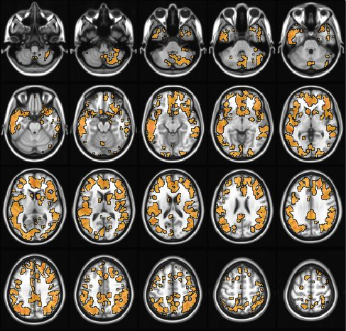
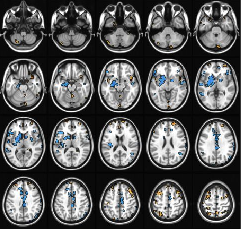
Voxel-wise group comparisons revealed a significant reduction in CBF throughout various cortical regions in individuals suffering from Long COVID (Fig 1). Conversely, an elevation in arterial transit time (ATT) was predominantly observed in the subcortical area and midline cortical region (Fig 2), with a significance level of corrected p<0.01. The regions of interest (ROIs) were determined by identifying the clusters that exhibited a significant disparity in CBF or ATT between Long COVID patients and the control group. Remarkably, at a significance level of p<0.05, the averaged CBF values of the ROIs displayed positive associations with cognitive assessments, encompassing Trial 1 (r=.477), short delay (r=.480), long delay (r=.474), and visual reproduction (r=.464). On the contrary, ATT exhibited negative correlations with these cognitive measures (r=-.483, -.466, -.463, -.469, respectively). Intriguingly, the Long COVID patients demonstrated a distinct relationship between age and regional CBF reduction (r=-.473, p=0.006), as well as ATT elevation (r=.681, p<0.001). Conversely, the control group only exhibited a non-significant trend in this regard.Conclusions
Our initial findings indicate that there may be a fundamental mechanism involving neurovascular abnormalities that contribute to the neurocognitive impairments observed in individuals experiencing Long COVID during the aging process. Our data also suggest that ATT could be an important physiological measure in Long COVID and related neurodegeneration research. However, it is imperative to conduct further investigations in order to validate these results, especially in a larger and more longitudinally designed sample. In light of the emerging concerns regarding the susceptibility of older patients with Long COVID to subsequently developing Alzheimer's disease (AD), the outcomes obtained from research conducted in this particular domain will undoubtedly carry significant clinical implications. Therefore, it is crucial to expand the scope of this research to fully understand the intricate relationship between neurovascular abnormalities, Long COVID, and the potential development of AD in aging individuals.Acknowledgements
This project was supported by NIH grant R21AG077746-01A1 (to Y.W.)References
1. Perlis RH, et al. Prevalence and Correlates of Long COVID Symptoms Among US Adults. JAMA Netw Open. 2022;5(10):e2238804.
2. Subramanian A, et al. Symptoms and risk factors for long COVID in non-hospitalized adults. Nat Med. 2022;28(8):1706-1714.
3. Monje M, Iwasaki A. The neurobiology of long COVID. Neuron. 2022;110(21):3484-3496.
4. Wang L, et al. Association of COVID-19 with New-Onset Alzheimer's Disease. J Alzheimers Dis 2022 20220729. DOI: 10.3233/JAD-220717.
5. Cohen AD, et al. Longitudinal Reproducibility of MR Perfusion Using 3D Pseudocontinuous Arterial Spin Labeling With Hadamard-Encoded Multiple Postlabeling Delays. Journal of magnetic resonance imaging : JMRI 2020; 51: 1846-1853. 2019/12/01. DOI: 10.1002/jmri.27007.
Figures