2510
Diffusion tensor imaging of multiple sclerosis patients with progression independent of relapse activity show increased white matter damage1Translational Imaging in Neurology (ThINk) Basel, Department of Biomedical Engineering, Faculty of Medicine, University Hospital Basel and University of Basel, Basel, Switzerland, 2Department of Neurology, University Hospital Basel, Basel, Switzerland, 3Research Center for Clinical Neuroimmunology and Neuroscience Basel (RC2NB), University Hospital Basel and University of Basel, Basel, Switzerland, 4Department of Health Sciences, University of Genova, Genova, Italy, 5Department of Clinical Research, University Hospital Basel, University of Basel, Basel, Switzerland, 6Clinical Outcomes in Research (CORe), University of Melbourne, Melbourne VIC, Australia, 7Department of Clinical Research, University Hospital Basel, University of Basel, Basel, Basel, Switzerland
Synopsis
Keywords: Multiple Sclerosis, Neurodegeneration, White matter, PIRA
Motivation: Progression independent of relapse activity (PIRA) is the most frequent manifestation of disability accumulation in multiple sclerosis (MS), but the mechanisms leading to PIRA are currently unknown.
Goal(s): To investigate the link between PIRA and white matter degeneration in people with MS.
Approach: To compare the integrity of normal-appearing white matter (NAWM) between patients with MS who experienced PIRA versus stable patients using diffusion tensor imaging (DTI) measures from a clinical-compatible protocol.
Results: Patients with PIRA exhibited significant differences in DTI-derived measures compared to stable patients: reduced fractional anisotropy and increased mean and radial diffusivity in NAWM.
Impact: This study sheds light on the relationship between progression independent of relapse activity (PIRA) and white matter degeneration in people with multiple sclerosis. The results have important implications for understanding the mechanisms of disability progression in relapsing-remitting multiple sclerosis.
Introduction
Multiple sclerosis (MS) is a chronic disease of the central nervous system, characterized by inflammatory, demyelinating, and neurodegenerative processes1. People with relapsing-remitting MS may acquire disability due to focal neuroinflammatory events - resulting in relapses2 - but also through progression independent of relapse activity (PIRA)3. PIRA has been associated with increased rates of brain and cortical atrophy4 along with cervical spinal cord atrophy and a greater prevalence of chronic active lesions. The association between PIRA and changes of white matter (WM) is unknown.Diffusion Tensor Imaging (DTI) quantifies the magnitude and direction of water diffusion in biological tissues5,6. DTI-derived measures, like fractional anisotropy (FA), axial diffusivity (AD), radial diffusivity (RD), and mean diffusivity (MD), have been related to demyelination and axonal injury7,8.
Methods
We selected 258 patients with relapsing-remitting MS from the Swiss MS Cohort9 study with an MRI scan at 3T including fluid attenuated inversion recovery (FLAIR, resolution: 1x1x1mm3), magnetization prepared rapid gradient-echo (MPRAGE, resolution: 1x1x1mm3) and single-shell diffusion-weighted magnetic resonance imaging (DW-MRI, resolution: 1.8x1.8x1.8mm3; 20 b-value=1000 s/mm2 and 10 b-value=0 s/mm2), and a 4-year clinical follow-up. We identified patients with at least one PIRA event (defined as an Expanded Disability Status Scale (EDSS) increase of ⩾1.5 if baseline EDSS=0; ⩾1.0 if 1.0-5.5; ⩾0.5 if >5.5, confirmed after ⩾ 6 months, in the absence of relapses10).DW-MRI were preprocessed using tools from FSL11 and MRtrix312 performing the following steps: denoising and correction of ringing artifacts, eddy current distortions, misaligning artifacts, and bias field. We generated maps of the DTI-derived measures FA, AD, MD, and RD, of normal-appearing WM (NAWM). White matter lesions were semi-automatically segmented from T2 hyperintensities on FLAIR.
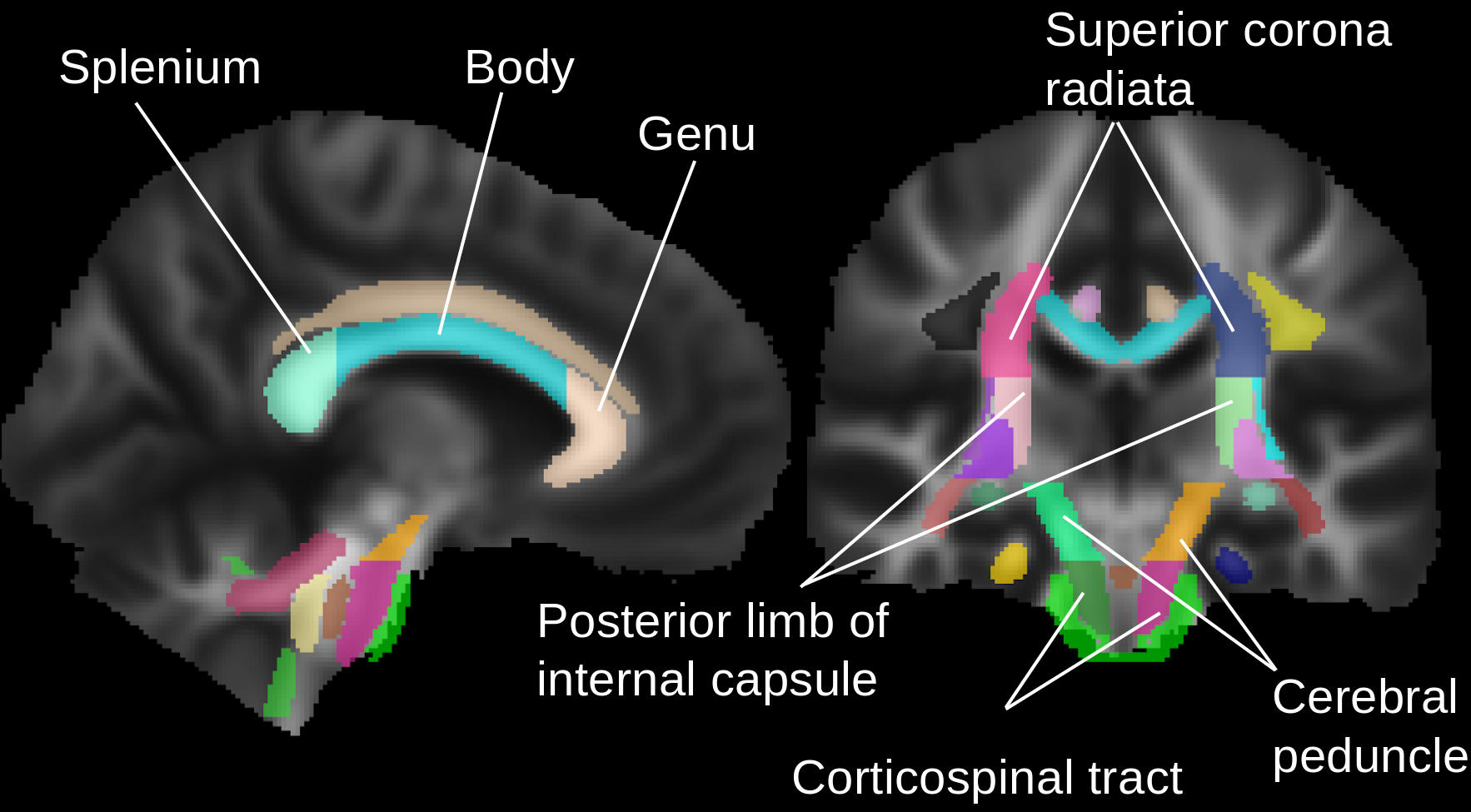
We conducted two studies. First, we used the JHU DTI-based WM atlas13 to assess the microstructural properties of the corpus callosum (CC) and motor tracts (MTs), see selected regions of interest (ROI) in Figure 1. We compared DTI-derived measures on NAWM between patients with PIRA (n=39, 74% female; mean±SD age 50.3±12.0y) and stable patients (n=219, 63% female; 48.0±11.2y). To account for the differences in the groups, we computed propensity-score weights (using age, sex, disease duration, T2-lesion volume, presence of relapses in the last 2 years, and treatment as criteria) that we used in a linear model to compare the groups (using age, sex, disease duration, T2-lesion volume, presence of relapses in the last 2 years, and treatment as covariates). p-values were adjusted for multiple comparisons with the false-discovery rate.
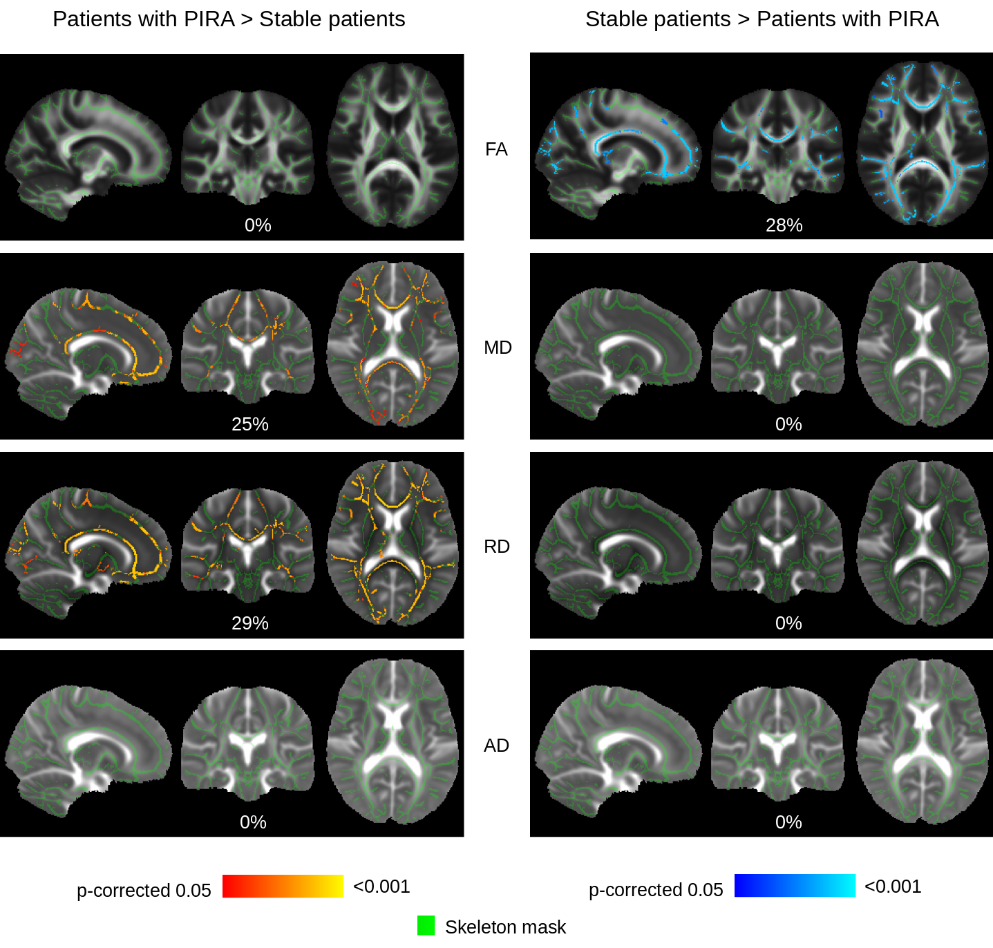
Second, we used Tract-Based Spatial Statistics (TBSS)14 to perform voxelwise cross-subject statistical analyses15 of the DTI-derived measures. TBSS projects subjects' FA data onto a mean FA tract skeleton; by creating a distance map which was used to project MD, RD, and AD into the tract skeleton. We also projected T2-lesion maps to the tract skeleton, to exclude lesional regions, to perform the analyses on the NAWM. We used age, sex, disease duration, lesion volume, presence of relapses in the last 2 years, and treatment as covariates. We used Threshold-Free Cluster Enhancement16 and corrected for multiple comparisons.
Results
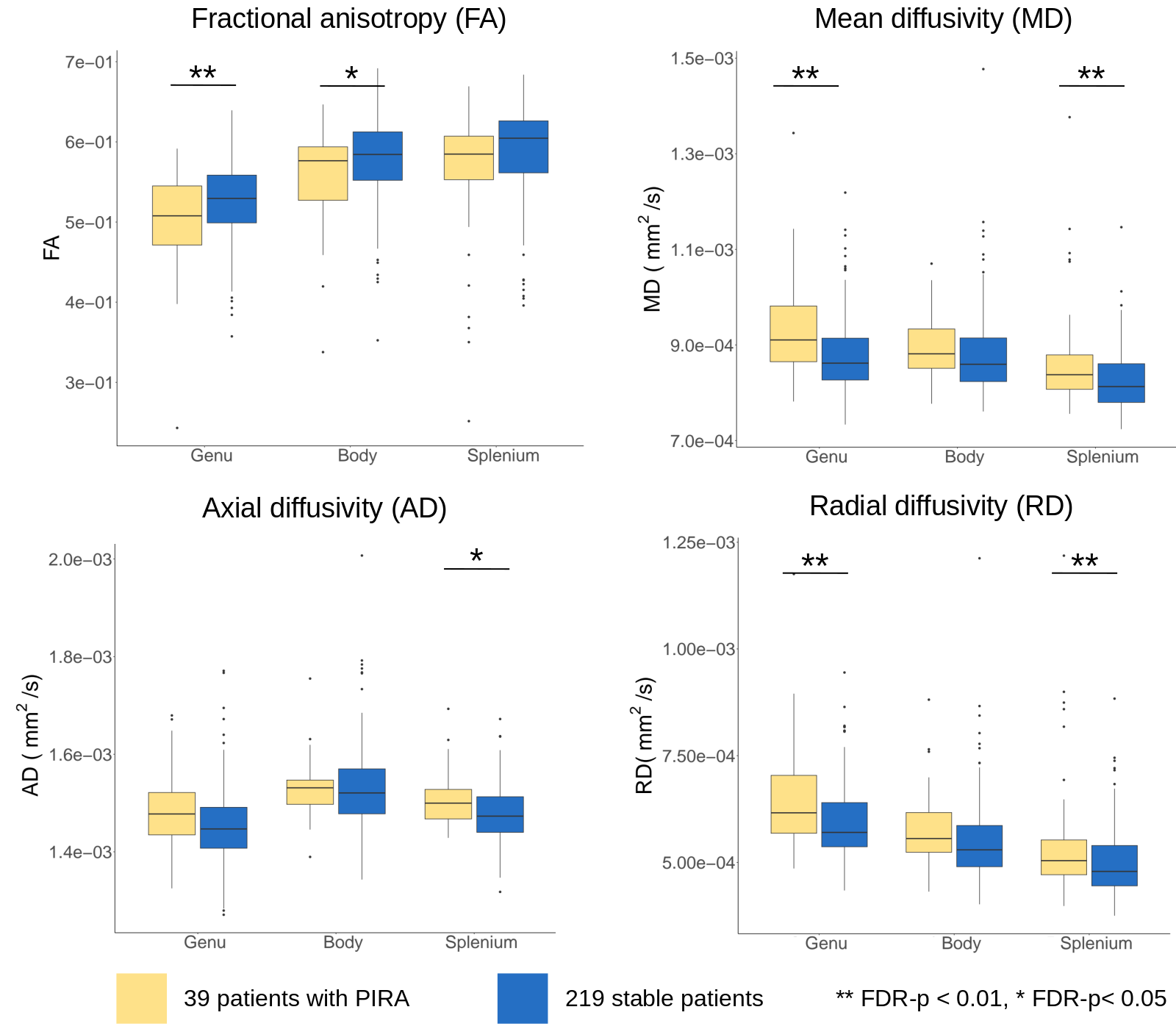
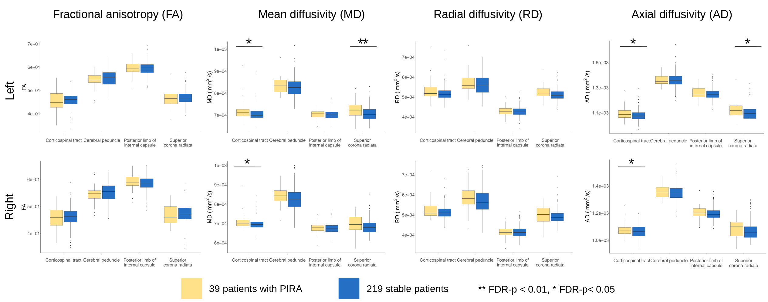
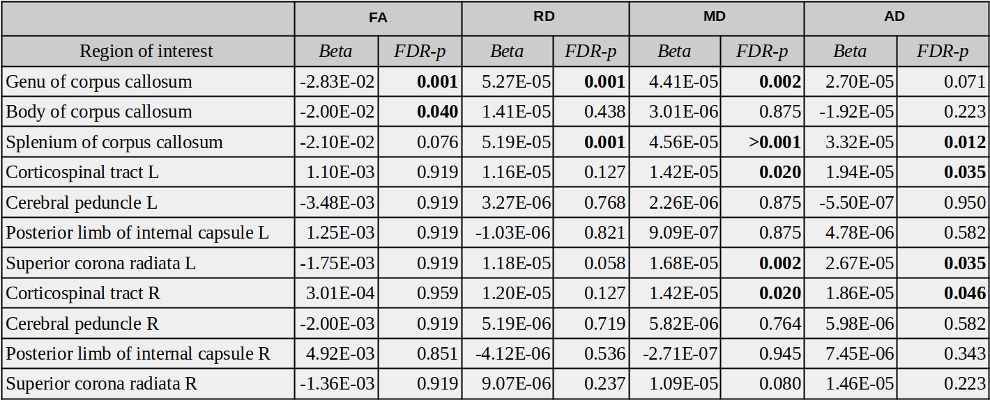
Table 1 shows the results of the ROI analysis. In patients with PIRA, we observed a reduction of FA in the body of CC (p<0.05) and in the genu (p<0.01) and an increase of MD (p<0.01) and RD (p<0.01) in the genu compared to stable patients. We also observed an increase of MD (p<0.01), RD (p<0.01), and AD (p=0.01) in the splenium (Figure 2). We observed a trend toward a decrease in FA and an increase in RD, MD, and AD in MTs; MD and AD were increased in the left (p=0.02 and p=0.03) and right (p=0.02 and p=0.04) corticospinal tract and in the left superior corona radiata (p<0.01 and p=0.03), see Figure 3.In the voxelwise analysis of the TBSS we observed similar trends: 28% of the voxels in the skeleton presented significantly higher FA values in stable patients compared to patients with PIRA. Patients with PIRA presented significantly greater values in 25% of the voxels of the skeleton for MD, and in 29% of the voxels for RD (Figure 4).
Conclusions
Patients with PIRA showed alterations compatible with increased degeneration in parts of the CC and in MTs compared to patients without PIRA. Similar results were obtained on the voxelwise analysis performed on the mean FA tract skeleton, suggesting Wallerian degeneration17 as a possible contributor to the development of disability accumulation independent of relapse activity.Acknowledgements
No acknowledgement found.References
- Stadelmann C, Wegner C, Brück W. Inflammation, demyelination, and degeneration: recent insights from MS pathology. Biochim Biophys Acta. 2011;1812 (2):275-282. doi:10.1016/j.bbadis.2010.07.007.
- Lublin FD, Baier M, Cutter G. Effect of relapses on development of residual deficit in multiple sclerosis. Neurology. 2003;61(11):1528-1532. doi:10.1212/01.WNL.0000096175.39831.21.
- Müller J, Cagol A, Lorscheider J, et al. Harmonizing Definitions for Progression Independent of Relapse Activity in Multiple Sclerosis: A Systematic Review. JAMA Neurol. Published online October 02, 2023. doi:10.1001/jamaneurol.2023.3331
- Cagol A, Schaedelin S, Barakovic M, et al. Association of Brain Atrophy With Disease Progression Independent of Relapse Activity in Patients With Relapsing Multiple Sclerosis. JAMA Neurol. 2022;79(7):682–692.
- Basser, P. J., Mattiello, J., and LeBihan, D. MR diffusion tensor spectroscopy and imaging. Biophysical journal, 1994;66(1):259–267. S0006-3495(94)80775-1[PII].
- Pierpaoli, C., Jezzard, P., Basser, P. J., Barnett, A., and Di Chiro, G. Diffusion tensor MR imaging of the human brain. Radiology, 1996;201(3):637–648.PMID: 8939209.
- Sun, S. W., Liang, H. F., Le, T. Q., Armstrong, R. C., Cross, A. H., & Song, S. K. Differential sensitivity of in vivo and ex vivo diffusion tensor imaging to evolving optic nerve injury in mice with retinal ischemia. NeuroImage, 2006;32(3), 1195–1204. S1053-8119(06)00540-4.
- Kolasa, M, Hakulinen, U, Brander, A, et al. Diffusion tensor imaging and disability progression in multiple sclerosis: A 4-year follow-up study. Brain Behav. 2019; 9:e01194. https://doi.org/10.1002/brb3.1194
- Disanto G, Benkert P, Lorscheider J, Mueller S, Vehoff J, Zecca C, Ramseier S, Achtnichts L, Findling O, Nedeltchev K, Radue EW, Sprenger T, Stippich C, Derfuss T, Louvion JF, Kamm CP, Mattle HP, Lotter C, Du Pasquier R, Schluep M, Pot C, Lalive PH, Yaldizli Ö, Gobbi C, Kappos L, Kuhle J; SMSC Scientific Board. The Swiss Multiple Sclerosis Cohort-Study (SMSC): A Prospective Swiss Wide Investigation of Key Phases in Disease Evolution and New Treatment Options. PLoS One. 2016 Mar 31;11(3):e0152347. doi: 10.1371/journal.pone.0152347. PMID: 27032105; PMCID: PMC4816556.
- Kappos L, Butzkueven H, Wiendl H, et al. Tysabri® Observational Program (TOP) Investigators. Greater sensitivity to multiple sclerosis disability worsening and progression events using a roving versus a fixed reference value in a prospective cohort study. Mult Scler. 2018;24 (7):963-973. doi:10.1177/1352458517709619.
- Jenkinson M., Beckmann C.F., Behrens T.E., Woolrich M.W., Smith S.M.. FSL. NeuroImage, 2012;62:782-90.
- Tournier J-D, Smith R, Raffelt D, Tabbara R, Dhollander T, Pietsch M, et al. MRtrix3: a fast, flexible and open software framework for medical image processing and visualisation. Neuroimage 2019;202:116137.
- Mori et al., MRI Atlas of Human White Matter. Elsevier, Amsterdam, The Netherlands (2005)
- S.M. Smith, M. Jenkinson, H. Johansen-Berg, D. Rueckert, T.E. Nichols, C.E. Mackay, K.E. Watkins, O. Ciccarelli, M.Z. Cader, P.M. Matthews, and T.E.J. Behrens. Tract-based spatial statistics: Voxelwise analysis of multi-subject diffusion data. NeuroImage, 31:1487-1505, 2006.
- Winkler AM, Ridgway GR, Webster MA, Smith SM, Nichols TE. Permutation inference for the general linear model. NeuroImage, 2014;92:381-397.
- Smith S.M., Nichols T.E. Threshold-free cluster enhancement: addressing problems of smoothing, threshold dependence and localisation in cluster inference NeuroImage, 2009:44 (1), pp. 83-98
- Ciccarelli O, Werring DJ, Barker GJ, et al. A study of the mechanisms of normal-appearing white matter damage in multiple sclerosis using diffusion tensor imaging--evidence of Wallerian degeneration. J Neurol. 2003;250(3):287-292. doi:10.1007/s00415-003-0992-5
Figures