2509
Enlarged choroid plexus in relapsing-remitting multiple sclerosis may lead to brain structural changes through glymphatic impairment1Tongji Hospital, Tongji Medical College, Huazhong University of Science and Technology, Wuhan, China
Synopsis
Keywords: Multiple Sclerosis, Multiple Sclerosis
Motivation: The enlarged choroid plexus (CP) in relapsing-remitting multiple sclerosis (RRMS) patients may further cause glymphatic function change and brain tissue damage.
Goal(s): To explore potential links between structural brain changes in RRMS patients and CP volume as well as glymphatic function.
Approach: Brain structural volume was obtained by Freesurfer segmentation. The diffusion tensor image analysis along the perivascular space (DTI-ALPS) index was used to assess the function of glymphatic system.
Results: In both cross-sectional and longitudinal studies, RRMS patients had increased CP volume and decreased DTI-ALPS index. Impaired glymphatic clearance partially mediates the effects of CP enlargement on brain structural changes.
Impact: Enlarged CP and impaired glymphatic system in RRMS patient may cause brain tissue damage. Using CP as a target for interventions may be beneficial to improve glymphatic system function and become a promising treatment strategy.
Introduction
The choroid plexus (CP) involves in the maintenance of neural tissues and the regulation of immune cells as well as neuronal repair[1]. Previous studies have demonstrated that CP volume may be a marker of disease activity in multiple sclerosis (MS)[2; 3]. The "waste clearance" system of the central nervous system (CNS) is defined as the glymphatic system[4]. In MS patients, immune cell infiltration in the perivascular space may interfere with normal glymphatic function. Ineffective clearance of toxic wastes could maintain a pro-inflammatory state and further trigger neuronal injury. Recently, diffusion tensor image analysis along the perivascular space (DTI-ALPS) has provided a noninvasive method for assessing the function of the glymphatic system[5-8].Abnormal cerebrospinal fluid (CSF) secretion and prolonged CSF morphological remodeling may affect glymphatic circulation[9; 10]. Pro-inflammatory factors in CSF may drive the inflammatory response through glymphatic system, thereby further leading to brain tissue damage. The present study aims to explore the changes of CP volume and glymphatic system in relapsing-remitting MS (RRMS) patients, both cross-sectionally and longitudinally, and their potential associations with brain structural changes.
Methods
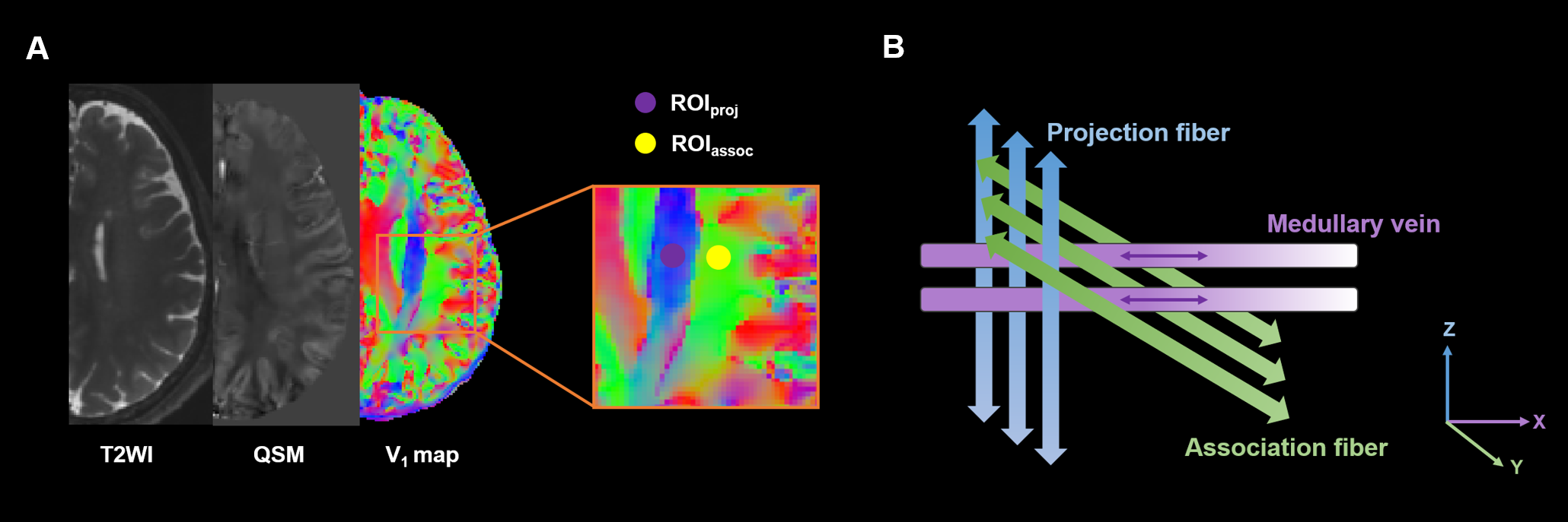
Sixty-five RRMS patients and 48 healthy controls (HC) underwent 3D T1WI, 3D T2 FLAIR, 3D multiple echo gradient echo (mGRE) and DTI. Twenty of RRMS patients underwent a follow-up MRI with a mean interval of 1.29±0.70 years. The brain structural volume was segmented by Freesurfer (package 7.3.2) and expressed as the ratio to total intracranial volume (TIV) multiplied by 1000. Quantitative susceptibility imaging (QSM) was reconstructed from the mGRE sequence[11]. Referring to the QSM, the slice where the veins perpendicular to the lateral ventricles was selected. Two regions of interest were placed in the projection fibers and the associative fibers respectively (Figure 1A). Then, DTI-ALPS index was calculated as the ratio of x-axis diffusivities in projection fiber and in association fiber over the y-axis diffusivities in projection fiber and z-axis diffusivities in association fiber (Figure 1B)[12]. The analysis of covariance and the Wilcoxon signed-rank test were used to compare the differences in MRI metrics. Model-based mediation analysis was performed to explore the possible mediation effect of DTI-ALPS index between CP and brain structural change.Results
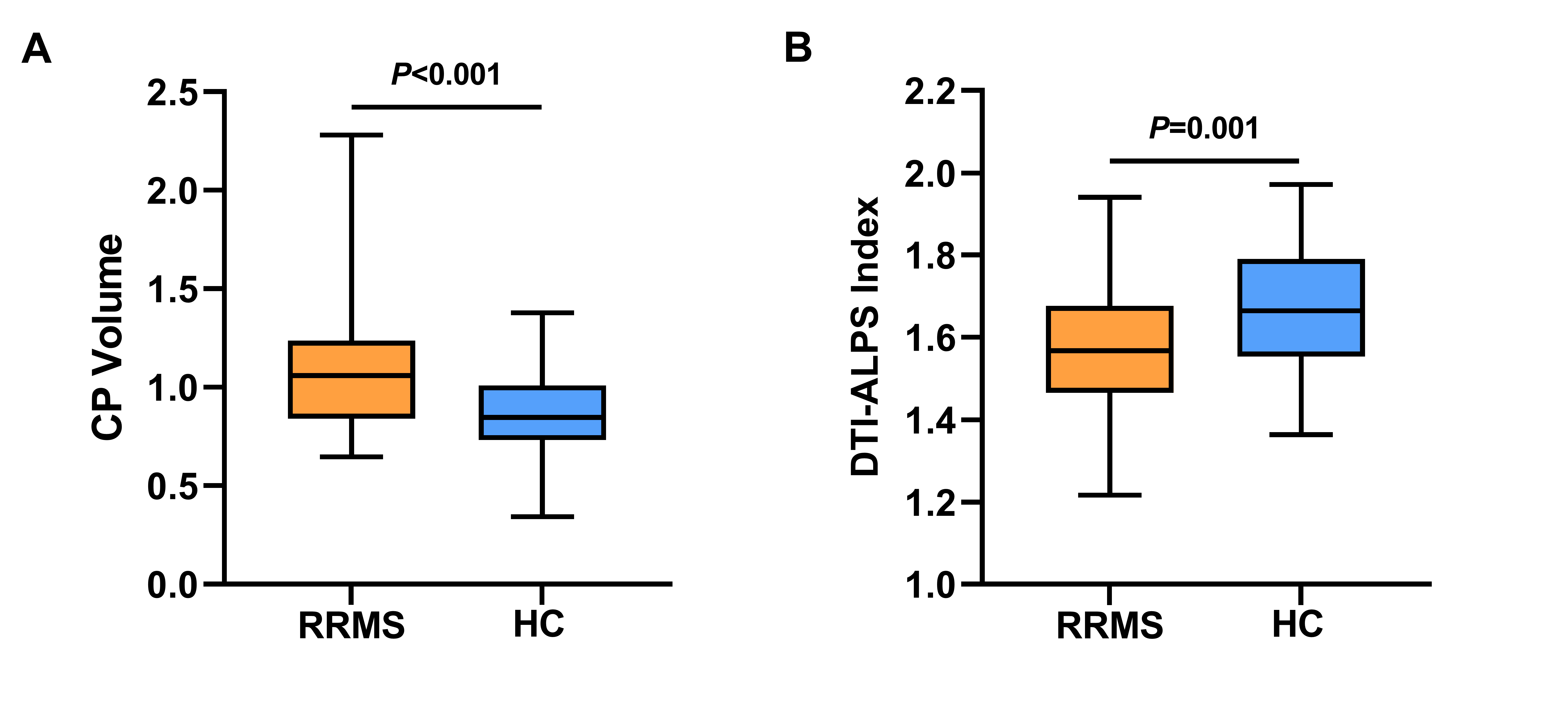
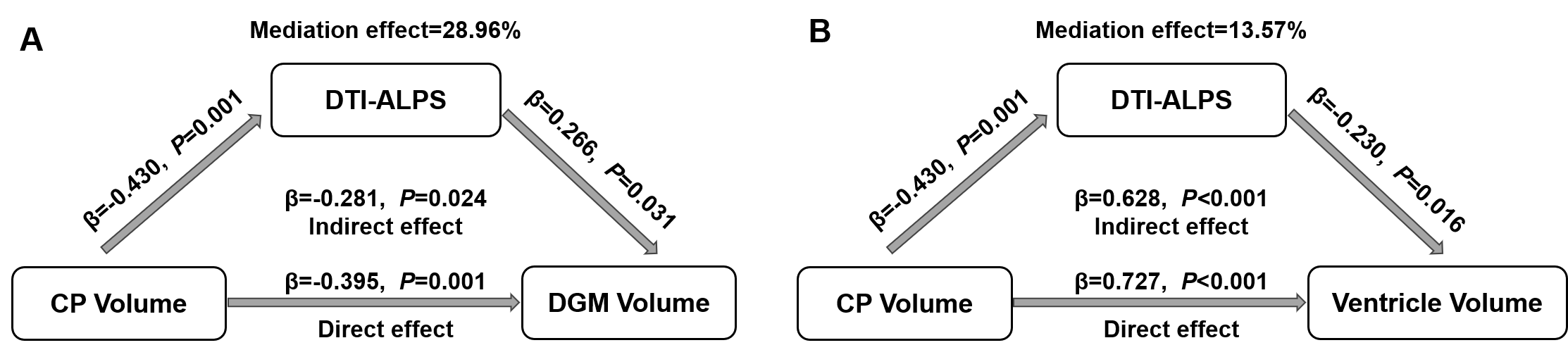
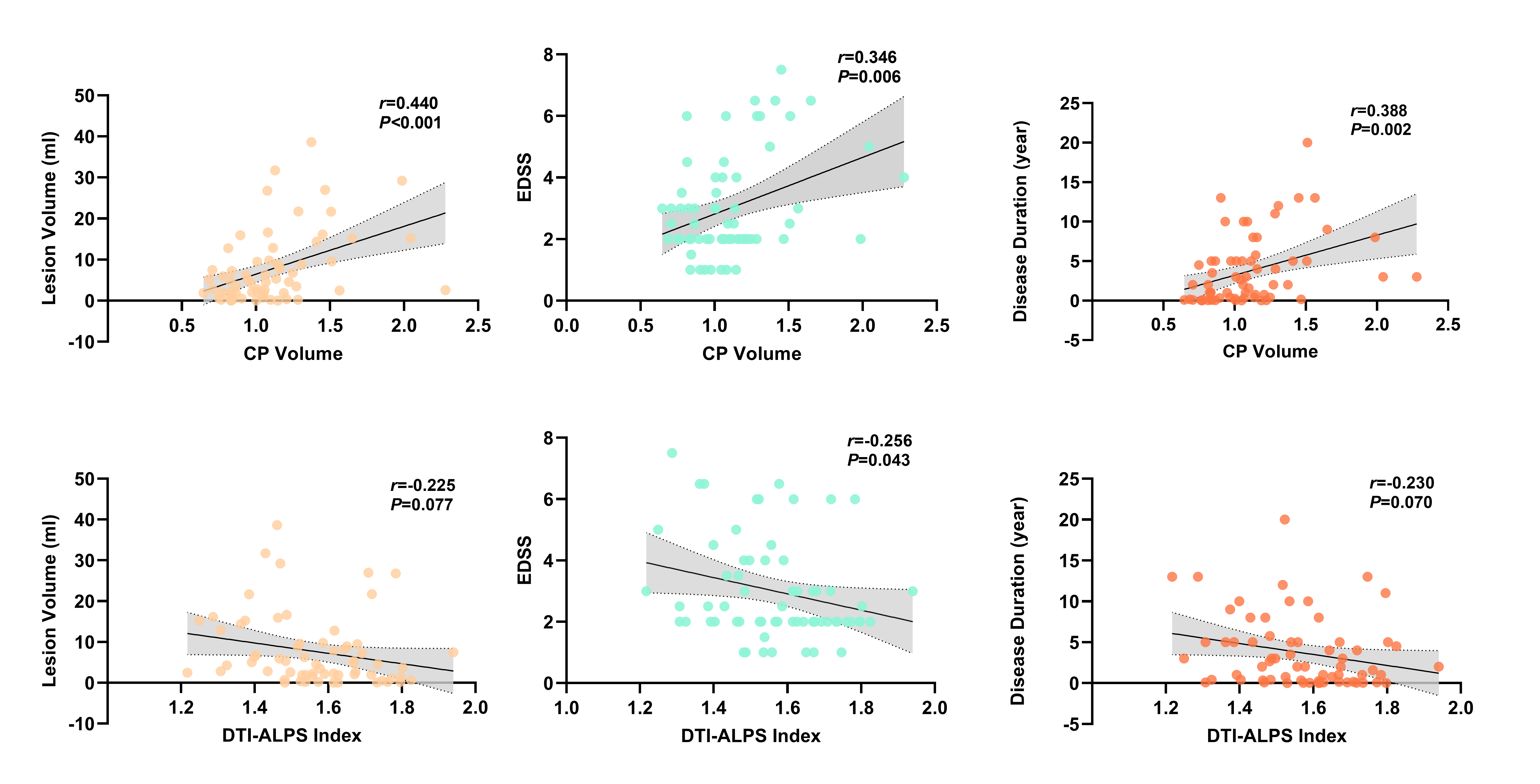
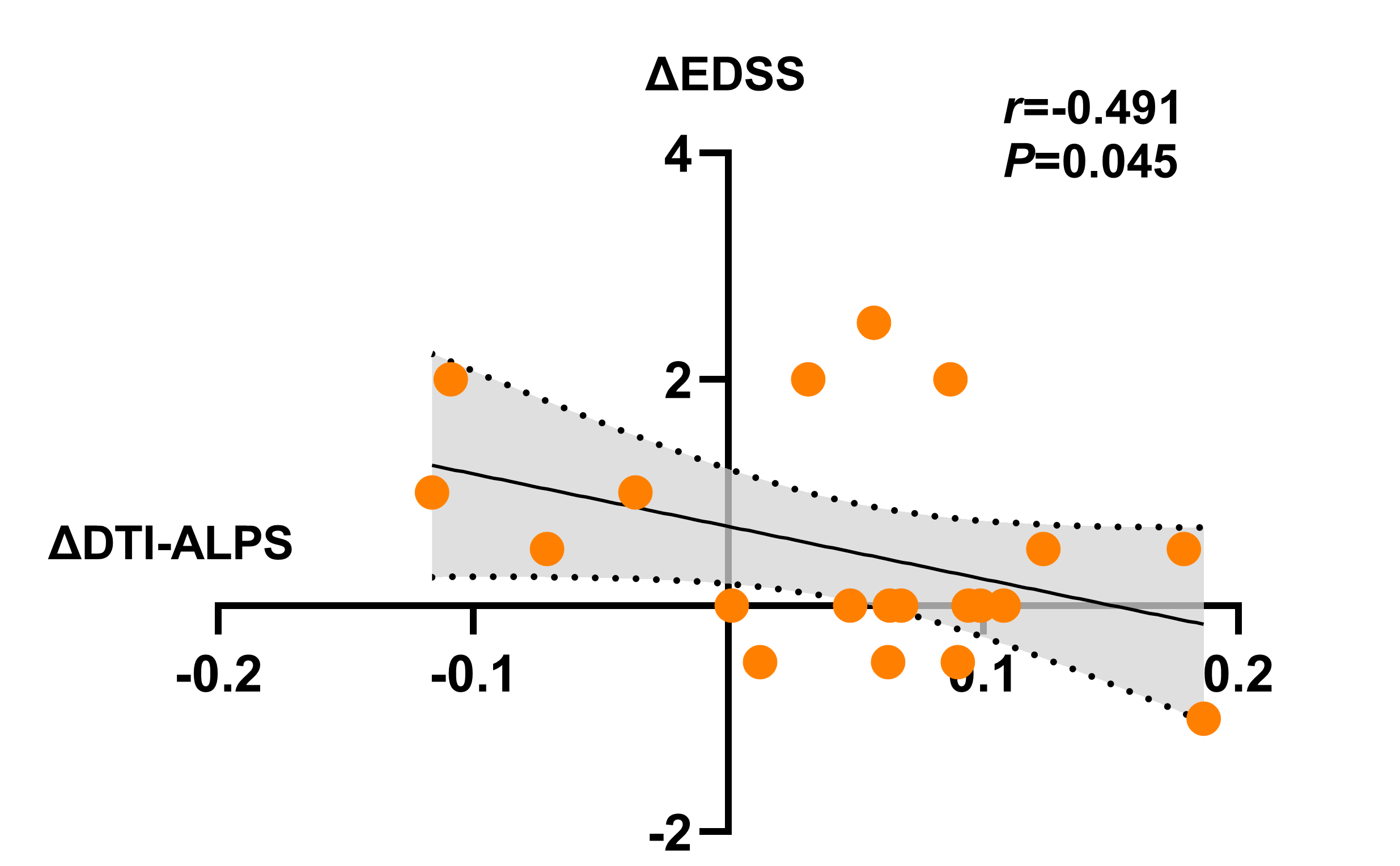
Compared to HC, CP volume in RRMS was significantly increased (P<0.001), and DTI-ALPS index was significantly decreased (P=0.001, Figure 2). The volumes of white matter, thalamus, putamen and pallidus were significantly decreased in RRMS (P<0.05), and the volumes of lateral ventricle and third ventricle were increased (P<0.05). Mediation analysis showed DTI-ALPS index partially mediated the association between CP enlargement and deep gray matter (DGM) volume (combination of thalamus, putamen and pallidum volumes) in RRMS after adjusting for sex and age (Figure 3A), and between CP enlargement and ventricle volume (combination of third ventricle and fourth ventricle volumes) (Figure 3B). In the longitudinal study, CP volume at follow-up was significantly increased compared to that at baseline (P=0.015), and DTI-ALPS index at follow-up was significantly decreased (P=0.016).CP volume and DTI-ALPS index were also significantly correlated with Expanded Disability Status Scale (EDSS) (r=0.346, P=0.006, r=-0.256, P=0.043, Figure 4). Notably, the variation of DTI_ALPS index during the follow-up period were significantly and negatively correlated with the variation of EDSS (r=-0.491, P=0.045, Figure 5).
Discussion
Previous studies have proposed various hypotheses to explain the increased volume of the CP in MS patients. The activation and infiltration of immune cells[13], immune proliferation and migration of endothelial cells[14], increased permeability of the blood-CSF barrier[15], and oxidative stress[16] are all considered as potential contributing factors. Therefore, there may be a correlation between CP volume and the degree of brain tissue inflammation in MS. Due to the accumulation of inflammatory factors in the CSF and the obstruction of fluid outflow, it may affect the CSF-brain interstitial fluid exchange dependent on aquaporins 4 in the foot process of astrocytes, which may have an impact on the function of the glymphatic system. Li et al found that patients with higher CP volume had more significant impaired glymphatic clearance by glymphatic MRI[17].We speculate that CP enlargement may lead to further brain damage through dysfunction of the glymphatic system, especially DGM atrophy and enlarged ventricles. Impairment of the glymphatic pathway may lead to inefficient clearance of toxic proteins and proinflammatory factors, which could diffuse from CSF to periventricular white matter, forming an inflammatory environment, leading to chronic periventricular tissue damage and demyelination[18]. The stimulation of long-term inflammatory response may cause brain structural changes.
Conclusion
CP enlargement and glymphatic impairment may be closely related to the atrophy of DGM and ventricular enlargement in RRMS, and may be a potential indicator for predicting disability progression.Acknowledgements
This work was supported by the National Natural Science Foundation of China [grant numbers U22A20354 and 81730049].References
1 Dani N, Herbst RH, McCabe C et al (2021) A cellular and spatial map of the choroid plexus across brain ventricles and ages. Cell 184:3056-3074.e3021
2 Ricigliano VAG, Morena E, Colombi A et al (2021) Choroid Plexus Enlargement in Inflammatory Multiple Sclerosis: 3.0-T MRI and Translocator Protein PET Evaluation. Radiology 301:166-177
3 Manouchehri N, Stuve O (2021) Choroid plexus volumetrics and brain inflammation in multiple sclerosis. Proc Natl Acad Sci U S A 118
4 Taoka T, Naganawa S (2020) Neurofluid Dynamics and the Glymphatic System: A Neuroimaging Perspective. Korean J Radiol 21:1199-1209
5 Cacciaguerra L, Carotenuto A, Pagani E et al (2022) Mri Evaluation of Perivascular Space Abnormalities in Neuromyelitis Optica. Ann Neurol. 10.1002/ana.26419IF: 11.2 Q1
6 Kamagata K, Andica C, Takabayashi K et al (2022) Association of MRI Indices of Glymphatic System With Amyloid Deposition and Cognition in Mild Cognitive Impairment and Alzheimer Disease. Neurology 99:e2648-2660
7 Si X, Guo T, Wang Z et al (2022) Neuroimaging evidence of glymphatic system dysfunction in possible REM sleep behavior disorder and Parkinson's disease. NPJ Parkinsons Dis 8:54
8 Toh CH, Siow TY (2021) Factors Associated With Dysfunction of Glymphatic System in Patients With Glioma. Front Oncol 11:744318
9 Damkier HH, Brown PD, Praetorius J (2013) Cerebrospinal fluid secretion by the choroid plexus. Physiol Rev 93:1847-1892
10 Choi JD, Moon Y, Kim HJ, Yim Y, Lee S, Moon WJ (2022) Choroid Plexus Volume and Permeability at Brain MRI within the Alzheimer Disease Clinical Spectrum. Radiology 304:635-645
11 Liu Z, Spincemaille P, Yao Y, Zhang Y, Wang Y (2018) MEDI+0: Morphology enabled dipole inversion with automatic uniform cerebrospinal fluid zero reference for quantitative susceptibility mapping. Magn Reson Med 79:2795-2803
12 Taoka T, Masutani Y, Kawai H et al (2017) Evaluation of glymphatic system activity with the diffusion MR technique: diffusion tensor image analysis along the perivascular space (DTI-ALPS) in Alzheimer's disease cases. Jpn J Radiol 35:172-178
13 Rodríguez-Lorenzo S, Konings J, van der Pol S et al (2020) Inflammation of the choroid plexus in progressive multiple sclerosis: accumulation of granulocytes and T cells. Acta Neuropathol Commun 8:9
14 Vercellino M, Votta B, Condello C et al (2008) Involvement of the choroid plexus in multiple sclerosis autoimmune inflammation: a neuropathological study. J Neuroimmunol 199:133-141
15 Ayub M, Jin HK, Bae JS (2021) The blood cerebrospinal fluid barrier orchestrates immunosurveillance, immunoprotection, and immunopathology in the central nervous system. BMB Rep 54:196-202
16 Campbell GR, Mahad DJ (2012) Clonal expansion of mitochondrial DNA deletions and the progression of multiple sclerosis. CNS Neurol Disord Drug Targets 11:589-597
17 Li Y, Zhou Y, Zhong W et al (2023) Choroid Plexus Enlargement Exacerbates White Matter Hyperintensity Growth through Glymphatic Impairment. Ann Neurol 94:182-195
18 Klistorner S, Barnett MH, Parratt J, Yiannikas C, Graham SL, Klistorner A (2022) Choroid plexus volume in multiple sclerosis predicts expansion of chronic lesions and brain atrophy. Ann Clin Transl Neurol 9:1528-1537
Figures