2508
Improved Myelin Content Estimates from Clinical Selective Inversion Recovery Scans via Anisotropic Filtering1Barrow Neurological Institute, Phoenix, AZ, United States
Synopsis
Keywords: Multiple Sclerosis, Multiple Sclerosis
Motivation: Traditionally, Selective Inversion Recovery (SIR) images require long scan times for sufficient SNR, while shorter clinical scans yield low SNR images with noisy pool-size-Ratio (PSR) maps, leading to potential inaccuracies.
Goal(s): An advanced filtering method has been used to improve the precision and accuracy of PSR maps from lower SNR scans.
Approach: An advanced filtering method has been used to improve the precision and accuracy of PSR maps from lower SNR scans.
Results: Initial results demonstrate that this method produces PSR maps comparable to longer, higher SNR scans from shorter clinical scans.
Impact: The implementation of nonlinear anisotropic filtering methods significantly improves the practicality of SIR imaging in a clinical setting, offering quick, accurate myelin content assessments (without blurring tissue boundaries like linear filters) for applications in MS.
Introduction
SIR is a quantitative magnetization transfer (qMT) method that provides information on myelination from conventional inversion recovery sequences1,2. More specifically, SIR provides estimates of the macromolecular pool-size-ratio (PSR), which relates to myelin content changes and disease severity in multiple sclerosis (MS)3,4. In contrast to more common pulsed saturation qMT, SIR does not require independent estimates of ΔB0, B1+, and T1. This results in a method that is easy to implement, making SIR well-suited multi-site/vendors studies. One challenge with SIR is long scan times. To overcome this, we optimized SIR sampling schemes and acquisitions 5,6; however, scan times for whole-brain SIR can often exceed 10 minutes even with these optimized approaches. For many clinical scenarios, this remains challenging and methods are needed to reduce scan times, while maintaining sufficient signal-to-noise ratio (SNR)7. PSR, in particular, has a harmful noise sensitivity that results in biased estimates in low SNR regimes. While linear smoothing techniques can remove noise, these approaches blur spatial information, which may limit our ability to detect focal changes within small lesions. Nonlinear anisotropic smoothing methods have been shown to improve the reliability of diffusion MRI while preserving structural boundaries8. As a result, the primary objective herein is to explore the performance of a nonlinear anisotropic filtering method in clinically relevant SIR scans (7.5-minute whole-brain scan) relative to PSR maps derived from 50% longer scans with higher SNR.Method
MRI data were acquired in six subjects (2 RRMS patients: 2 female, age: 50-54 y.o.; 4 healthy controls: 1 female, age: 23-36 y.o.) using a 3.0-T Ingenia MRI scanner and 32-channel head coil. Clinical SIR data were collected at four optimized combinations of inversion (15, 15, 278, 1007 ms) and predelay times (684, 4171, 2730, and 10 ms) and a 3D turbo spin-echo (TSE) readout, with a field of view (FOV) of 210×210×90 mm3, resolution=2.25 mm isotropic, TE=66 ms, TSE factor = 22, CS-SENSE factor=6, slice oversampling factor=2.1 and scan time≈7.5 minutes. For comparison, higher SNR SIR data acquired using the same parameters except: FOV=210×210×119 mm3, slice oversampling factor=2.3, and scan time≈11.2 minutes. The low SNR SIR images underwent anisotropic diffusion filtering9, with number of iterations=15, kappa (controls edge preservation) = 5 to balance maintaining tissue boundaries against noise reduction, and time step (i.e., convergence speed) = 0.01 to ensure stability in the diffusion process. PSR was then estimated (with Sm=0.83 and kmf=12.5 s-1)5,6 for clinical (low SNR) data with and without the filter. In addition, PSR maps derived from the longer scans (high SNR) using the same approach for comparison. FreeSurfer was used to extract white matter masks for 12 regions of interest (ROIs), focusing primarily on periventricular regions known to be preferentially affected in MS. The mean values were computed for each ROI to compare high and low SNR images, and paired t-tests were used to compare the high SNR SIR data to lower SNR data before and after filtering.Results
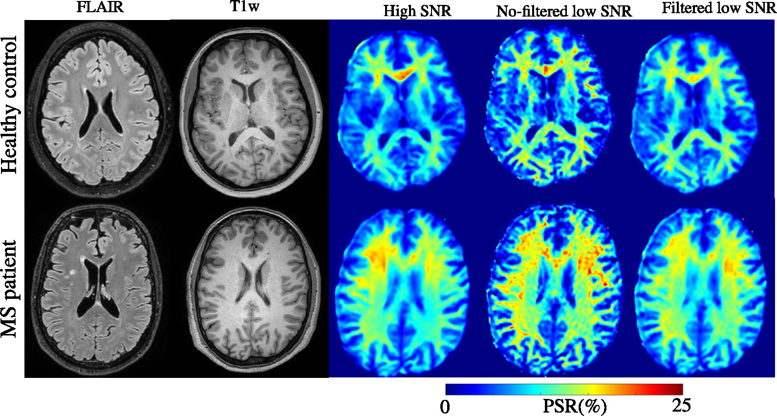
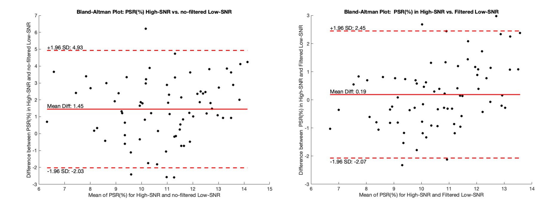
T1-weighted images, FLAIR images, and PSR map calculated from high and low SNR data for a representative control and patient are shown in Figure 1. From the unfiltered low SNR images, a noisy PSR map is observed with systematically overestimated PSR values throughout white matter regions (spurious areas with red hues). After filtering, the clinical PSR maps were comparable to PSR maps from the longer, higher SNR SIR scan without significant loss of boundary information. Bland-Altman plots are shown in Figure 2 to quantitatively compare PSR values calculated from higher SNR with both clinical PSR values before and after filtering. Of note, narrower limits of agreement were observed for the filtered clinical PSR values, suggesting improved consistency after filtering was applied. Statistical tests further substantiated these findings. No significant difference was observed between high SNR and filtered clinical measurements (p=0.17), whereas systematic differences were observed in the clinical data prior to filtering (p=0.01). These results suggest that anisotropic diffusion filtering reduces the systematic bias in PSR estimates introduced by noise, enhancing the reliability of the PSR measurements.Discussion and Conclusion
This study shows that anisotropic diffusion filtering can improve SNR in SIR images while maintaining boundary information, thus enabling determination of PSR maps from shorter, clinically viable scans. Future studies will expand on these findings by exploring the impact of varying filter parameters on different SNR levels and across varied tissue types along with investigating the impact of filtering on scan-rescan reliability. This is a critical step in developing PSR as a biomarker, as it dictates the minimum change needed to reliably detect a true biological change due to progression or treatment response.Acknowledgements
We thank NIH R21 NS125535 and National MS Society RG-2111-3872 for support. We would also like to thank our MRI Tech Sharmeen Maze.References
1. Crescenzo, Francesco, Damiano Marastoni, Anna Isabella Pisani, Agnese Tamanti, Caterina Dapor, Annalisa Colombi, Alessandro Brillo et al. "The Prognostic Value of White-Matter Selective Double Inversion Recovery MRI Sequence in Multiple Sclerosis: An Exploratory Study." Diagnostics 11, no. 4 (2021): 686.
2. Bagnato, Francesca, Simon Hametner, Giulia Franco, Siddharama Pawate, Subramaniam Sriram, Hans Lassmann, John Gore, Seth E. Smith, and Richard Dortch. "Selective inversion recovery quantitative magnetization transfer brain MRI at 7T: clinical and postmortem validation in multiple sclerosis." Journal of Neuroimaging 28, no. 4 (2018): 380-388.
3. Dortch, R.D., Li, K.E., Gochberg, D.F., Welch, E.B., Dula, A.N., Tamhane, A.A., Gore, J.C. and Smith, S.A., 2011. Quantitative magnetization transfer imaging in human brain at 3 T via selective inversion recovery. Magnetic resonance in medicine, 66(5), pp.1346-1352.
4. Wang, P., Sisco, N., Yoo, W., Borazanci, A., Karis, J. and Dortch, R., 2023. Rapid whole‐brain myelin imaging with selective inversion recovery and compressed SENSE. Magnetic resonance in medicine, 89(3), pp.1041-1054.
5. Dortch, R.D., Bagnato, F., Gochberg, D.F., Gore, J.C. and Smith, S.A., 2018. Optimization of selective inversion recovery magnetization transfer imaging for macromolecular content mapping in the human brain. Magnetic resonance in medicine, 80(5), pp.1824-1835.
6. Cronin, M.J., Xu, J., Bagnato, F., Gochberg, D.F., Gore, J.C. and Dortch, R.D., 2020. Rapid whole-brain quantitative magnetization transfer imaging using 3D selective inversion recovery sequences. Magnetic resonance imaging, 68, pp.66-74.
7. Weishaupt, D., Köchli, V.D., Marincek, B., Weishaupt, D., Köchli, V.D. and Marincek, B., 2003. Factors affecting the signal-to-noise ratio.
8. Moraschi, M., Hagberg, G.E., Di Paola, M., Spalletta, G., Maraviglia, B. and Giove, F., 2010. Smoothing that does not blur: effects of the anisotropic approach for evaluating diffusion tensor imaging data in the clinic. Journal of Magnetic Resonance Imaging: An Official Journal of the International Society for Magnetic Resonance in Medicine, 31(3), pp.690-697.
9. Parker, G.J., Schnabel, J.A., Symms, M.R., Werring, D.J. and Barker, G.J., 2000. Nonlinear smoothing for reduction of systematic and random errors in diffusion tensor imaging. Journal of Magnetic Resonance Imaging: An Official Journal of the International Society for Magnetic Resonance in Medicine, 11(6), pp.702-710.
Figures