2504
Linking inter-individual differences in brain structure to post-COVID fatigue1School of Future Technology, Xi’an Jiaotong University, Xi'an, China, 2Department of Medical Imaging, The First Affiliated Hospital of Xi’an Jiaotong University, Xi'an, China, 3Department of Medical Imaging, The Affiliated Hospital of Northwest University·Xi’an No.3 Hospital, Xi'an, China, 4GE HealthCare MR Research, Beijing, China, Beijing, China
Synopsis
Keywords: Neuroinflammation, COVID-19
Motivation: Identify individuals at risk of developing post-COVID fatigue
Goal(s): Propose a "vulnerability" hypothesis of post-COVID fatigue
Approach: A multi-center, prospective, longitudinal study
Results: The GMV in frontal-limbic regions may serve as potential neural underpinnings that identify individuals at risk of developing post-COVID fatigue
Impact: Our findings emphasized the essential role of frontal-limbic system in the development of post-COVID fatigue, which will improve public health interventions by allowing for the early identification of individuals at risk of persistent post-COVID fatigue and facilitating subsequent neuromodulation treatment.
Methods: The present study is a multi-center, prospective, longitudinal study which conducted across nine hospitals. We used structural magnetic resonance imaging and comprehensive neuropsychological assessments in a large and well-defined mildly SAS-COV2 infected cohort (COV+), and contemporary control group (COV-) in the acute phase of the disease (Clinical trial: NCT05745805). All MRI scans were carried out using 3 Tesla scanners. The nine centres used various MRI systems and suppliers. To manage non-biological differences caused by varied multi-centre MRI scanners and imaging acquisition procedures, ComBat harmonisation method was applied in all datasets. T1-weighted 3- dimensional images from the acute phase underwent processing using the Computational Anatomy Toolbox (CAT12) within Statistical Parametric Mapping 12. The regional grey matter volume (GMV) was extracted according to the Human Brainnetome Atlas. Voxel-wise differences in GMV between COV+ and COV- groups during the acute phase were analyzed using independent t-tests, while age, sex, education years, and TIV were considered as confounding variables.
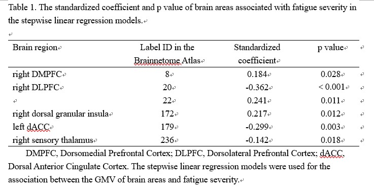
Results: Individuals who experienced mild COVID-19 symptoms (COV+) underwent neuropsychological evaluations (n = 335) and MRI scans (n = 271) within a month of their infection, with 191 (70.5%) being followed up at 3 months. Additionally, 67 healthy controls (COV-) went through the same protocol for recruitment purposes. No changes were observed in the total intracranial volume, grey matter volume, white matter volume, or cerebrospinal fluid volume between the COV+ and COV- group when analyzed for global tissue volume differences at baseline. The GMV of the right DLPFC only had an indirect effect on anxiety through the mediating factor of fatigue, however, no direct effect of DLPFC GMV on anxiety was observed. Furthermore, the mediation model demonstrated that tiredness had a substantial mediating influence on the bond between GMV in the left dACC and anxiety. Specifically, individuals with a lesser GMV in the right DLPFC during the initial assessment showed increased levels of fatigue symptoms 3 months following the infection.
Discussion: Functional integration between cortical and subcortical structures, including the prefrontal and cingulate cortex, basal ganglia, and thalamus, is believed to be a crucial neural circuitry in Multiple Sclerosis (MS) patients who experience fatigue4-5. Therefore, our research provides novel findings indicating that individual differences in GMV, specifically in the cortical structures (DLPFC and dACC), known as the frontal-limbic network, may serve as early neurobiological markers of fatigue susceptibility linked to COVID-19 infection.
Conclusion: We present new evidence concerning the neuroanatomical foundation of vulnerability to fatigue and underscore the significance of fatigue as a significant connector originating from GMV in frontal-limbic areas to co-occurring neuropsychiatric symptoms in the initial stage of recuperation following infection. Our discoveries demonstrate the role of the frontal-limbic system in predisposing individuals to experience post-COVID fatigue.
Acknowledgements
We would like to thank all patients and healthy volunteers that participated in the study.References
1. Ceban F, Ling S, Lui LMW, Lee Y, Gill H, Teopiz KM, et al. (2022): Fatigue and cognitive impairment in Post-COVID-19 Syndrome: A systematic review and meta-analysis. Brain Behav Immun, 101: 93-135.
2. Ortelli P, Ferrazzoli D, Sebastianelli L, Engl M, Romanello R, Nardone R, et al. (2021): Neuropsychological and neurophysiological correlates of fatigue in post-acute patients with neurological manifestations of COVID-19: Insights into a challenging symptom. J Neurol Sci, 420: 117271
3.Heine J, Schwichtenberg K, Hartung TJ, Rekers S, Chien C, Boesl F, et al. (2023): Structural brain changes in patients with post-COVID fatigue: a prospective observational study. EClinicalMedicine, 58: 101874.
4. Derache N, Grassiot B, Mézenge F, Emmanuelle Dugué A, Desgranges B, Constans JM, et al. (2013): Fatigue is associated with metabolic and density alterations of cortical and deep gray matter in Relapsing-Remitting-Multiple Sclerosis patients at the earlier stage of the disease: A PET/MR study. Mult Scler Relat Disord, 2: 362-9.
5. Pravatà E, Zecca C, Sestieri C, Caulo M, Riccitelli GC, Rocca MA, et al. (2016): Hyperconnectivity of the dorsolateral prefrontal cortex following mental effort in multiple sclerosis patients with cognitive fatigue. Mult Scler, 22: 1665-75.
Figures