2502
Diffusion Basis Spectrum Imaging in Midlife Obesity: Associations with Abdominal Adipose Tissue1Washington University in St. Louis, Saint Louis, MO, United States, 2Missouri state university, Columbia, MO, United States, 3Department of Computer Science, State University of New York at Binghamton, Binghamton, NY, United States, 4University of California San Diego, La Jolla, CA, United States
Synopsis
Keywords: Neuroinflammation, Alzheimer's Disease
Motivation: Whether midlife obesity and abdominal adiposity contributes to neuroinflammation, is key to Alzheimer disease (AD) prevention.
Goal(s): We aimed to investigate the association between obesity and abdominal visceral and subcutaneous adipose tissue (VAT and SAT) and neuroinflammation.
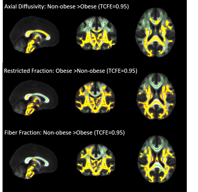
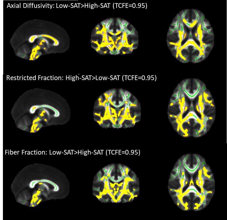
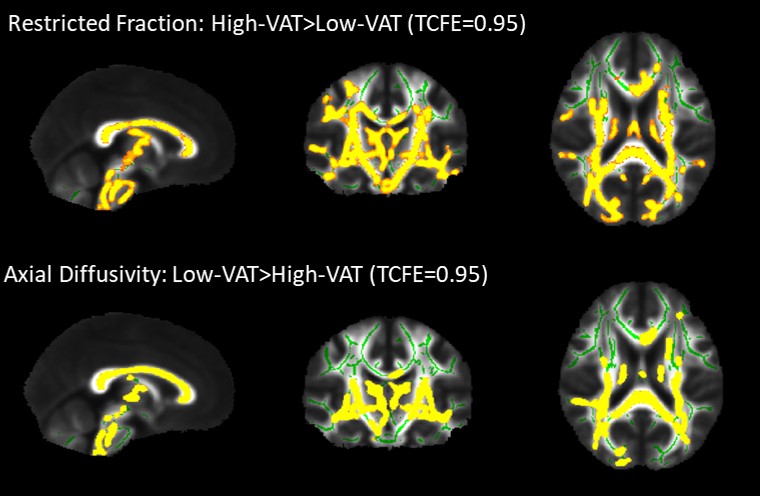
Approach: For this aim, we performed brain and abdominal MRI scans to compare inflammation cellularity, edema, and axonal density using diffusion basis spectrum imaging (DBSI), between the obese vs. non-obese, the high- vs. low-VAT and high- vs. low-VAT groups.
Results: A widespread higher inflammation cellularity and a lower axonal density was observed in the obese vs. non-obese, high-SAT vs. low-SAT, and high-VAT vs. low-VAT females.
Impact: Higher neuroinflammation and lower axonal density in females with obesity and higher abdominal fat, highlights the sex-specific role of midlife abdominal obesity in neuroinflammation and neurodegeneration, which prompts future studies to target body fat for modifying neuroinflammation and AD prevention.
Purpose
Obesity and abdominal adiposity in midlife are shown to increase the risk of Alzheimer disease1. However, it is not clear whether midlife adiposity is associated with increased macrophage infiltration in the brain and neuroinflammation2. Herein, we aimed to look into the associations of obesity, as evidenced by body mass index (BMI) of 30 kg/m2 or higher, and abdominal visceral and subcutaneous fat with brain histology, using diffusion basis spectrum imaging (DBSI) analysis3.Methods
In total, 54 cognitively normal middle-aged subjects (50.46 ± 6.19 years, male: 21 (38.9%), obesity: 32 (59.3 %), BMI: 32.18 ± 6.99 kg/m2) underwent brain and abdominal 3T MRI. Abdominal visceral and subcutaneous adipose tissue (VAT, SAT) were semi-automatically segmented using VOXel Analysis Suite (Voxa). A diffusion spectrum imaging scheme with a total of 98 diffusion samplings was acquired 3. Diffusion basis spectrum imaging maps including fractional anisotropy (FA, overall integrity), axial diffusivity (AD, axonal injury), radial diffusivity (RD, myelin loss), restricted fraction (RF, inflammation cellularity), hindered fraction (HF, extracellular edema), and fiber fraction (FF, axonal density) maps were calculated using in-house software scripted in MATLAB and Statistics Toolbox Release. FMRIB Software Library (FSL) was used to perform eddy current and movement correction and to remove non-brain tissue and DBSI-derived maps were processed using a tract-based spatial statistics (TBSS) pipeline to allow for whole-brain white matter voxel-wise analyses. Using the Randomize tool from FSL, the difference between obese vs. non-obese groups, high-VAT vs. low-VAT groups, and high-SAT vs. low-SAT groups were investigated for each DBSI-derived white matter skeleton, with age and sex as covariates, and a threshold of 0.05 for false-discovery rate. The sex differences were further investigated.Results
Lower FF and AD and higher RF in widespread areas of the white matter skeleton, were observed in the obese compared to the non-obese group, as well as in the high-SAT compared to the low-SAT group. Lower AD and higher RF were observed for the high-VAT group compared to the low-VAT group. All of these differences were only significant in females, not males, but higher RF in obese vs. non-obese was observed both in males and females.Conclusion
Our data support lower axonal density and fiber integrity, as well as higher inflammation cellularity in cognitively normal, middle-aged obese female individuals. While high VAT is only associated with higher brain inflammation cellularity, high SAT is associated with lower axonal density, as well. Consistent with this, higher inflammation cellularity and lower fiber integrity have been observed in younger obese individuals4. Overall, our data suggest the differential role of visceral and subcutaneous abdominal fat in promoting neuroinflammation and axonal damage, and thereby Alzheimer disease.Acknowledgements
This study was funded by the National Institute of Health (NIH), under the project number 1RF1AG072637-01 entitled “Neuroinflammation and Alzheimer's Disease Imaging Biomarkers in Midlife Obesity” and was performed at Washington University in Saint Louis. Enrollment and assessment of Alzheimer Disease Research Center (ADRC) participants were funded by these projects: Alzheimer’s Disease Research Center (ADRC, P30AG066444), Antecedent Biomarkers for Alzheimer Disease: The Adult Children Study (ACS, P01AG026276), and Healthy Aging and Senile Dementia (HASD, P01AG003991).References
1 Li J, Joshi P, Ang TFA, Liu C, Auerbach S, Devine S, et al. (2021). Mid- to Late-Life Body Mass Index and Dementia Risk: 38 Years of Follow-up of the Framingham Study. Am J Epidemiol, 190:2503-2510.
2 Litwiniuk A, Bik W, Kalisz M, Baranowska-Bik A (2021). Inflammasome NLRP3 Potentially Links Obesity-Associated Low-Grade Systemic Inflammation and Insulin Resistance with Alzheimer's Disease. Int J Mol Sci, 22.
3 Wang Y, Wang Q, Haldar JP, Yeh FC, Xie M, Sun P, et al. (2011). Quantification of increased cellularity during inflammatory demyelination. Brain, 134:3590-3601.
4 Samara A, Murphy T, Strain J, Rutlin J, Sun P, Neyman O, et al. (2020). Neuroinflammation and White Matter Alterations in Obesity Assessed by Diffusion Basis Spectrum Imaging. Front Hum Neurosci,13:464.
Figures