2497
Altered topological properties of morphological and functional brain networks in patients with NMOSD: An individual-based study1Department of Radiology, The First Affiliated Hospital of Nanchang University, Nanchang, China, 2Queen Mary School, Nanchang University, Nanchang, China, 3Department of Biomedical Engineering, College of Engineering, Peking University, Beijing, China, 4Spin Imaging Technology Co Ltd, Nanjing, China, 5MR Research, GE Healthcare, Beijing, China, 6Neuroimaging Laboratory, Jiangxi Medical Imaging Research Institute, Nanchang, China
Synopsis
Keywords: Neuroinflammation, Neuroinflammation
Motivation: Alterations of brain structure and function in patients with neuromyelitis optica spectrum disorder (NMOSD) are not fully understood.
Goal(s): To assess the alteration of individual brain network topological properties and their clinical significance in NMOSD.
Approach: 18 NMOSD patients and 22 healthy controls were enrolled. Individual morphological (MBNs) and functional brain networks (FBNs) were created and compared.
Results: The results revealed compensatory increases in local network properties in NMOSD to maintain overall stability. The MBNs showed more significant changes and stronger correlations with clinical information than FBNs.
Impact: Our findings provided insights into NMOSD's complex neurological mechanisms from a brain network perspective and revealed the clinical significance of MBN and FBN in patients with NMOSD.
Introduction
Neuromyelitis optica spectrum disorder (NMOSD) is a rare autoimmune disease affecting the central nervous system, primarily seen in young Asian adults, especially females [1]. NMOSD is characterized by severe optic neuritis, extensive transverse myelitis, and common lesion locations in the brain [1,2]. Patients often experience relapsing symptoms and potential neurological impairments [3]. However, the brain alterations and plasticity mechanisms of NMOSD patients are not fully understood. Magnetic resonance imaging (MRI) is not only vital for early NMOSD detection but also an essential tool to investigate the underlying mechanisms. This study employed region similarity methods to create single-subject MBNs based on MRI and used graph theory for network analysis. Resting-state functional brain networks (FBNs) were also analyzed for comparison. Correlation analysis sought to identify relationships between network properties and clinical information. Understanding how pathological factors alter topological properties of brain networks may help explain clinical symptoms in NMOSD patients, shedding light on this complex disorder's underlying mechanisms.Materials and Method
18 NMOSD patients and 22 healthy controls (HCs) were enrolled. The disability status, cognitive function, and fatigue impact were assessed. Brain images including high-resolution T1-weighted and resting-state fMRI scans were acquired for each participant. Here, individual MBNs were constructed using interregional Kullback-Leibler divergence-based similarity, while FBNs were built using Pearson correlation [4]. Then, we compared the topological properties of both MBNs and FBNs and conducted partial correlation analysis to explore associations between changes in network properties and clinical variables.Results
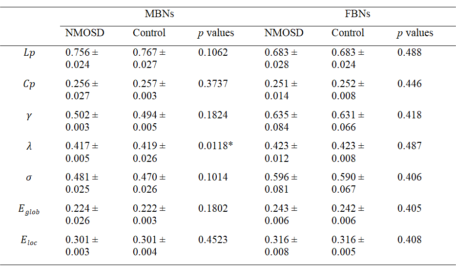
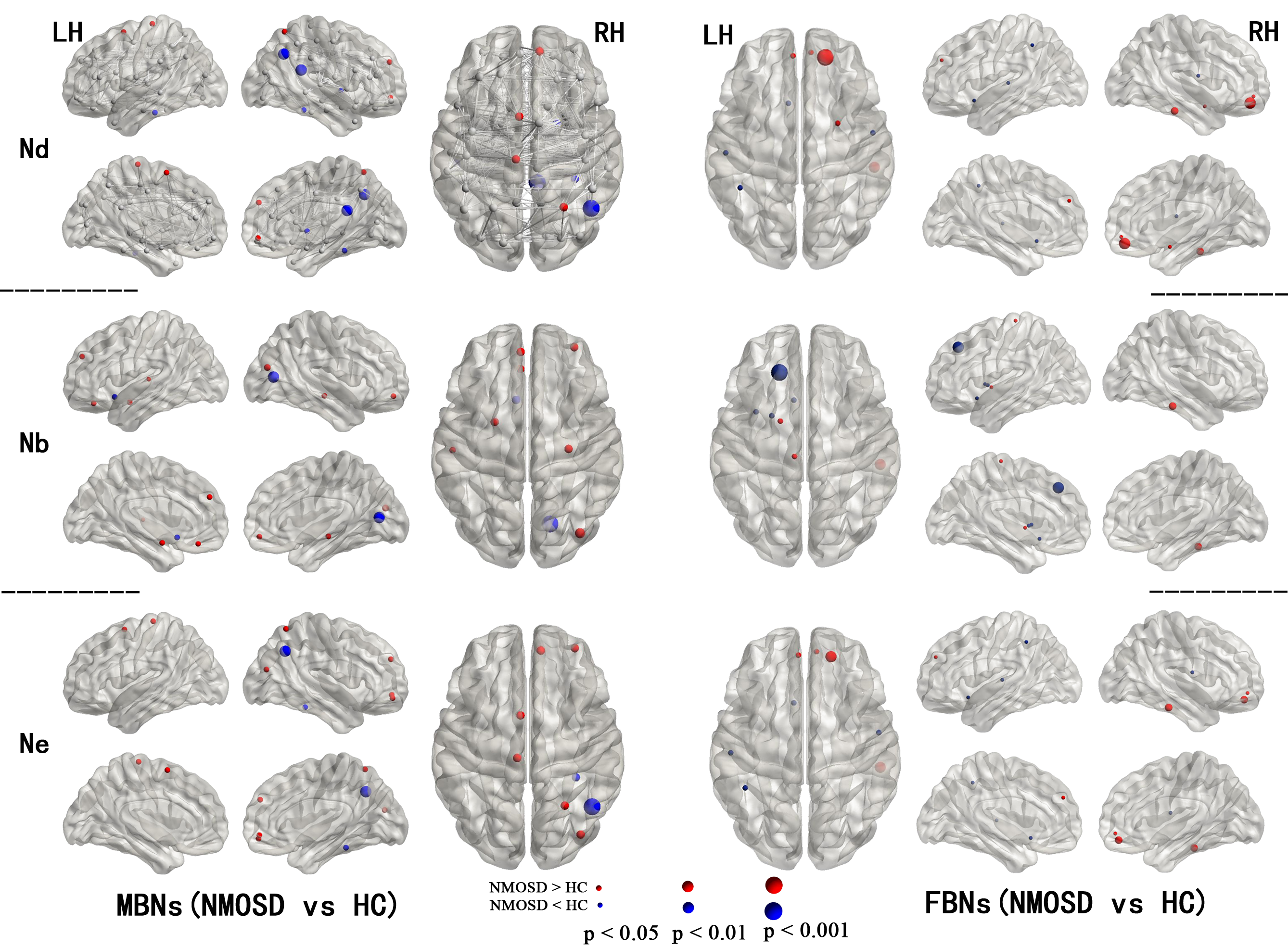
NMOSD patients included in the study had an average disease duration of 25.7 months, with mild disability (average EDSS score of 1.05) and mild cognitive impairment (mean MMSE score of 26.65).Both NMOSD and HCs exhibited small-world properties in MBNs and FBNs. No significant differences were found in global properties of FBNs between the two groups. However, NMOSD patients showed a significant decrease in λ in MBNs compared to HCs (P = 0.0118, FDR corrected), while other global properties of MBNs remained similar (Table 1).Significant differences in nodal properties were observed between NMOSD patients and HCs (P < 0.05, FDR corrected). NMOSD patients exhibited decreased Nodal betweenness (Nb) in most brain regions, except for the left olfactory cortex (Figure 1). Additionally, MBNs showed more compensatory increases in nodal properties.
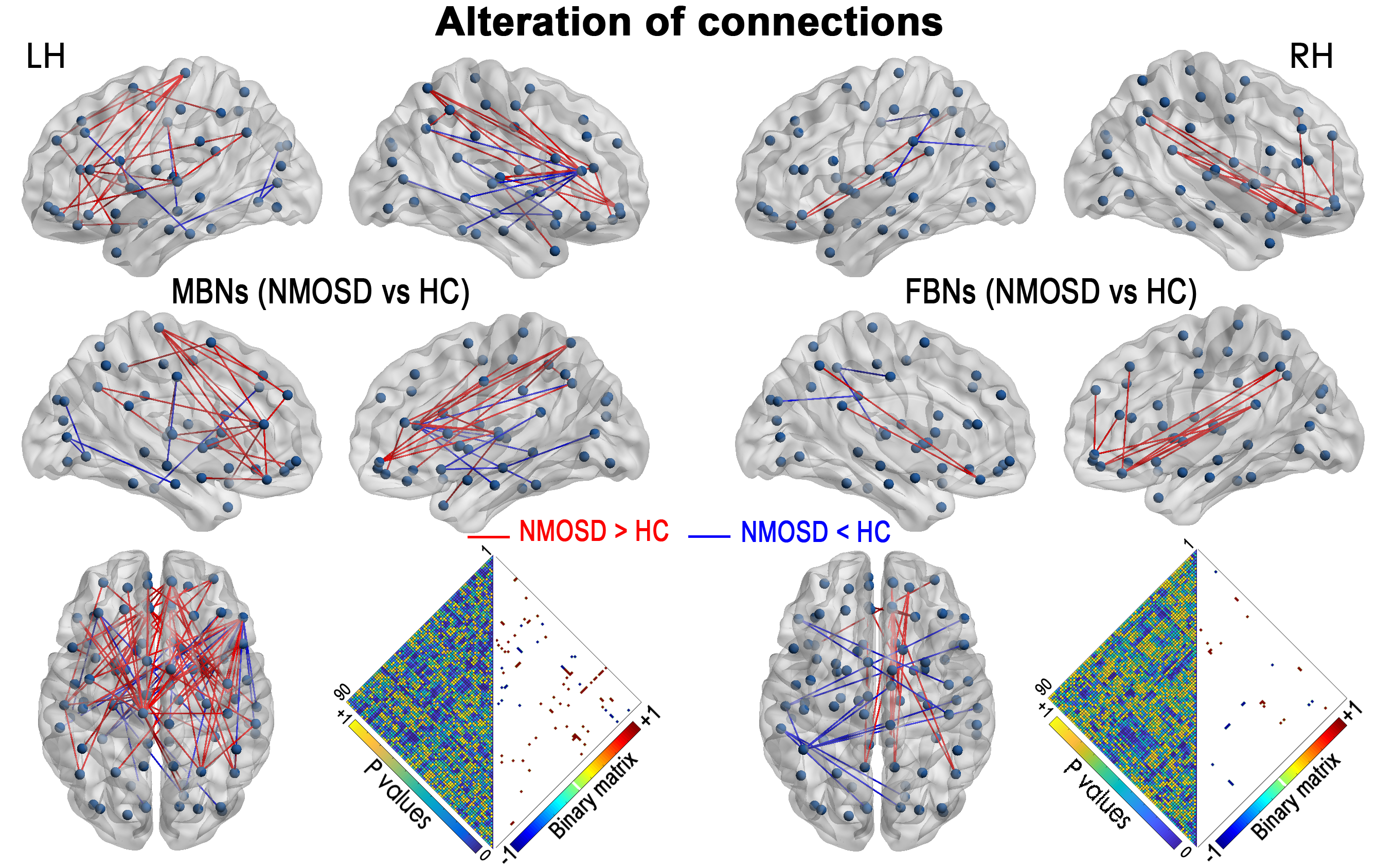
Both MBNs and FBNs in NMOSD patients exhibited significant alterations in network connectivity compared to HCs (P < 0.01, NBS corrected). MBNs showed a higher number of both increased and decreased connections compared to FBNs (Figure 2).
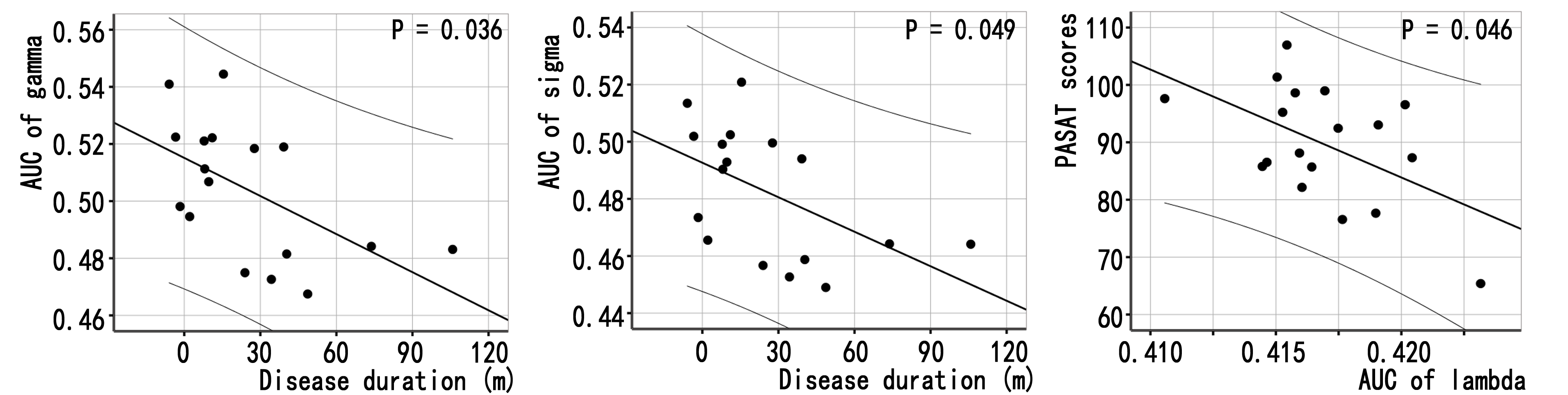
Correlations between global network properties of MBNs and clinical variables were observed (Figure 3). No significant correlations were found between global properties of FBNs and clinical variables.
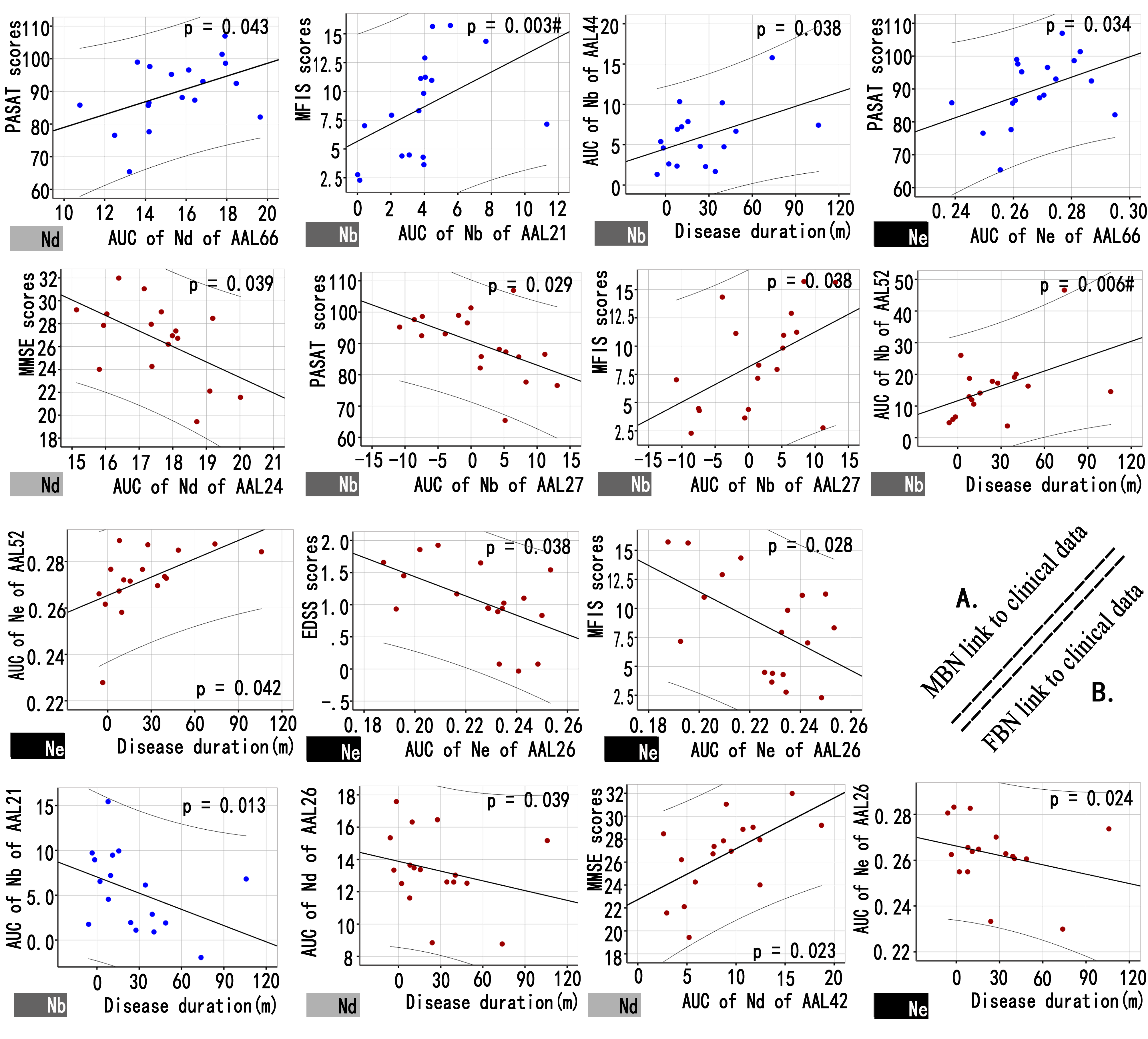
Figure 4 demonstrated significant correlations between nodal properties of MBNs or FBNs and clinical variables (P < 0.05/6).
Discussion and Conclusion
In this study, we investigated alterations in MBNs and FBNs of NMOSD and their relationship with clinical information. Our results showed: (1) NMOSD patients have reduced global efficiency in MBNs, and it was correlated with cognitive impairments; (2) both increased and decreased nodal properties of MBNs and FBNs were observed, indicating compensatory mechanisms; (3) Compared to FBNs, MBNs show better correlations with clinical information.NMOSD patients exhibited a decrease in the global efficiency of interregional information integration in MBNs, which was correlated with PASAT scores. This decrease suggests reduced information flow efficiency within the brain network, potentially contributing to cognitive impairments common in NMOSD. These findings align with NMOSD patients' experience of cognitive impairments and slower processing speed. However, such alterations were not observed in FBNs.
Nodal properties in NMOSD patients were found to decrease in several brain regions, primarily associated with white matter lesions and network disconnection. These changes corresponded with clinical functional impairments, such as fatigue and mild cognitive impairment. Notably, NMOSD patients also displayed compensatory increasement of nodal properties mainly in the medial frontal, motor, visual association, and default mode network. The increased nodal properties of medial frontal network were correlated with cognitive and fatigue assessments. The motor and visual association network showed evidence of compensation in response to functional decline. The default mode network was correlated with disability level and fatigue.
The findings suggest that NMOSD patients with mild disabilities exhibit increased local network properties as a compensatory mechanism to uphold overall stability. Moreover, individual brain morphological networks exhibit a higher degree of changes in NMOSD patients and display stronger associations with clinical variables compared to functional networks.
Acknowledgements
Funding
This work was supported by Innovation and Entrepreneurship Training Program for College Students of Jiangxi province [202310403075]; and Jiangxi’s double thousand plan [jxsq2023201039].
References
[1] Wingerchuk DM et al. Neurology 85:177-189, 2015.
[2] Clarke L et al. Clin Exp Immunol. 206:251-265, 2021.
[3] Carnero Contentti E et al. Mult Scler Relat Disord. 19:73-78, 2018.
[4] Wang H et al. Brain Behav. 6:e00448, 2016.
Figures