2493
Reproducibility of the quantification of cerebral perfusion using multi-delay arterial spin labeling MRI at 5T1Peking Union Medical College Hospital, Beijing, China, 2United Imaging Research Institute of Intelligent Imaging, Beijing, China, 3Handan Central Hospital, Handan, China
Synopsis
Keywords: Blood Vessels, Arterial spin labelling, ultra-high field, reproducibility
Motivation: To develop and optimize multi-delay pCASL at ultra-high field 5T MR system and further assess the reproducibility of this technique for the quantifications of cerebral perfusion.
Goal(s): Develop a whole-brain multi-delay pCASL imaging protocol with good reproducibility at 5T MRI.
Approach: Optimize labeling gradient parameters for the field inhomogeneities, scan 8 healthy volunteers and test the reproducibility of cerebral blood flow(CBF) and arterial transit time(ATT).
Results: We first achieved whole-brain multi-delay pCASL imaging at ultra-high field with prolonged post-labeling delays. CBF showed excellent reproducibility in all brain regions, especially in subcortical region. ATT showed excellent reproducibility in anterior brain regions.
Impact: Our findings enable a reliable quantitative analysis of whole-brain perfusion from multi-delay pCASL at ultra-high field with good reproducibility, offering a promising advancement in the effective and accurate diagnosis for neurological diseases.
Background and objectives
Pseudo-continuous arterial spin labeling (pCASL) at ultra-high field could benefit from increased SNR and prolonged T1, but suffer from field inhomogeneities, increased SAR and limited coverage. Here, we present 3D whole-brain GRASE-based pCASL with multiple post-labeling delays (PLDs) at 5T MR system. We aimed to optimize the labeling parameters and further assess the reproducibility of this multi-delay pCASL for the quantifications of cerebral perfusion.Methods
The MR imaging was performed on a 5T MR scanner (uMR Jupiter, United Imaging Healthcare, China) with a 2Tx/48Rx head coil. First, pCASL labeling parameters including average gradient (Gavg), maximum gradient (Gmax) and their ratio (Gratio=Gmax/Gavg) were optimized for the field inhomogeneities. Three parameter sets (labeling 1: Gavg=1&Gratio=10; labeling 2: Gavg=0.6&Gratio=10; labeling 3: Gavg=0.3&Gratio=9) were chosen and characterized in 3 healthy subjects. Other pCASL parameters included: transverse, GRASE, TE/TR 16.3/6485ms, resolution 2.5*2.5*4mm3, FOV 200*200*128mm3, labeling time 1800ms, PLD 1800ms, average 4, scan time 2:56. The optimal gradient setting was employed on the following experiments.Then, 8 adult healthy volunteers (6 females, 26.4±3.0 years old) were scanned twice on the same 5T MR scanner. Calibration was set to invalid between the within-session scans. The labeling plane was planned using a TOF scout image, located at the level of V3 segment of the vertebral arteries (VA) and perpendicular to both VA and internal carotid arteries. A total of 7 PLDs was conducted including 500, 1000, 1500, 2000, 2500, 3000 and 3500ms, with the average time of 1, 1, 1, 1, 2, 2, 2. Other parameters including: transverse, GRASE, TE/TR 13.9/6089ms, resolution 3.5*3.5*4mm3, FOV 224*224*128 mm3, labeling time 1800ms, scan time 6:39. High-resolution anatomical images of T1-MPRAGE were also collected for image registration, with parameters as below: sagittal, TE/TR 3.4/9.4ms, TI 1050ms, resolution 0.7*0.7*0.7mm3, FOV 256*220*182 mm3, flip angle 9º, ACS factor 3, scan time 4:07.
Perfusion parameters (cerebral blood flow [CBF] and arterial transit time [ATT]) were analyzed on uOmnispace.MR (United Imaging Healthcare, China), co-registered to T1W images on SPM12 and resliced to MNI space. Quantitative analyses were conducted by segmenting total gray matter to frontal, parietal, temporal, occipital, limbic lobes and subcortical region. Within-subject coefficient of variation (wsCV) and intraclass correlation coefficient (ICC) were calculated to assess the reproducibility and reliability of multi-delay ASL in GM and 6 regions of interest (ROIs). Also, correlation coefficient plots and Bland-Altman plots were used to evaluate the measurement agreements between scans.
Results
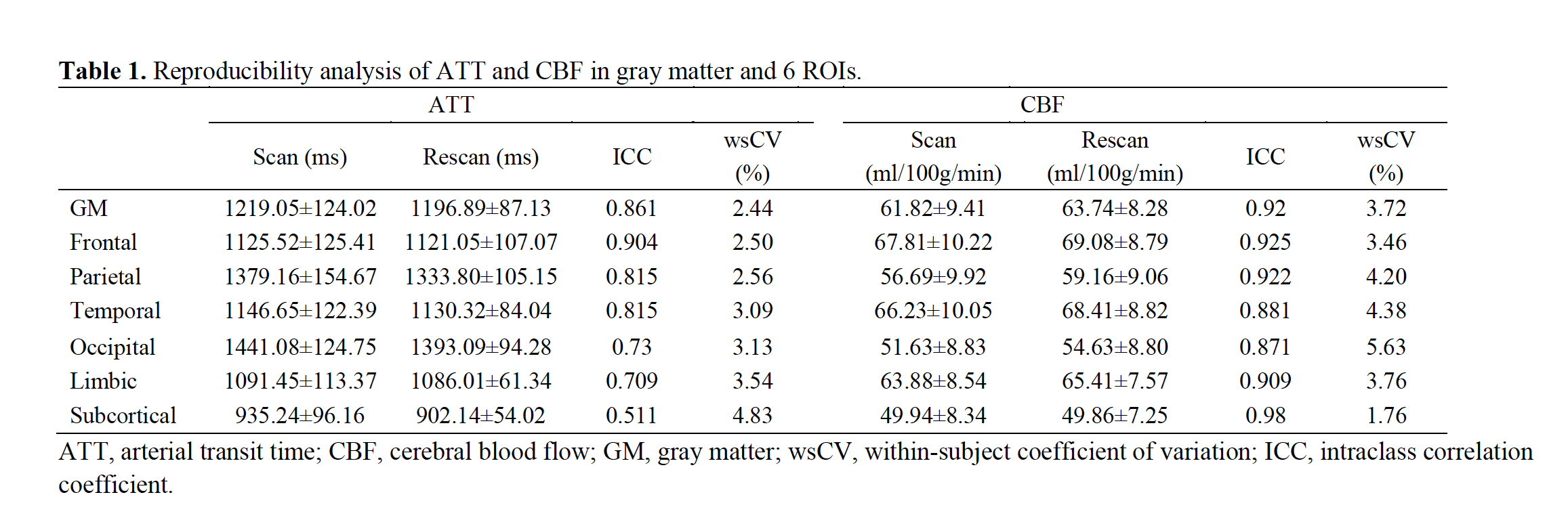
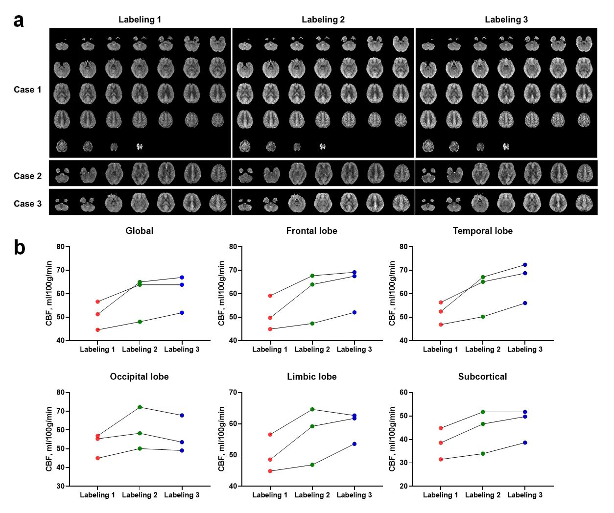
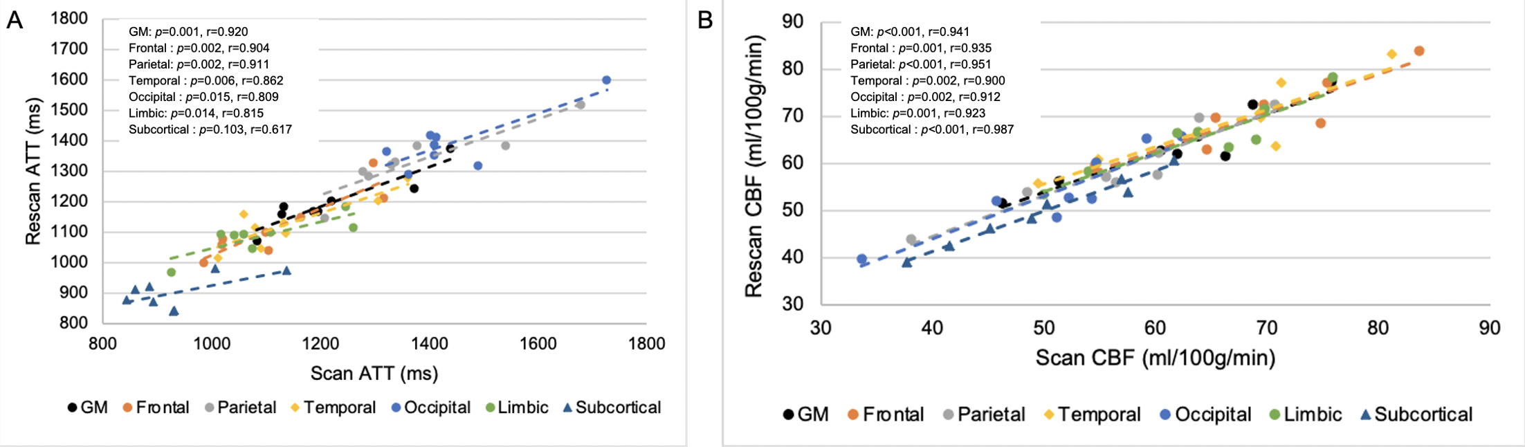
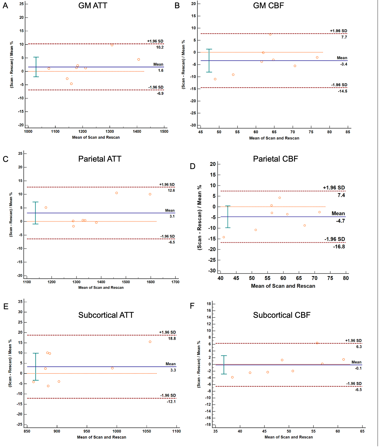
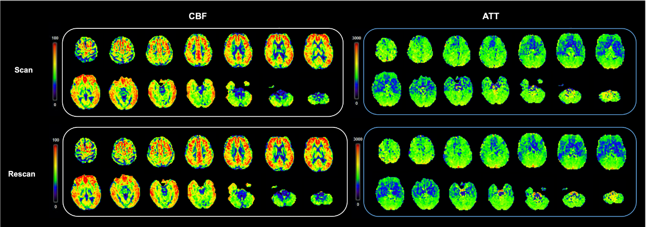
CBF images between 3 labeling sets from three subjects are shown in Figure 1a. Comparisons across different brain regions revealed that labeling 3 had the highest CBF values, indicating the highest labeling efficiency even in the B0 and B1 inhomogeneities (Figure 1b).The reproducibility and reliability of multi-delay ASL are shown in Table 1. ATT showed excellent reliability in GM, frontal, parietal and temporal lobes with ICCs higher than 0.8. CBF showed excellent reliabilities in total GM and all ROIs. ICCs of CBF were higher than that of ATT in GM and all ROIs. Both ATT and CBF had excellent wsCV (less than 5%). Importantly, CBF in subcortical region had the highest ICC and the lowest wsCV (0.98 and 1.76, respectively). In GM and almost all ROIs, the correlation analysis showed strong test-retest agreements (Figure 2). Similarly, subcortical CBF showed the highest correlation (p<0.001, r=0.987). In Bland-Altman analysis, the plots were randomly distributed without dependency within 95% confidence intervals (Figure 3). A representative case is shown in Figure 4.Discussion
We first achieved whole-brain multi-delay pCASL imaging at ultra-high field with prolonged PLDs. The longest PLD was set as 3500ms which was difficult to measure at 3T and would contribute to the diagnosis of cerebrovascular diseases a lot. We found that the reproducibility of CBF was excellent in all brain regions, especially in subcortical region, which may benefit from the increased SNR and prolonged T1 at ultra-high field. Moreover, the reproducibility of ATT performed better in anterior brain regions than occipital, limbic lobes and subcortical region. It may be due to the tortuous movement, highly anatomical variation and tiny diameter of vertebrobasilar artery and penetrating branches [1]. This result was in line with previous ASL studies [2,3].Conclusion
We have shown that a whole-brain multi-delay pCASL imaging at ultra-high field can be used to quantify ATT and CBF in almost all brain regions with good reproducibility, especially in subcortical region. This ongoing study is enrolling more volunteers and patients with cerebrovascular diseases to further strengthen the findings.Acknowledgements
We thank all the volunteers who participated in the study.References
[1] Chng SM, Petersen ET, Zimine I, Sitoh YY, Lim CC, Golay X. Territorial arterial spin labeling in the assessment of collateral circulation: comparison with digital subtraction angiography. Stroke. 2008 Dec;39(12):3248-54.
[2] Wu B, Lou X, Wu X, Ma L. Intra- and interscanner reliability and reproducibility of 3D whole-brain pseudo-continuous arterial spin-labeling MR perfusion at 3T. J Magn Reson Imaging. 2014 Feb;39(2):402-9.
[3] Lin T, Qu J, Zuo Z, Fan X, You H, Feng F. Test-retest reliability and reproducibility of long-label pseudo-continuous arterial spin labeling. Magn Reson Imaging. 2020 Nov;73:111-117.
Figures