2491
Multi-delay Arterial Spin Labeling Magnetic Resonance Imaging of Cerebral Blood Flow in Patients With Vertebrobasilar dolichoectasia1China-Japan Friendship Hospital, Beijing, China, 2MR Research China, Beijing, China
Synopsis
Keywords: Blood Vessels, Arterial spin labelling
Motivation: Vertebrobasilar dolichoectasia (VBD) poses a risk to cerebral blood flow and can lead to ischemic strokes. Understanding its impact on blood flow and identifying predictive factors are crucial.
Goal(s): This study aims to assess differences in CBF and other parameters between VBD patients and controls. We also investigate the influence of extracranial blood flow and predict PCI risk factors.
Approach: 32 VBD patients and 32 controls underwent Multi-delay-ASL and Doppler ultrasound. We analyzed CBF, aCBV, ATT, and blood flow velocities.
Results: VBD patients exhibited lower CBF and aCBV in both circulations and longer ATT. Bilateral occipital and cerebellar ATTs predicted PCI.
Impact: This study enhances our understanding of VBD's impact on cerebral blood flow and identifies predictors of posterior circulation ischemia. It underscores the importance of maintaining extracranial blood flow, aiding in early detection and prevention of ischemic strokes in VBD patients.
Introduction
The assessment of CBF is crucial in the evaluation of vertebrobasilar dolichoectasia disease. This study analyzed differences in the mCBF, aCBV and ATT of the anterior and posterior circulations between patients with VBD and control subjects. We also investigated whether the blood flow velocity of the extracranial carotid/vertebral arteries can influence mCBF, and predicted risk factors for PCI in VBD patients.Methods
A total of 32 patients with VBD and 32 healthy subjects were prospectively enrolled in the study. All patients were diagnosed with VBD by magnetic resonance angiography (MRA). All subjects underwent multi-delay arterial spin labeling (Multi-delay-ASL) and carotid and vertebral Doppler ultrasound examinations. CereFlow software was used for post-processing to obtain mCBF, aCBV and ATT. The comparison of mCBF, aCBV and ATT in the anterior circulation (frontal lobe, parietal lobe, and insula) and posterior circulation (occipital lobe, cerebellum) between the control and study groups was conducted. The peak systolic velocity (PSV), end diastolic velocity (EDV), mean PSV (mPSV), and mean EDV (mEDV) of the extracranial internal carotid/vertebral arteries were compared between two groups. The factors associated with mCBF in the two groups was explored. Receiver operating characteristic curve (ROC) was used to predict the potential of PCI in VBD patients.Results
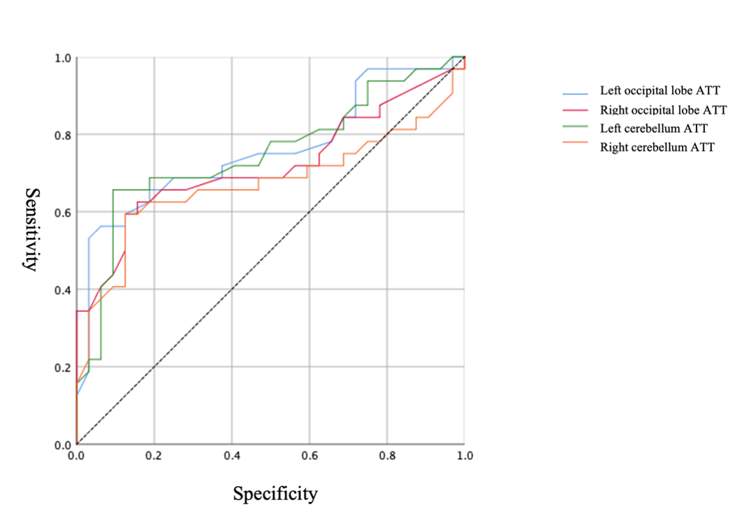
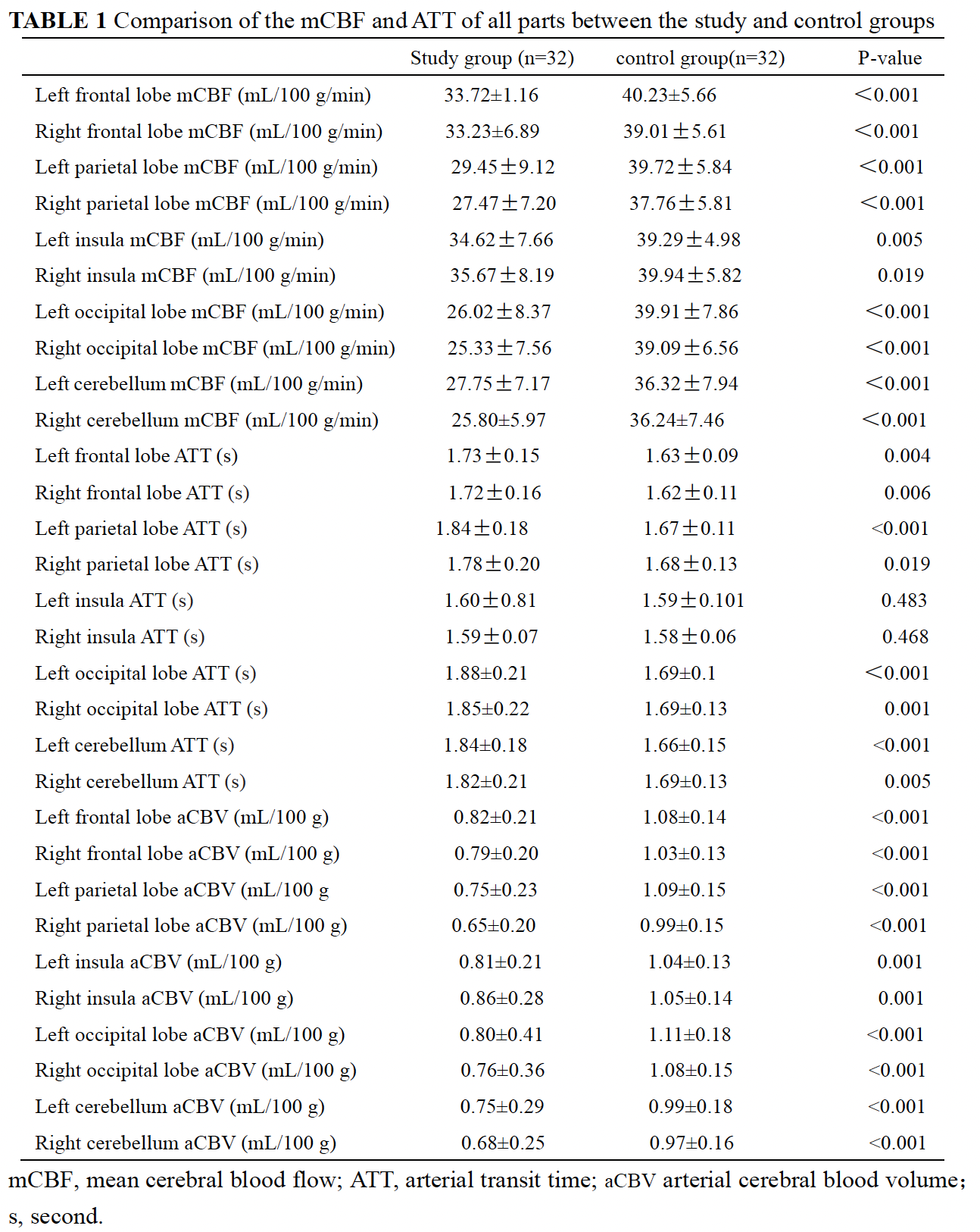
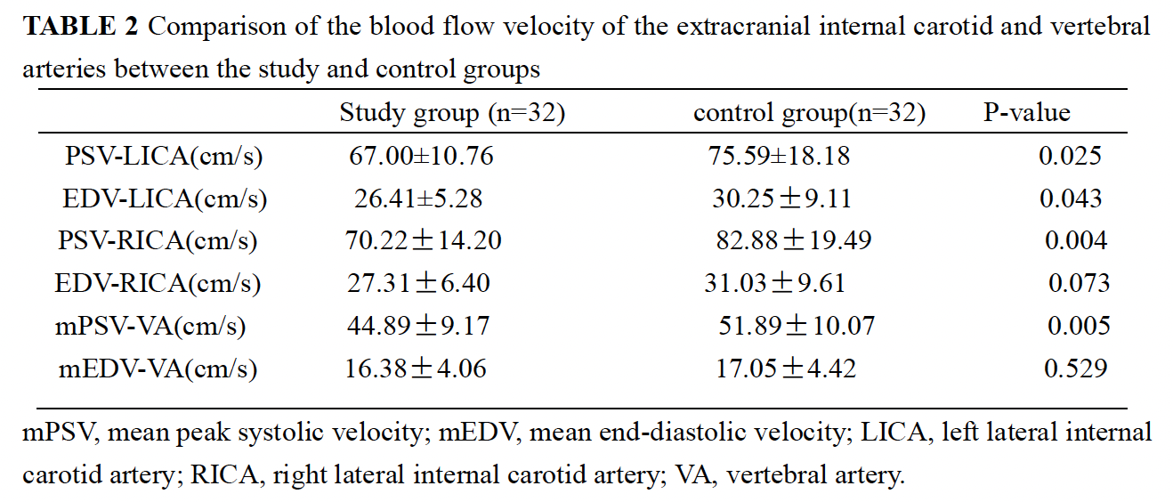
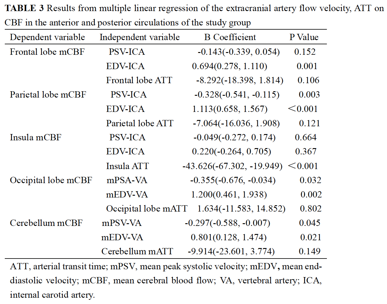
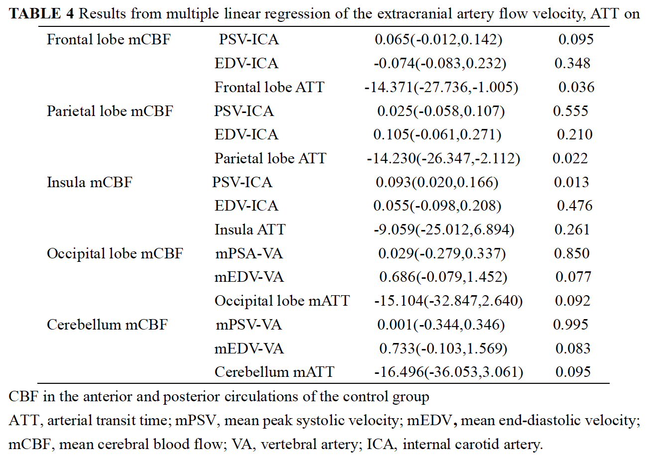
The mCBFs and aCBVs of the anterior and posterior circulations in the study group were lower than those of the control group. The ATTs in the patient group were all significantly longer than those of the control group (p < 0.05), except for the insula in the control group. Except for the mEDV of the vertebral arteries, the blood flow velocity of the extracranial internal carotid/vertebral arteries differed significantly between the control and patient groups (p < 0.05). The mPSV and mEDV of the vertebral arteries correlated with mCBF of the occipital lobes and the cerebellum in the study group, respectively (p < 0.05). ROC analysis revealed that bilateral occipital lobe and cerebellar ATTs were risk factors for predicting PCI in VBD patients.Discussions
In patients with VBD, the mCBFs and CBVs values of both the anterior circulation and posterior circulation were significantly lower than those of normal healthy people, and the ATT values of the were longer than healthy people. Dolichoectasia of vertebrobasilar can result in changes in cerebral blood flow distribution, which are closely related to the structure of vessel walls and the hemodynamics across the dolichoectasia. There are significant correlations between ATT and mCBF. As the mCBF value decreased, the ATT increased. The relationship between ATT and CBF in patients with VBD is of great importance in the posterior circulation. The blood flow velocity in the vessels of the posterior circulation is lower. Because the blood vessel walls vasodilation and become thinning, which slows down blood flow and results in a low perfusion state. In this situation, compensatory elongation of ATT will help to maintain local perfusion in a steady state. The adjustment of ATT may reflect slow flow compensating for a disrupted blood supply through one of the proximal arteries. In the patients with VBD, the mPSV and mEDV of the vertebral arteries correlated with mCBF of the occipital lobes and the cerebellum, but this phenomenon was not observed in the controls. We suggest that mCBF of the posterior circulation in patients with VBD is dolichoectasia to the change in the upstream blood supply. In the pathological state, patients with VBD seem to have a declined ability to cope with the reduced blood inflow, suggesting a vulnerability to the decrease in perfusion. Our study supports the idea that maintenance of extracranial blood flow is of great significance in the preservation of intracranial hemodynamics, especially those of the posterior circulation. In this study showed that bilateral occipital lobe and cerebellar ATTs were risk factors for predicting PCI in VBD patients. Multi-delay-ASL facilitated quick measurement of ATT by low-resolution time coding technology, and choosing of a reasonable PLD based on this known ATT, thereby allowing a more accurate mCBF measurement to be obtained. VBD leads to slow blood flow and decreased perfusion in posterior circulation. The low-perfusion state leads to significantly prolonged ATT, which may a potential risk factor for occurrence of PCI.Conclusions
VBD leads to insufficient perfusion in the anterior and posterior circulation regions, and prolonged ATT in both occipital lobes and cerebellum can predict the occurrence of ischemic stroke in the posterior circulation. Extracranial blood flow may influence intracranial hemodynamics in the posterior circulation in patients with VBD. The maintenance of extracranial blood flow is of great significance in the preservation of intracranial hemodynamics.Acknowledgements
No acknowledgement found.References
[1] Park PSW, Yogendrakumar V, Yassi N, Campbell BCV. Persistent Posterior Fossa Hypoperfusion in the Setting of Vertebrobasilar Dolichoectasia. Stroke. 53(7): e276-e28, 2022.
[2] Zhang DP, Ma QK, Zhang JW, Zhang SL, Lu GF, Yin S. Vertebral artery hypoplasia, posterior circulation infarction and relative hypoperfusion detected by perfusion magnetic resonance imaging semiquantitatively. J Neurol Sci 368:41-46, 2016.
[3] Peng YF, Zhang HL, Zhang DP, Zhao M, Zhang SL, Yin S. Perfusion by delayed time to peak in vertebrobasilar dolichoectasia patients with vertigo. Ann Clin Transl Neurol 5(12):1562-1573, 2018.
[4] Förster A, Kerl HU, Wenz H, Mürle B, Habich S, Groden C. Fluid Attenuated Inversion Recovery Vascular Hyperintensities Possibly Indicate Slow Arterial Blood Flow in Vertebrobasilar Dolichoectasia. J Neuroimaging. 25(4):608-13, 2015.
[5] Li W, Feng Y, Lu W, Xie X, Xiong Z, Jing Z, Cai X, Huang L. Evaluating the morphological changes of intracranial arteries and whole-brain perfusion in undetermined isolated vertigo. J Neurol Sci.15: 370:70-77, 2016.
[6] Zheng T, Tang W, Shan Y, Guo R, Gao Y, Tian C, Liu L, Sun L, Liu W, Zhou Z, Jin Y, Duan S, Han B, Fan Y, Zhu X, Liu Z. Studying the imaging features and infarction mechanism of vertebrobasilar dolichoectasia with high-resolution magnetic resonance imaging. Brain Pathol. 33(2): e13135, 2023.
Figures