2489
Age-related assessment in a subset of SHIP data of intracranial vessel geometry from TOF MRI data1Department of Medical Physics, Institute of Physics, University of Greifswald, Greifswald, Germany, 2Department of Radiology, Northwestern University Feinberg School of Medicine, Chicago, IL, United States, 3Institute for Community Medicine, University Medicine Greifswald, Greifswald, Germany, 4Institute for Diagnostic and Interventional Radiology, Pediatric and Neuroradiology, University Medical Center Rostock, Rostock, Germany, 5Institute of Diagnostic Radiology and Neuroradiology, University Medicine Greifswald, Greifswald, Germany
Synopsis
Keywords: Blood Vessels, Blood vessels
Motivation: Intracranial vessels undergo age-related changes with increased risk of vascular pathologies such as atherosclerosis, or intracranial aneurysms. The occurrence probability of these pathologies could be indicated based on an altered vascular geometry.
Goal(s): The application of a geometric analysis to the vascular system of healthy volunteers of two age groups to study age-related changes in the geometry of intracranial arteries.
Approach: The analysis involved labeled centerline determination of intracranial arteries and calculating geometric parameters.
Results: Our study provided quantitative insights into age-related changes in intracranial artery geometry. Future research with larger, diverse samples can enhance the understanding and enable ML-based analyses.
Impact: The objective was to examine age-related changes in women of intracranial arteries by performing a geometric analysis on healthy volunteers. The quantitative evaluation enhances the comprehension of how intracranial artery geometry evolves with age.
Motivation
Intracranial vessels are subject to aging resulting in arteriosclerotic changes1. They predispose to vascular pathologies, including stenoses and aneurysms2, which are evident in basal cerebral arteries (Circle of Willis, CoW)3, and have a higher incidence in women4. A study by Żyłkowski et al5 focused on correlating age-related geometric changes in vessels with the incidence of aneurysms. They could show that the average curvature of the bifurcation of the middle cerebral artery (MCA) increased with age, but that torsion (rotation of vessel) decreased. Choudhry et al6 demonstrated that length and tortuosity increased with age, but not the vessel diameter. Based on these findings, we investigated age-related vessel geometry in a subset of the SHIP (Study of Health In Pomerania) MRI cohort7. In this centerline-based analysis, the vascular geometry of CoW arteries was quantified, including variants of MCA branching. We determined vessel curvature for all spatial directions and its magnitude, vessel radius, length and tortuosity8. We hypothesized that due to the higher incidence of arteriosclerosis in older women, they express vessels with higher curvature and tortuosity while maintaining vessel diameter. According to5, we expect a change in the spatial directions of curvature.Methods
Time-of-flight (ToF) MR angiograms of the CoW vessels of N=60 participants (women, 45.20 ± 19.2 years) were analyzed. All datasets were acquired at 1.5T, segmented9, and transformed to MNI space to ensure a standardized coordinate system and anatomical identification of brain structures10,11. The centerlines were determined for the C1-C6 segments of the ICA and the M1 and M2 segments of the MCA using MATLAB12. Segments were defined manually and confirmed by an experienced board-certified neuroradiologist. ICA segments definitions were: C1- cervical segment, C2 -horizontal petrous segment, C3- lacerum segment, C4- cavernous segment, C5- clinoid and supraclinoidal segment, and C6 - carotid terminus13. M1 was defined as the horizontal part of the MCA and M2 was the insular part13. M2 was subdivided into the anterior (M2A), middle (M2B), and posterior (M2C) branches. Enrolled subjects were divided into two groups: 1) 20-29 years (N1=30, 25.9 ± 2.2 years) and 2) 60-69 years (N2=30, 63.9 ± 2.52 years). The curvature was:\[ curvature=\left\{% \begin{array}{ll} G & \hbox{, r=0} \\ D & \hbox{, norm(D)=0} \\ G'/r^2 & \hbox{, else.} \\ \end{array}% \right. \]
based on three adjacent centerline points forming a triangle with vector G from the center of the circle to one of the vertices, its transposed vector G', radius r and normal vector D (relative to triangle)14. In this study, we evaluated the median curvature, median vessel radius, tortuosity (ratio of arterial segment length and Euclidean distance of vessel endpoints), and vessel length. Both groups were compared for each hemisphere separately by using the Mann-Whitney-U test.
Results
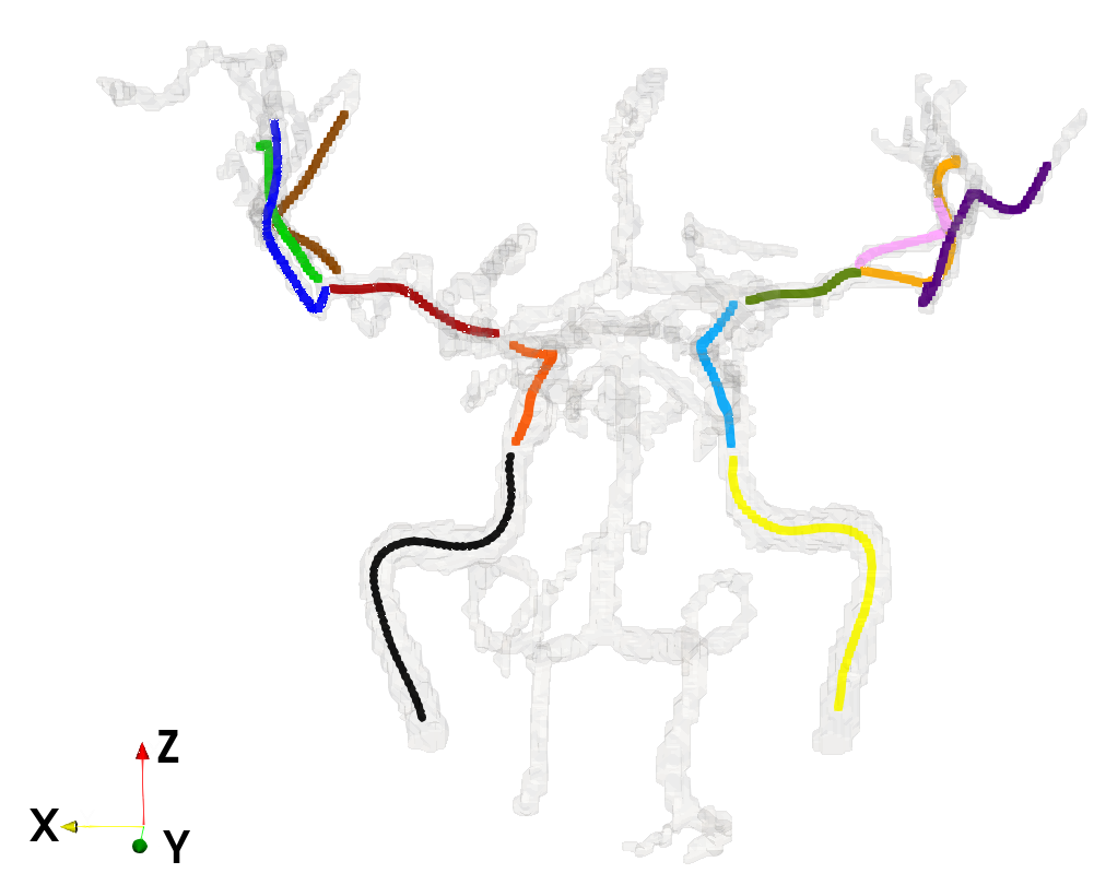
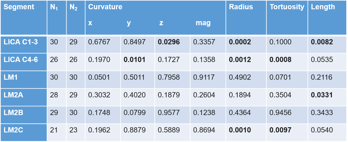
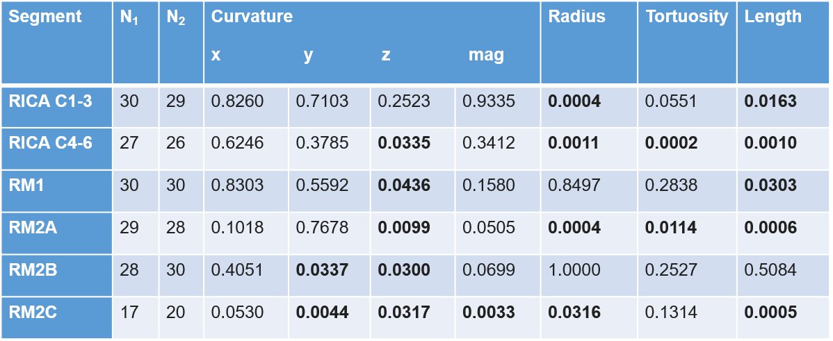
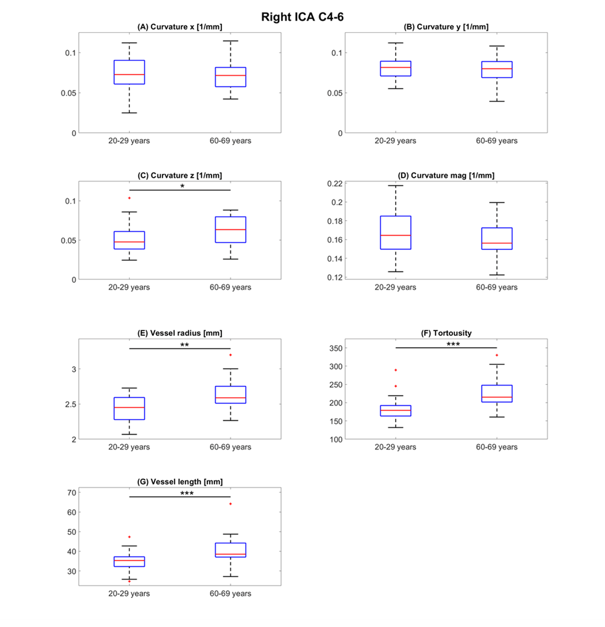
Figure 1 shows a 62-year-old subject with labeled centerlines. Tables 1 and 2 summarize all results of both hemispheres. Figure 2 lists evaluated parameters for each vessel with color-coded significance levels. The x-component of the median curvature shows no changes with increasing age, whereas the y-component decreased in the left ICA C4-6 and right M2B and M2C. The z-component decreased in the left ICA C1-3, increased in the right ICA C4-6, and decreased in the right M1 segment as well as the entire right M2 branch. The curvature magnitude decreased only in the right M2C. The vessel radius showed an increase in left and right ICA segments C1-3 and C4-6 and a decrease in M2C of both hemispheres. Tortuosity increased in the left and right ICA C4-6 and the right M2A and decreased in the left M2C. The vessel length increased in all segments on the right hemisphere, except for M2B. On the left, the vessel length increased in the ICA C1-3 and M2A. Figure 3 shows results of the right ICA C4-6 of the right hemisphere. Summarized, the median of the z curvature component, median of the vessel radius, tortuosity, and vessel length were larger in older women in most vessel segments.Discussion and conclusion
Our method enabled quantitative assessment of age-related changes in geometry of intracranial arteries. The centerline-based analysis revealed changing vessel curvature, radius, length, and tortuosity of most investigated segments with increasing age, except for M2B of the left hemisphere. This could be due to the small number of subjects or the limited spatial resolution. Based on the results, future investigations are warranted including more subjects, adding more age groups as well as male subjects to achieve higher power, and allowing investigation of gender differences. Results could serve as ground truth for machine learning-based analyses.Acknowledgements
This work was funded by the National Institutes of Health (NIH 1R21NS122511-01, NIH 1R01HL149787) and the Käthe-Kluth junior research group of the University of Greifswald.
References
[1] Pauline Mouches et al. Human Brain Mapping. 2022;43:2554-2566.
[2] Wang et al. Cardiovas Dis J Int Traditional Chin Western Med. (2018) 16:1622–4.
[3] Brisman et al. 2006. N. Engl. J. Med. 355(9), 928-939.
[4] Fréneau et al. (2022). Front. Cardiovasc. Med. 9:815668.
[5] Żyłkowski et al. Journal of Anatomy. 2021;238:765-784.
[6] Choudhry et al. J. NeuroIntervent Surg 2016;8:536-540.
[7] John et al. Sozial-und Präventivmedizin 46(3), 186–194 (2001).
[8] Hyung Jun Kim et al. Journal of Stroke. 2021;23(2):213-222.
[9] Forkert et al. Magn. Reson. Imag. 31(2), 262-271(2013)
[10] Mouches et al. Nature. (2021)11:12236.
[11] Mazziotta et al. Philosophical Transactions of the Royal Society of London B: Biological Sciences,356(1412), 1293–1322.
[12] Vali et al. Magn. Reson. Med. 2019;82:749-762.
[13] Schünke et al. Prometheus LernAtlas der Anatomie: Kopf, Hals und Neuroanatomie. Stuttgart: Thieme; 2015.
[14] Are Mjaavatten (2023). Curvature of a 1D curve in a 2D or 3D space (https://www.mathworks.com/matlabcentral/fileexchange/69452-curvature-of-a-1d-curve-in-a-2d-or-3d-space), MATLAB Central File Exchange.
Figures