2487
Using compressed sensing-accelerated 4D flow MRI to observe venous sinus hemodynamics:Performance at different acceleration factors1Radiology, First Affiliated Hospital of Dalian Medical University, Dalian, China, 2Clinical & Technical Support, Philips Healthcare, Beijing, China
Synopsis
Keywords: Blood Vessels, Blood vessels, 4D flow MRI,compressed sensing,venous sinus
Motivation: Due to the small diameter of intracranial-venous sinus, higher spatial resolution is required when using 4D-flow MRI to observe hemodynamics, which greatly prolongs scan time.
Goal(s): Accelerating 4D-flow MRI of intracranial-venous sinus with compressed sensing (CS), and comparing the performance of CS acceleration factors (AFs).
Approach: The 4D-flow hemodynamic of straight sinus, superior sagittal sinus and transverse sinus with different AFs (SENSE4, CS4, CS6, CS8, and CS10) were compared in 8 healthy volunteers.
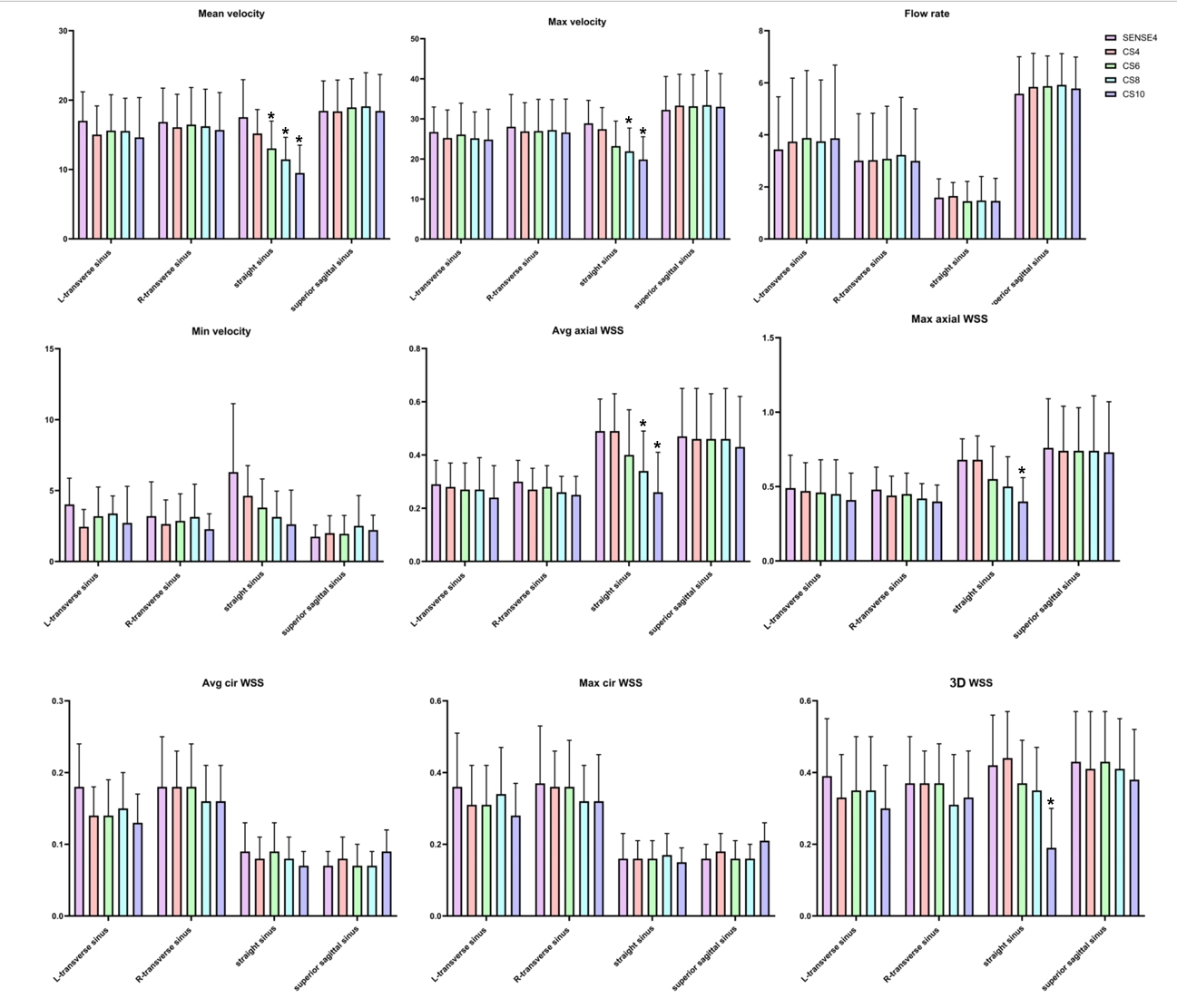
Results: Compared to the reference (SENSE4) scan, the hemodynamic for the straight sinus, several parameters were underestimated by CS6-CS10,while for the other sinuses obtained by CS4-CS10 showed no significant difference.
Impact: 4D flow MRI with acceleration by compressed sensing can be used as a efficient method to evaluate venous sinus hemodynamics in routine clinical practice.
Introduction
Three-dimensional time-resolved phase-contrast MRI, also called 4D flow MRI, has developed rapidly with its utility as an investigational tool expanding into a wider clinical role in the recent years1. With three-directional velocity encoding, 4D flow MRI can provide rich hemodynamic information, such as wall shear stress (WSS), and give new insights into disease pathophysiology that could not be obtained through other existing imaging methods2-3. However, these benefits come at the expense of long acquisition time, especially when observing intracranial vessels, due to the requirement on high spatial resolution. Compressed sensing (CS), based on random under-sampling of medical imaging data with natural sparsity, can greatly shorten the scan time under the premise of ensuring image quality4. This study aims to evaluate the performance of 4D flow MRI with CS acceleration factors (AFs) to fasten the acquisition of 4D flow MRI and evaluated the stability of hemodynamic parameters at different AFs.Methods
8 healthy volunteers were recruited (4 males, mean age: 25.13±1.46 years). All participants were scanned using a 3.0T MR scanner (Ingenia CX, Philips Healthcare, Best, the Netherlands) equipped with a 32-channel head coil. 4D flow MRI were acquired with different CS AFs (CS4, CS6, CS8, and CS10), and the control group was acquired with SENSE AF=4. The other imaging parameters were: repetition time (TR)/echo time (TE) = 3.0/5.1 ms; flip angle = 8°; FOV = 180×180×46 mm3; acquisition voxel size = 1.6×1.6×1.6 mm3; reconstruction voxel size = 0.8×0.8×0.8 mm3; and VENC = 60 cm/s. The scan time of SENSE4, CS4, CS6, CS8, CS10 groups were 7min24s, 5min47s, 3min52s, 2min54s and 2min22s, respectively. All 4D flow MRI scans were acquired with retrospective ECG-triggering (with 20 retrospectively constructed cardiac phases). For time-resolved flow quantification (including flow rate, mean/max/min velocity, 2D average/max axial WSS, 2D average/max circle WSS, 3D WSS) at three measurement planes (total of twelve planes) orthogonal to the midline at straight sinus, superior sagittal sinus and bilateral transverse sinus on the segmented volume were derived from 4D flow MRI using the commercial software (cvi42, version 5.14, Circle Cardiovascular Imaging, Canada). The hemodynamics of each venous sinus was described by the mean of the three planes. Data analyses were performed using IBM SPSS Statistics 26.0, a Shapiro–Wilk test was used to evaluate normality. An ANOVA or Kruskal-Wallis H test was used to compare 4D flow MRI scans with different AFs, and an LSD-t or Dunn-Bonferroni test for post hoc comparisons. P < 0.05 were statistically significant.Results
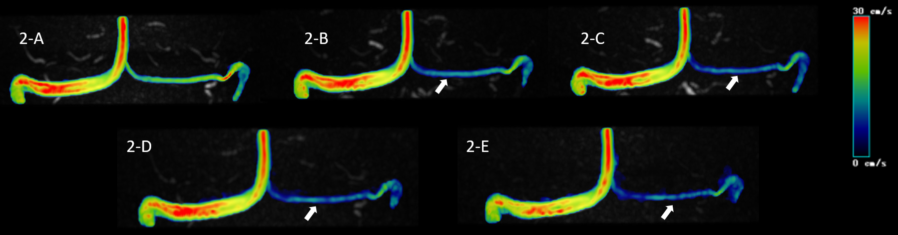
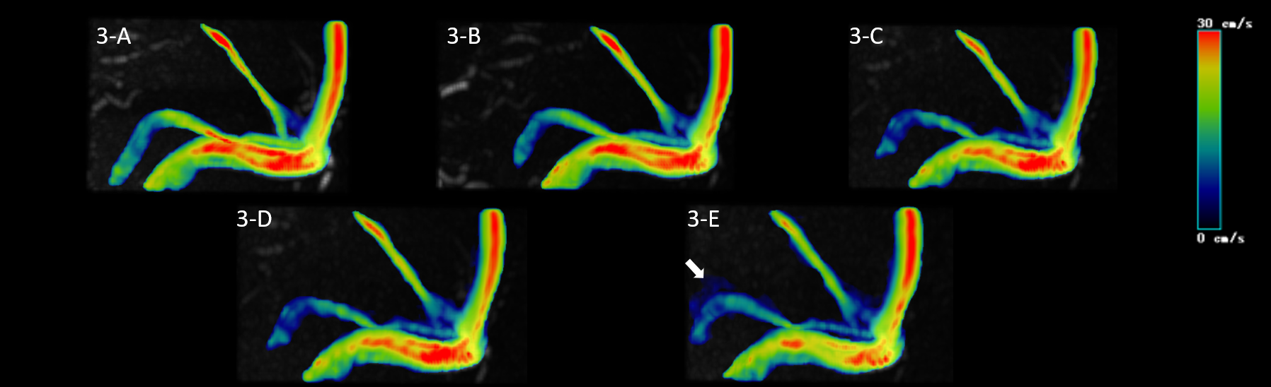
No significant difference in all flow quantifications obtained for transverse sinus and superior sagittal sinus, as well as flow rate, min velocity, 2D max circle WSS and 2D average circle WSS for straight sinus was observed among 4D flow MRI scans with different AFs (P > 0.05). While, for straight sinus, the mean velocity measured by CS6, CS8 and CS10, the max velocity and 2D average axial WSS measured by CS8 and CS10, and the 2D max axial WSS and 3D WSS measured by CS10 groups were significantly underestimated in comparison with the reference (SENSE4) scan (P < 0.05). (Figure 1-3)Discussion
This study showed that there was no significant difference among flow quantifications obtained by 4D flow scans with different AFs in transverse sinus and superior sagittal sinus. However, in straight sinus, compared with the reference (SENSE4) scan, the mean velocity in CS6, CS8 and CS10 groups, the max velocity and 2D average axial WSS in CS8 and CS10 groups, the 2D max axial WSS and 3D WSS in CS10 groups were all underestimated. Besides, with the increase of AFs, this underestimation became more obvious, which is similar to the previous studies5-6. We speculate that due to the smaller diameter of straight sinus, more accurate quantification is needed, resulting in the underestimation of multiple hemodynamic indicators under high CS acceleration. Therefore, an appropriate CS acceleration factor is needed to strike a balance between shorter scan times and the need for accurate hemodynamic quantification. In this study, no significant difference in the hemodynamic parameters for all three venous sinuses was observed between the CS4 group and the control group, therefore, CS4 (with a scan time reduction of 1min37s) could be considered as the optimal acceleration factor.Conclusion
Compared to the reference (SENSE4) scan, the hemodynamic parameters for superior sagittal sinus and transverse sinus obtained by CS4-CS10 showed no significant difference, while for the straight sinus, several parameters were underestimated by CS6-CS10. CS4 can be used as a optimal acceleration factor for 4D flow MRI in the observation of intracranial venous sinus with the premise of stable hemodynamic parameters.Acknowledgements
No acknowledgement found.References
[1] Pathrose A, Ma L, Berhane H, et al. Highly accelerated aortic 4D flow MRI using compressed sensing: Performance at different acceleration factors in patients with aortic disease[J]. MAGNET RESON MED. 2020; 85 (4): 2174-2187.
[2] Fatehi Hassanabad A, Burns F, Bristow MS, et al. Pressure drop mapping using 4D flow MRI in patients with bicuspid aortic valve disease: A novel marker of valvular obstruction[J]. Magn Reason Imaging. 2020; 65:175–182.
[3] Jarvis K, Pruijssen JT, Son AY, et al. Parametric hemodynamic 4D flow MRI maps for the characterization of chronic thoracic descending aortic dissection. J Magn Reson Imaging. 2020; 51:1357–1368.
[4] Craft, J, Li, Y, Nashta, NF, et al. Comparison between compressed sensing and segmented cine cardiac magnetic resonance: a meta-analysis[J]. BMC Cardiovasc Disord. 2023; 23 (1): 473.
[5] Ma LE, Markl M, Chow K, et al. Aortic 4D flow MRI in 2 minutes using compressed sensing,respiratory controlled adaptive k-space reordering, and inline reconstruction[J]. Magn Reson Med.2019; 81:3675–3690.
[6] Neuhaus E, Weiss K, Bastkowski R, Koopmann J, Maintz D, Giese D. Accelerated aortic 4D flow cardiovascular magnetic resonance using compressed sensing: applicability, validation and clinical integration[J]. J Cardiovasc Magn Reson. 2019; 21:65.
Figures