2486
Highly accelerated time-resolved 4D MRA using stack-of-stars golden-angle radial acquisition and subtraction-based subspace reconstruction1Department of Radiology, Northwestern University, Chicago, IL, United States, 2Center for Advanced Imaging Innovation and Research (CAI2R), Department of Radiology, New York University Grossman School of Medicine, New York, NY, United States
Synopsis
Keywords: Blood Vessels, Image Reconstruction
Motivation: ASL-based time-resolved 4D MRA potentially suffers from temporal blurring when accelerating image acquisition by exploiting temporal correlations.
Goal(s): To develop a robust 4D MRA reconstruction framework that enables a very high acceleration rate while preserving good temporal fidelity.
Approach: We developed a fast low-rank subspace high-resolution 4D MRA (Flash-4D-MRA) that combines SOS golden-angle radial sampling with joint subtraction-based self-calibrated low-rank subspace and magnitude-subtraction sparsity constraint to achieve an ultra-high temporal resolution. Each 4D MRA data was reconstructed with four high acceleration rates.
Results: Dynamic MRA images were successfully reconstructed using Flash-4D-MRA with higher acceleration rates without compromising temporal fidelity.
Impact: Flash-4D-MRA allows for the delineation of cerebral dynamic flow with good image quality and temporal fidelity at an ultra-high temporal resolution, which could be a potentially useful non-contrast 4D MRA technique in clinical applications to characterize fast-flow events.
Introduction
Non-contrast enhanced 4-dimensional MR angiography (4D MRA) based on ASL has gained increasing attention due to its completely non-invasive nature and capability of delineating dynamic blood flow with high spatial and temporal resolution. Recent advancements include accelerating 4D MRA acquisition by employing stack-of-stars (SOS) golden-angle radial acquisition combined with constrained reconstruction techniques, significantly reducing its acquisition time. However, temporal blurring is a potential challenge when harnessing temporal correlations in 4D MRA data1,2. Previous work has demonstrated that ASL subtraction between control and label images serves as an effective constraint term, offering the advantage of improved temporal dynamics without compromising image quality3. This work aims to develop a fast low-rank subspace high-resolution 4D MRA termed Flash-4D-MRA that combines SOS golden-angle radial sampling with joint subtraction-based self-calibrated low-rank subspace and magnitude-subtraction sparsity constraint to achieve an ultra-high temporal resolution of 15 ms/frame while preserving temporal fidelity without any loss of image quality.Method
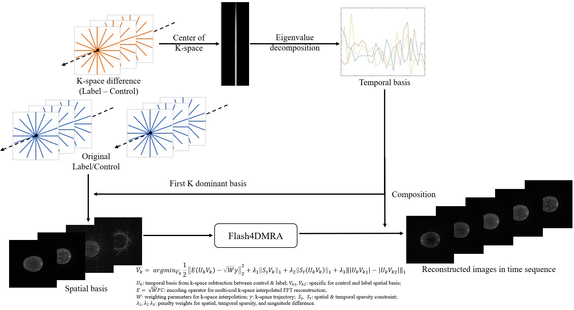
Theory of Flash-4D-MRAThe Flash-4D-MRA sequence is composed of pulsed ASL and SOS golden-angle radial bSSFP acquisition, as previously developed4. The reconstruction workflow of Flash-4D-MRA is shown in Figure 1. A self-calibrated low-rank subspace reconstruction is employed. Specifically, k-space subtraction between label and control is performed in the first step to enhance the ASL signal difference. The temporal basis is then extracted and estimated from the subtracted k-space center with eigenvalue decomposition. This step helps eliminate the influence of tissue components that dominate the image signal and contribute to the temporal feature; thus, the temporal feature of the subtraction basis mainly contains blood dynamics. The first K dominant temporal basis components $$$U_k \in C^{T\times K}$$$ are then picked up and decomposed with label and control datasets termed spatial basis $$$V_{kl} \in C^{K\times N^2}$$$, $$$V_{kc} \in C^{K\times N^2}$$$ individually to reduce the matrix size and accelerate the speed of reconstruction. In the following step, the spatial and temporal sparsity are used for total variation constraint. The label and control dataset are formed together to reconstruct simultaneously, and a magnitude difference constraint between label and control images represented by an L1 norm total variation fashion is employed to further improve the image quality. The optimized penalty weights for spatial, temporal total variation and magnitude subtraction are $$$\lambda_S = 0.0008$$$; $$$\lambda_T = 0.001$$$; $$$\lambda_{ms} = 0.0001$$$. To demonstrate the effectiveness of k-space subtraction on the proposed low-rank subspace reconstruction framework, another temporal basis was also estimated from control and label k-space data individually for comparison.
MRI experiments
Flash-4D-MRA datasets were collected on a Siemens Prisma 3T MR scanner using a 20-channel head coil with the following imaging parameters: FOV =256x256 mm2; voxel size=1x1x1.5 mm3, flip angle = 30o; 64 slices with slice under-sampling factor of 2, in-plane radial spokes = 500, the total scan time of 4 minutes and 40 seconds. Each Flash-4D-MRA dataset was reconstructed with the proposed low-rank subspace reconstruction with subtraction-based temporal basis and individual temporal basis with 3, 5,10, and 20 radial spokes of each frame corresponding to 15 ms/frame, 25 ms/frame, 50 ms/frame, and 100 ms/frame.
Results and Discussion
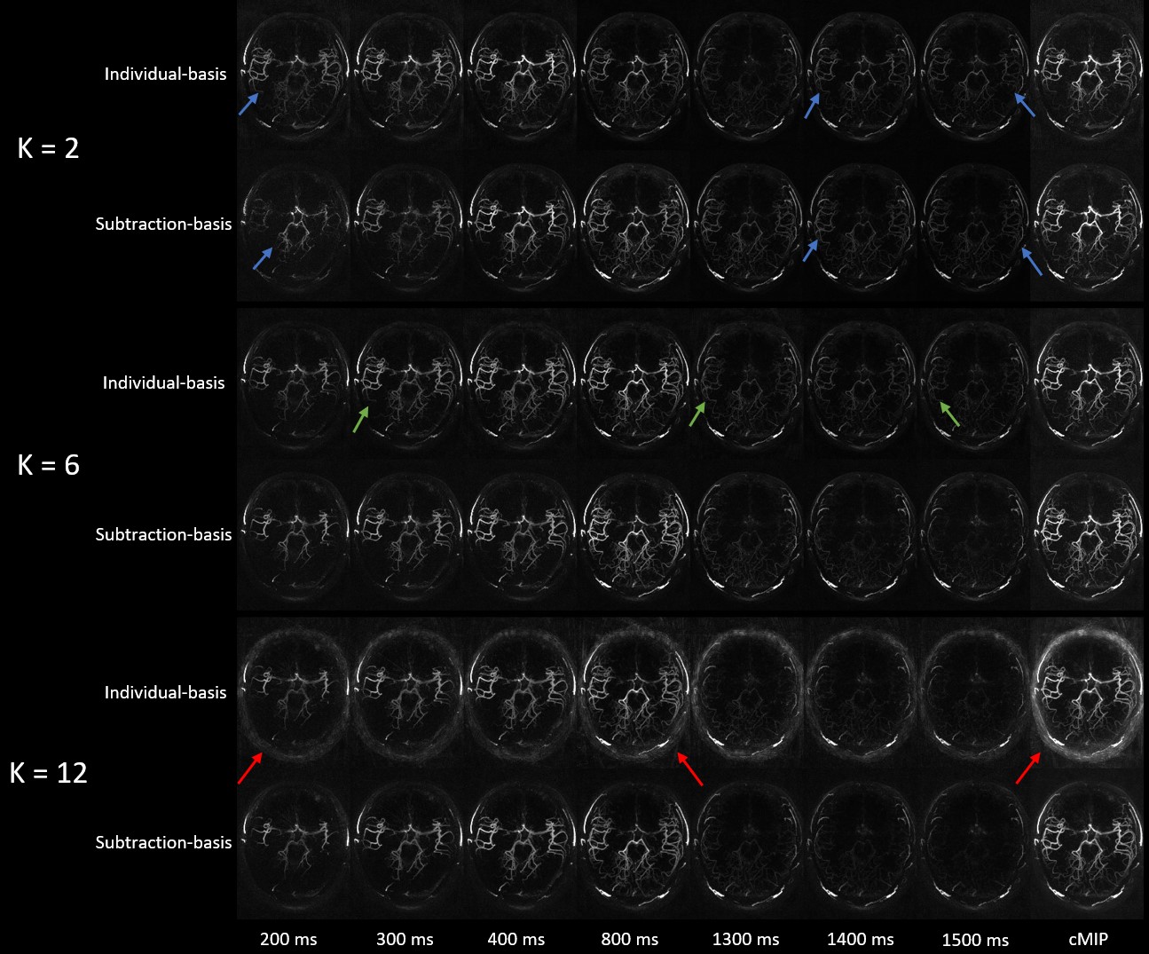
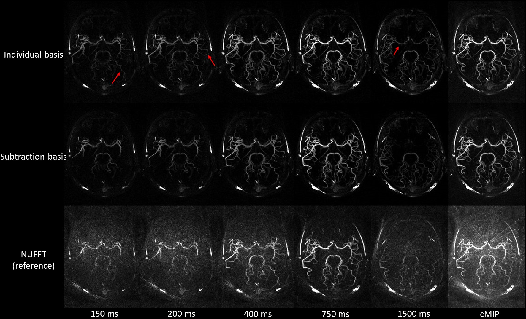
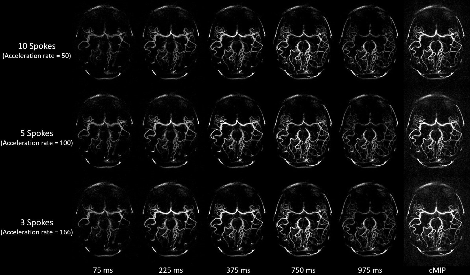
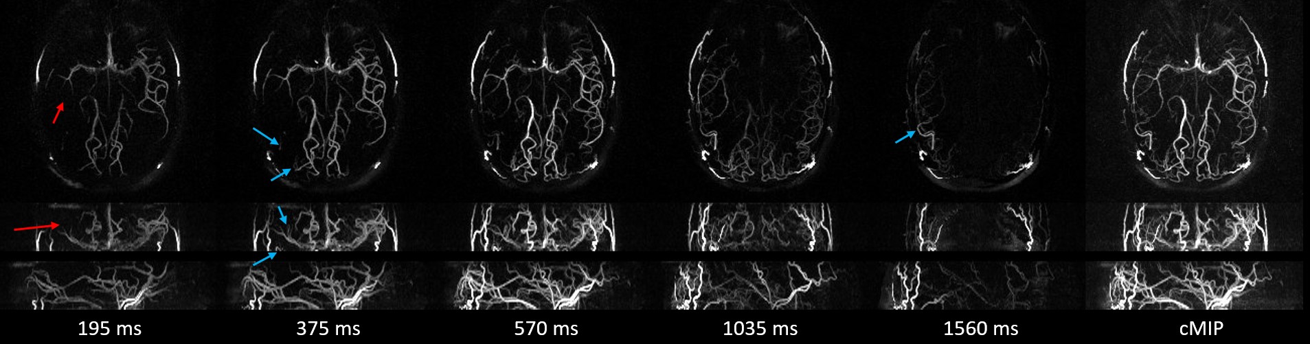
Figure 2 displays several frames of 4D MRA MIP and collapsed MIP (cMIP) images across all frames with different K using subtraction-based and individual temporal basis methods with 20 spokes/frame, respectively. As expected, the noise level intensified as K increased and temporal blurring showed up when K decreased significantly. However, the subtraction basis mitigated temporal burring across different Ks. K=6 was selected as the optimal K in the Flash-4D-MRA reconstruction, which offers good image quality without visible temporal blurring while significantly reducing computational demands. Figure 3 presents another 4D MRA case reconstructed using subtraction-based and individual temporal basis methods with 10 spokes per frame (50 ms/frame), respectively. NUFFT images serve as a reference. Temporal blurring reflected as earlier filling and late drainage was obvious using individual temporal basis. In contrast, the subtraction basis retained temporal fidelity without apparent temporal blurring. Figure 4 showcases another Flash-4D-MRA case with 10, 5, and 3 spokes per frame. Flash-4D-MRA demonstrated reliable performance even with a very high acceleration rate. Figure 5 illustrates a patient case with steno-occlusive disease in the right MCA. Dynamic blood flow alternations on the lesion side were clearly depicted using Flash-4D-MRA.Conclusion
This work presents Flash-4D-MRA, an advanced 4D MRA technique that combines SOS golden-angle radial sampling with joint subtraction-based self-calibrated low-rank subspace and magnitude-subtraction sparsity constraint, offering good image quality even at very high acceleration rates while preserving temporal fidelity. Flash-4D-MRA holds significant promise as a rapid dynamic MRA technique for clinical applications.Acknowledgements
This work was partly supported by National Institute of Health (NIH) grants R01NS118019, RF1AG072490, and BrightFocus Foundation A20201411S.References
1. Song HK, Yan L, Smith RX, Xue Y, Rapacchi S, Srinivasan S, Ennis DB, Hu P, Pouratian N, Wang DJ. Noncontrast enhanced four‐dimensional dynamic MRA with golden angle radial acquisition and K‐space weighted image contrast (KWIC) reconstruction. Magnetic resonance in medicine. 2014 Dec;72(6):1541-51.
2. Feng L, Grimm R, Block KT, Chandarana H, Kim S, Xu J, Axel L, Sodickson DK, Otazo R. Golden‐angle radial sparse parallel MRI: combination of compressed sensing, parallel imaging, and golden‐angle radial sampling for fast and flexible dynamic volumetric MRI. Magnetic resonance in medicine. 2014 Sep;72(3):707-17.
3. Zhou Z, Han F, Yu S, Yu D, Rapacchi S, Song HK, Wang DJ, Hu P, Yan L. Accelerated noncontrast‐enhanced 4‐dimensional intracranial MR angiography using golden‐angle stack‐of‐stars trajectory and compressed sensing with magnitude subtraction. Magnetic resonance in medicine. 2018 Feb;79(2):867-78.
4. Yan L, Wang S, Zhuo Y, Wolf RL, Stiefel MF, An J, Ye Y, Zhang Q, Melhem ER, Wang DJ. Unenhanced dynamic MR angiography: high spatial and temporal resolution by using true FISP–based spin tagging with alternating radiofrequency. Radiology. 2010 Jul;256(1):270-9.
Figures