2483
Automatic segmentation of lenticulostriate arteries from 7T contrast-enhanced MR angiography in patients with cerebral small vessel disease1Department of Clinical Neurosciences, University of Cambridge, Cambridge, United Kingdom, 2Genomics Research Centre, Human Technopole, Milan, Italy, Milan, Italy, 3Faculty of Computer Science, Otto von Guericke University Magdeburg, Magdeburg, Germany, 4Atkinson Morley Regional Neuroscience Centre, St George’s University Hospitals NHS Foundation Trust, London, United Kingdom, 5Wolfson Brain Imaging Centre, Department of Clinical Neurosciences, University of Cambridge, Cambridge, United Kingdom
Synopsis
Keywords: Blood Vessels, Vessels
Motivation: 7T TOF MRA detects the lenticulostriate arteries (LSA), which perfuse important subcortical structures and are implicated in the pathogenesis of cerebral small vessel disease (SVD).
Goal(s): This study aimed to automatically segment LSAs from 7T TOF MRA for SVD patients, to facilitate studies of the arterial pathology of SVD.
Approach: We applied a state-of-the-art deep learning model “DS6” and a classical multi-scale Frangi filter pipeline to 7T contrast-enhanced TOF MRA scans from 8 SVD patients for LSA segmentation.
Results: Both approaches showed comparable and satisfactory performance with mean test dice score=0.74. DS6 was more robust but less sensitive to lower-intensity arteries.
Impact: We present an automatic pipeline for 3D segmentation of the lenticulstriate arteries (LSAs) from 7T TOF MRA. This will enable clinical studies to characterise LSA morphology in cerebral small vessel disease which will open new avenues to understand its pathophysiology.
Introduction
Cerebral small vessel disease (SVD) causes 25% of ischaemic strokes1 and damages small perforating arteries in the brain, including the lenticulostriate arteries (LSAs), which perfuse important subcortical structures.2 In recent decades, 7T MR angiography (MRA) has dramatically improved the in vivo visualisation of LSAs.3 However, many studies quantifying LSA morphology rely on manual labelling using 2D-maximum intensity projection (MIP) images,2,3 which is time-consuming and unreliable for evaluating 3D-morphology. Many vessel segmentation methods, including those using deep learning (DL),4 have been proposed.5,6 However, they were mostly developed on high-quality data from healthy subjects, and their performance on SVD patient data remains unclear. Our study applied a state-of-the-art DL model and a classical method based on Frangi filters5 for LSA segmentation on an SVD cohort.Methods
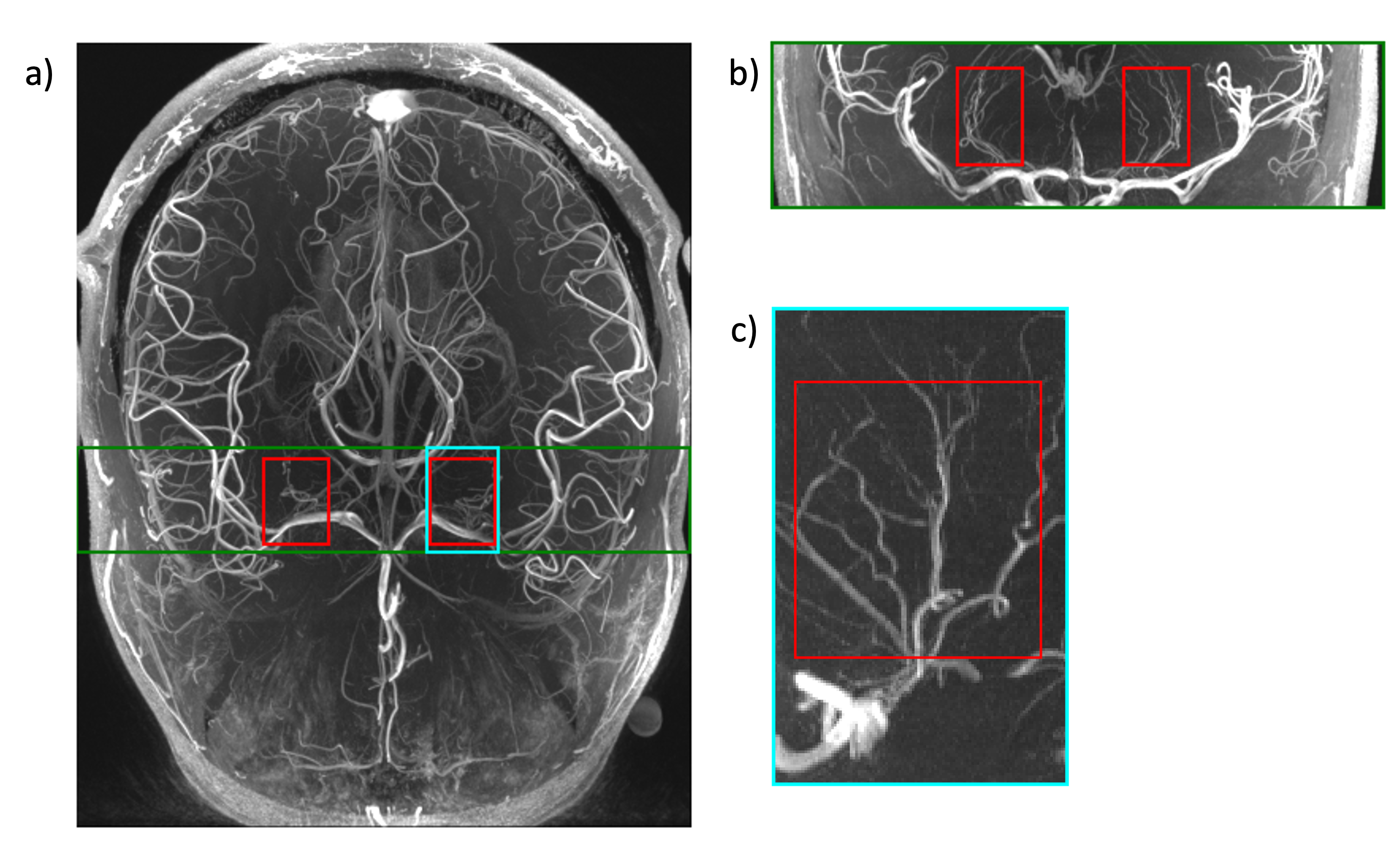
We used data from the Cambridge 7T Cerebral Small Vessel Disease (CamSVD) study, which is an ongoing study recruiting patients with lacunar stroke arising from SVD.3 Contrast-enhanced 7T Time-of-Flight (TOF) MRA was used for vessel segmentation. We randomly selected 8 subjects with lacunar stroke from the current cohort. For each subject, we defined and manually segmented a cubic region-of-interest (ROI) for each hemisphere, each measuring ∼65×100×100 voxels and including predominantly LSAs (Figure 1). These subjects were split into a development set (N=4; N=2 for training and N=2 for validation) and a test set (N=4). Given visible variance in image quality across subjects and to avoid the DL model learning incorrect representation, we used highest-quality images for training, and lowest-quality images for testing.We evaluated a novel DL model “DS6”, which uses a deformation-aware semi-supervised learning approach for vessel segmentation.4 DS6 was pre-trained and validated on 14 whole-brain non-contrast-enhanced 7T TOF MRA volumes from healthy volunteers in the StudyForrest dataset.7 We first tested this pre-trained model as-is on our development set. We also compared DS6 with other extended versions8 but found the original DS6 performed the best. Next, we fine-tuned DS6 using our training data, and the model state with the lowest validation loss was tested on the test set.
For comparison, we also applied the Multi‐Scale Frangi Diffusive Filter (MSFDF) Pipeline,5 which is based on vessel-enhancement filtering. We extended the pipeline with postprocessing methods including morphological closing and removal of small clusters. Optimal hyperparameters were selected based on highest average Dice score on the development set.
All models were evaluated on Dice similarity coefficient (DSC), Intersection over Union (IoU), sensitivity, positive predictive value (PPV).
Results and Discussion
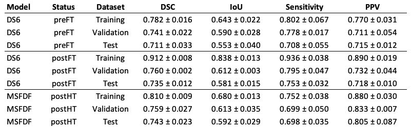
The evaluation results of the DS6 model pre- and post- fine-tuning (FT), and the tuned MSFDF pipeline on all three datasets are shown in Table 1. The preFT scores for DS6 demonstrated a commendable out-of-the-box efficacy of the method. Moreover, a pronounced disparity across the three sets is observable, which may be ascribed to variations in image quality. We then observed that fine-tuning DS6 improved the mean and stability of all metrics on the test set. This proves that DS6 can be fine-tuned even with very little new data to improve its performance for this cohort. Nevertheless, we plan to use more training data soon to reduce the overfitting we observed.Comparing the fine-tuned DS6 with MSFDF in testing, we found MSFDF achieved higher mean DSC and IoU, though the differences were insignificant. DS6 had higher sensitivity (0.753±0.032) than MSFDF (0.698±0.035). Whereas MSFDF had higher PPV (0.805±0.087) than DS6 (0.718±0.010), indicating more segmented voxels being truly vessels. Additionally, DS6 performed more stable than MSFDF on all metrics.
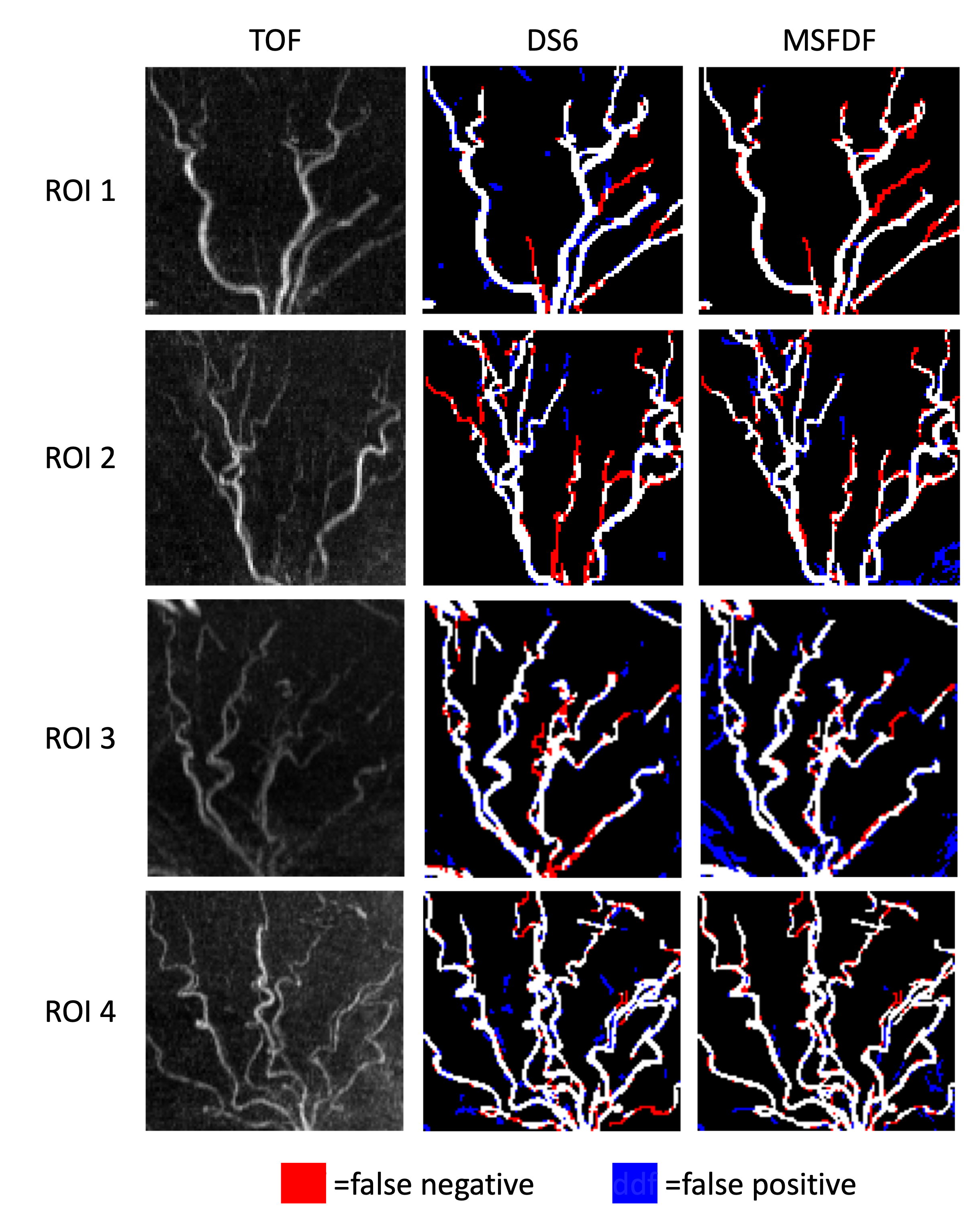
Finally, the segmentation by the fine-tuned DS6 and MSFDF pipeline for example test LSA ROIs are visualised in Figure 3. We observed in the right side of ROI 4 that both models delineated the vascular structure reasonably well despite pronounced background noise. However, both models suffered from over-segmentation where the bright noise pixels are more connected and resemble tubular structure. Moreover, MSFDF undersegmented the width of many branches, whereas DS6 missed more low-intensity LSA segments especially in ROI 2-3.
Conclusion
Our study applied a state-of-the-art DL method DS6, and classical method, MSFDF, for segmenting LSAs on 7T TOF MRA data from an SVD cohort. The fine-tuned DS6 and MSFDF models achieved a mean DSC of 0.735 and 0.743 respectively in testing, with DS6 exhibiting greater stability. Both approaches are feasible for semi-supervised analysis. We are continuing to train DS6 with larger data and regularisation techniques to mitigate overfitting, and we hope that it will enable 3D morphological analysis of LSAs for our SVD study, which has not previously been practical.Acknowledgements
We thank the clinical support provided by Dr Lupei Cai, and the computing facility support provided by Weizhe Lin. RL is funded by a PhD scholarship from Trinity College, Cambridge. This study was supported by the NIHR Cambridge Biomedical Research Centre (NIHR203312 and BRC-1215-20014). The views expressed are those of the authors and not necessarily those of the NIHR or the Department of Health and Social Care. The patient scans were funded by the Cambridge British Heart Foundation (BHF) Centre of Research Excellence (CRE, Centre Code: RE/18/1/34212) and a BHF project grant (PG/19/74/34670). H.S.M. was supported by an NIHR Senior Investigator award. For the purpose of open access, the authors have applied a CC-BY public copyright licence to any Author Accepted Manuscript version arising from this submission.References
- Wardlaw JM, Smith C, Dichgans M. Small vessel disease: mechanisms and clinical implications. The Lancet Neurology. 2019;18(7):684–696.
- Chen YC, Wei XE, Lu J, Qiao RH, Shen XF, Li YH. Correlation Between the Number of Lenticulostriate Arteries and Imaging of Cerebral Small Vessel Disease. Frontiers in Neurology. 2019;10(AUG):882.
- Osuafor CN, Rua C, Mackinnon AD, Egle M, Benjamin P, Tozer DJ, Rodgers CT, Markus HS. Visualisation of lenticulostriate arteries using contrast-enhanced time-of-flight magnetic resonance angiography at 7 Tesla. Scientific Reports 2022 12:1. 2022;12(1):1–9.
- Chatterjee S, Prabhu K, Pattadkal M, Bortsova G, Sarasaen C, Dubost F, Mattern H, de Bruijne M, Speck O, Nürnberger A. DS6, Deformation-Aware Semi-Supervised Learning: Application to Small Vessel Segmentation with Noisy Training Data. Journal of Imaging 2022, Vol. 8, Page 259. 2022;8(10):259.
- Bernier M, Cunnane SC, Whittingstall K. The morphology of the human cerebrovascular system. Human Brain Mapping. 2018;39(12):4962–4975.
- Seo SW, Kang CK, Kim SH, Yoon DS, Liao W, Wörz S, Rohr K, Kim YB, Na DL, Cho ZH. Measurements of lenticulostriate arteries using 7T MRI: new imaging markers for subcortical vascular dementia. Journal of the Neurological Sciences. 2012;322(1–2):200–205.
- Hanke M, Baumgartner FJ, Ibe P, Kaule FR, Pollmann S, Speck O, Zinke W, Stadler J. A high-resolution 7-Tesla fMRI dataset from complex natural stimulation with an audio movie. Scientific Data. 2014;1(1):1–18.
- Chatterjee S, Chintalapati K,
Radhakrishna C, Hudukula Ram Kumar SC, Sutrave R, Mattern H, Speck O,
Nürnberger A. Enhancing Vessel Continuity in Deep Learning based Segmentation
using Maximum Intensity Projection as Loss. ISMRM. 2023.
Figures