2482
Non-invasive Cerebral Arteriovenous Malformation Nidus Volume Measurement: Silent MRA Compared with TOF-MRA1Department of Neurosurgery, Xuanwu Hospital, Capital Medical University, Beijing, China, 2China International Neuroscience Institute, Beijing, China, 3Department of Radiology and Nuclear Medicine, Xuanwu Hospital, Capital Medical University, Beijing, China, 4Beijing Key Laboratory of Magnetic Resonance Imaging and Brain Informatics, Beijing, China, 5The National Center for Neurological Disorders, Xuanwu Hospital, Capital Medical University, Beijing, China
Synopsis
Keywords: Blood Vessels, Image Reconstruction, Cerebral arteriovenous malformations . Digital Subtraction Angiography. Magnetic resonance angiography . Nidus segmentation . Nidus volume
Motivation: Quantitative nidus volume measurement of cerebral arteriovenous malformation(CAVM)is of great important for the CAVM treatment management.
Goal(s): This study aims to investigate non-invasive nidus volume measurement of CAVM with silent MRA and explore the difference between silent MRA and TOF-MRA.
Approach: Nidus volume was measured by silent MRA, TOF-MRA, and digital subtraction angiography(DSA). Nidus volume measured by DSA was regarded as golden standard.
Results: Silent MRA can accurately measure the nidus volume of CAVM. There is a significant difference in nidus volume measurement between silent MRA and TOF-MRA, silent MRA also has advantage for CAVM patients treated by embolization.
Impact: As a non-invasive and contrast agent-free MRA imaging technology, silent MRA can be used as an effective and accurate imaging tool for nidus volume measurement and as a follow-up tool for nidus volume change evaluation after surgery, embolization, and radiotherapy.
INTRODUCTION
Accurate cerebral arteriovenous malformations (CAVM) nidus volume measurement is of essential importance for evaluating treatment strategy and neurological prognosis.This study aimed to compare the accuracy of silent MR angiography (MRA) for measuring CAVM nidus volume with time-of-flight MR angiography (TOF-MRA) and analyze the clinical factors that may affect the nidus volume measurement.METHODS
A total of 80 patients diagnosed with CAVM who received silent MRA, TOF-MRA, and digital subtraction angiography(DSA)examinations were retrospectively reviewed. A standardized nidus volume measurement protocol was used to measure the nidus volume by these three imaging tools.RESULTS
The mean nidus volume measured by 3D-DSA, silent MRA, and TOF-MRA was 9.073±7.667 ml, 9.55±7.814 ml, and 7.773±7.11ml, respectively. The mean nidus volume difference was 0.477±1.715 ml (P =0.069) and 1.30±2.0 0 ml (P=0.00) between 3D-DSA and silent MRA, 3D-DSA, and TOF-MRA, respectively. The linear correlation coefficient of the nidus volume was 0.976 (P<0.001) and 0.966 (P<0.001), respectively. For nidus overlap volume and volume discrepancy, Psilentol (0.775±0.109) was significantly higher than Ptofol (0.538±0.206)(P=0.00), and Ptofdis (0.196±0.192) was significantly higher than Psilentdis (0.114±0.147) (P=0.005). For both silent MRA and TOF MRA, the nidus overlap volume and volume discrepancy were significantly associated with previous embolization status.CONCLUSION
Nidus volume measurement by silent MRA is accurate and practical. Silent MRA can be used for nidus volume measurement and as a follow-up tool for nidus volume change evaluation after surgery, embolization, and radiotherapy.Acknowledgements
We are grateful to Doctor Juan Wei of GE Healthcare for technical support.References
1. Chen Ching-Jen, Ding Dale, Derdeyn Colin P, et al.(2020)Brain arteriovenous malformations: A review of natural history, pathobiology, and interventions. Neurology 95(20).
2. Solomon Robert A, Connolly E Sander.(2017)Arteriovenous Malformations of the Brain. The New England journal of medicine 376(19):1859-1866.
3. Derdeyn Colin P, Zipfel Gregory J, Albuquerque Felipe C, et al.(2017)Management of Brain Arteriovenous Malformations: A Scientific Statement for Healthcare Professionals From the American Heart Association/American Stroke Association. Stroke 48(8):e200-e224.
4. Chen K-K , Guo W-Y , Yang H-C , et al. (2017)Application of Time-Resolved 3D Digital Subtraction Angiography to Plan Cerebral Arteriovenous Malformation Radiosurgery. AJNR. American journal of neuroradiology 38(4):740-746.
5. Ozyurt Onur , Dincer Alp , Erdem Yildiz Mehmet , et al.(2017) Integration of arterial spin labeling into stereotactic radiosurgery planning of cerebral arteriovenous malformations. Journal of magnetic resonance imaging : JMRI 46(6):1718-1727.
6. Hasegawa Hirotaka , Hanakita Shunya , Shin Masahiro, et al.(2018)Integration of rotational angiography enables better dose planning in Gamma Knife radiosurgery for brain arteriovenous malformations. Journal of neurosurgery 129(Suppl1):17-25.
7. Turner Ryan C , Lucke-Wold Brandon P , Josiah Darnell , et al.(2016)Stereotactic radiosurgery planning based on time-resolved CTA for arteriovenous malformation: a case report and review of the literature. Acta neurochirurgica 158(8):1555-62.
8. Chenoune Y , Tankyevych O , Li F , et al.(2019)Three-dimensional segmentation and symbolic representation of cerebral vessels on 3DRA images of arteriovenous malformations. Computers in biology and medicine 115:103489.
9. Hamilton M G , Spetzler R F.(1994)The prospective application of a grading system for arteriovenous malformations. Neurosurgery 34(1):2-6; discussion 6-7.
10. Jiao Yuming , Wu Jun , Chen Xin , et al.(2018)Spetzler-Martin grade IV and V arteriovenous malformations: Treatment outcomes and risk factors for negative outcomes after surgical resection. Journal of clinical neuroscience : official journal of the Neurosurgical Society of Australasia 61.
11. Morgan Michael Kerin , Davidson Andrew Stewart , Assaad Nazih N A , et al.(2017) Critical review of brain AVM surgery, surgical results and natural history in 2017. Acta neurochirurgica 159(8):1457-1478.
12. Lopes Demetrius K , Moftakhar Roham , Straus David , et al.(2015)Arteriovenous malformation embocure score: AVMES. Journal of neurointerventional surgery 8(7):685-91.
13. Seymour Zachary A , Chan Jason W , Sneed Penny K , et al.(2019)Dose response and architecture in volume staged radiosurgery for large arteriovenous malformations: A multi-institutional study. Radiotherapy and oncology : journal of the European Society for Therapeutic Radiology and Oncology 144:180-188.
14. Meng Xiangyu, Gao Dezhi, Jin Hengwei .(2021)Factors Affecting Volume Reduction Velocity for Arteriovenous Malformations After Treatment With Dose-Stage Stereotactic Radiosurgery. Frontiers in oncology 11:769533.
15. Seymour Zachary A, Chan Jason W, McDermott Michael W , et al.(2021)Adverse radiation effects in volume-staged radiosurgery for large arteriovenous malformations: a multiinstitutional study. Journal of neurosurgery 136(2):503-511.
16. Li Weiyan , Wang Yuqiang , Lu Lantao , et al.(2021)The factors associated with obliteration following stereotactic radiosurgery in patients with brain arteriovenous malformations: a meta-analysis. ANZ journal of surgery 92(5).
17. Huang Yu-Jie, Jih-Tsun Lee, Tsair-Fwu Hsu, et al. (2017) Consistency between targets delineated by angiography, computed tomography, and magnetic resonance imaging in stereotactic radiosurgery for arteriovenous malformation. Stereotactic and Functional Neurosurgery: Official Journal of the World Society for Stereotactic and Functional Neurosurgery.
18. Yu C, Petrovich Z, Apuzzo M. L. J., et al. (2004). Study of magnetic resonance imaging-based arteriovenous malformation delineation without conventional angiography. Neurosurgery(4), 4.
19. Veeravagu A, Hansasuta A, Jiang B, et al. (2009). Volumetric analysis of intracranial arteriovenous malformations contoured for cyberknife radiosurgery with 3-dimensional rotational angiography vs computed tomography/magnetic resonance imaging. Neurosurgery, 73(2), 262-270.
20. Conti A, Pontoriero A, Giuseppe Faragò, et al. (2011). Integration of three-dimensional rotational angiography in radiosurgical treatment planning of cerebral arteriovenous malformations. International Journal of Radiation Oncology*Biology*Physics, 81(3).
21. Aoyama H, Shirato H, Katoh N, et al. (2005) Comparison of imaging modalities for the accurate delineation of arteriovenous malformation, with reference to stereotactic radiosurgery. International Journal of Radiation Oncology, Biology, Physics, 62(4), 1232-1238.
22. Tomura N, Saginoya T, Kokubun M, et al. (2019) Comparison of time-of-flight–magnetic resonance angiography from silent scan magnetic resonance angiography in depiction of arteriovenous malformation of the brain. Journal of Computer Assisted Tomography, 43.
23. Buis D R, Lagerwaard F J, Dirven C M F, et al. (2007) Delineation of brain avms on mr-angiography for the purpose of stereotactic radiosurgery. Int J Radiat Oncol Biol Phys, 67(1), 308-316.
24. Wowra B, Muacevic A, Tonn J C, et al. (2009) Obliteration dynamics in cerebral arteriovenous malformations after cyberknife radiosurgery: quantification with sequential nidus volumetry and 3-tesla 3-dimensional time-of-flight magnetic resonance angiography. Neurosurgery(2).
25. Moon J I, Baek H J, Ryu K H, et al. (2017) A novel non-contrast-enhanced mra using silent scan for evaluation of brain arteriovenous malformation: a case report and review of literature. other(45).
26. Tomura N, Saginoya T, Kokubun M, et al. (2019) Comparison of time-of-flight–magnetic resonance angiography from silent scan magnetic resonance angiography in depiction of arteriovenous malformation of the brain. Journal of Computer Assisted Tomography, 43.
27. Arai N, Akiyama T, Fujiwara K, et al. (2020) Silent mra: arterial spin labeling magnetic resonant angiography with ultra-short time echo assessing cerebral arteriovenous malformation. Neuroradiology, 62(4), 455-461.
28. Wu C X, Zang Z X, Hong T, et al. (2021) Signal intensity ratio of draining vein on silent mr angiography as an indicator of high-flow arteriovenous shunt in brain arteriovenous malformation. European Radiology, 1-10.
Figures
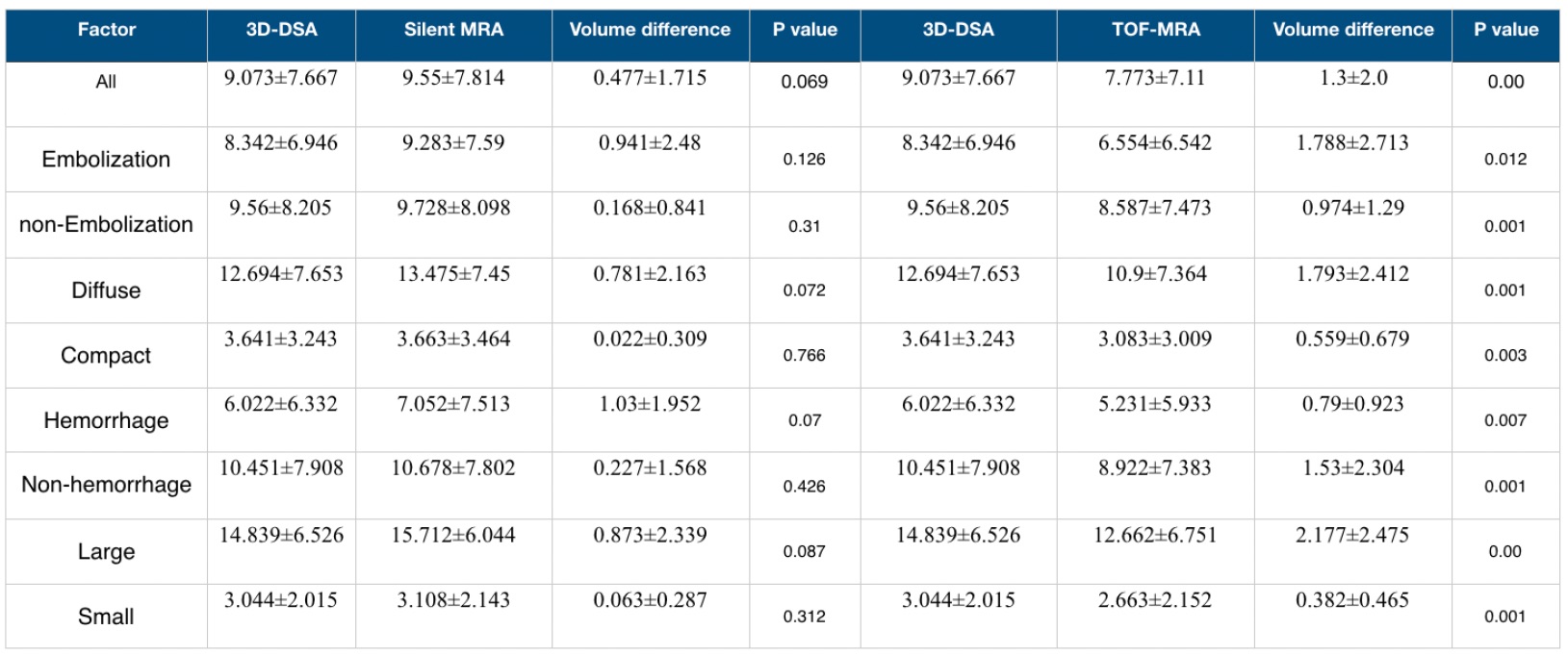
The comparison of nidus volume measurement results between 3D-DSA and silent MRA/ TOF-MRA
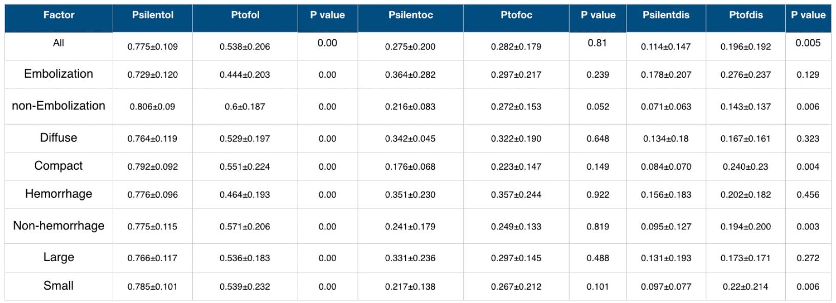
The comparison of overlapping, over-cover nidus volume and nidus volume differences in different imaging modalities
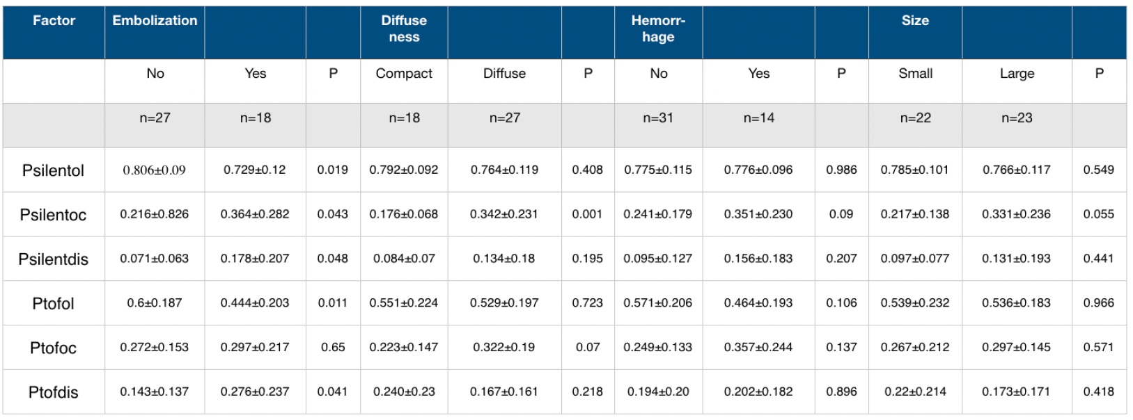
The influence of clinical factors on nidus volume measurement
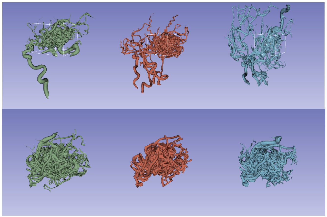
Standardized nidus volume measurement protocol proposed by our team. Green: 3D-DSA. Red: silent MRA. Blue: TOF-MRA.
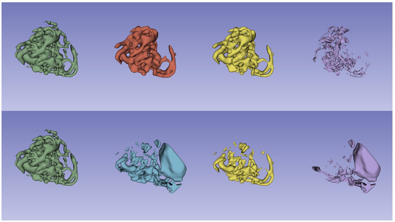
Illustration of a CAVM case measured by 3D-DSA, silent MRA, and TOF-MRA. Green: 3D-DSA. Red: silent MRA. Blue: TOF-MRA. Yellow: overlapping volume. Purple: over-cover volume.