2481
Echo-uT1RESS Sequence with Radial Stack-of-Stars Acquisition for Dynamic Contrast-Enhanced Dark Blood Imaging1The Bernard and Irene Schwartz Center for Biomedical Imaging, Department of Radiology, New York University Grossman School of Medicine, New York, NY, United States, 2Center for Advanced Imaging Innovation and Research (CAI2R), Department of Radiology, New York University Grossman School of Medicine, New York, NY, United States, 3Vilcek Institute of Graduate Biomedical Sciences, New York University Grossman School of Medicine, New York, NY, United States, 4Radiology, Northshore University HealthSystem, Evanston, IL, United States, 5Siemens Medical Solutions, New York, NY, United States
Synopsis
Keywords: Blood Vessels, Tumor, dark-blood DCE
Motivation: Scans with dark-blood contrast can improve the conspicuity of small metastases in contrast-enhanced brain examinations and may be useful for vessel-wall imaging.
Goal(s): To describe a novel sequence, called echo-uT1RESS, that overcomes the limitations of the frequently utilized T1 SPACE sequence.
Approach: The proposed stack-of-stars echo-uT1RESS sequence applies saturation-recovery preparation followed by 3D PSIF readout. Due to radial k-space acquisition, scans are motion-insensitive and can be acquired during free breathing. Dynamic contrast-enhanced images can be obtained through GRASP reconstruction.
Results: Evaluation in a small patient cohort demonstrated high conspicuity of enhancing lesions and vessel walls with reduced motion artifacts compared to conventional sequences.
Impact: The described radial echo-uT1RESS sequence offers improved lesion-to-background and dark-blood contrast. The sequence may help to increase the sensitivity for detecting small metastases and may find application in vessel-wall imaging. It is also compatible with dynamic imaging using GRASP reconstruction.
Introduction
Magnetization-prepared rapid gradient-echo (MP-RAGE) acquisition is a standard choice for contrast-enhanced imaging of the brain. It depicts both enhancing lesions and blood vessels with bright signal intensity. To improve the conspicuity of small metastases, it can be advantageous to instead utilize sequences with dark-blood properties, as also required for imaging vessel walls in the head and neck. So far, T1-weighted SPACE has been the primary sequence for this application.1 However, its limitations include sensitivity to respiratory motion and blurring from T2 modulation, which can be problematic for applications outside the head. Recently, the unbalanced T1 relaxation-enhanced steady-state (uT1RESS) sequence has been proposed, which suppresses intravascular blood using unbalanced weakly spoiled gradients while keeping the enhancing lesion bright.2,3In this work, we present an improved variant with dark-blood contrast, called echo-uT1RESS, which is based on 3D PSIF readout and radial stack-of-stars sampling. The sequence is motion-robust and, furthermore, supports dynamic contrast-enhanced (DCE) acquisition. First results are demonstrated for brain, neck, and prostate imaging at 3T.
Methods
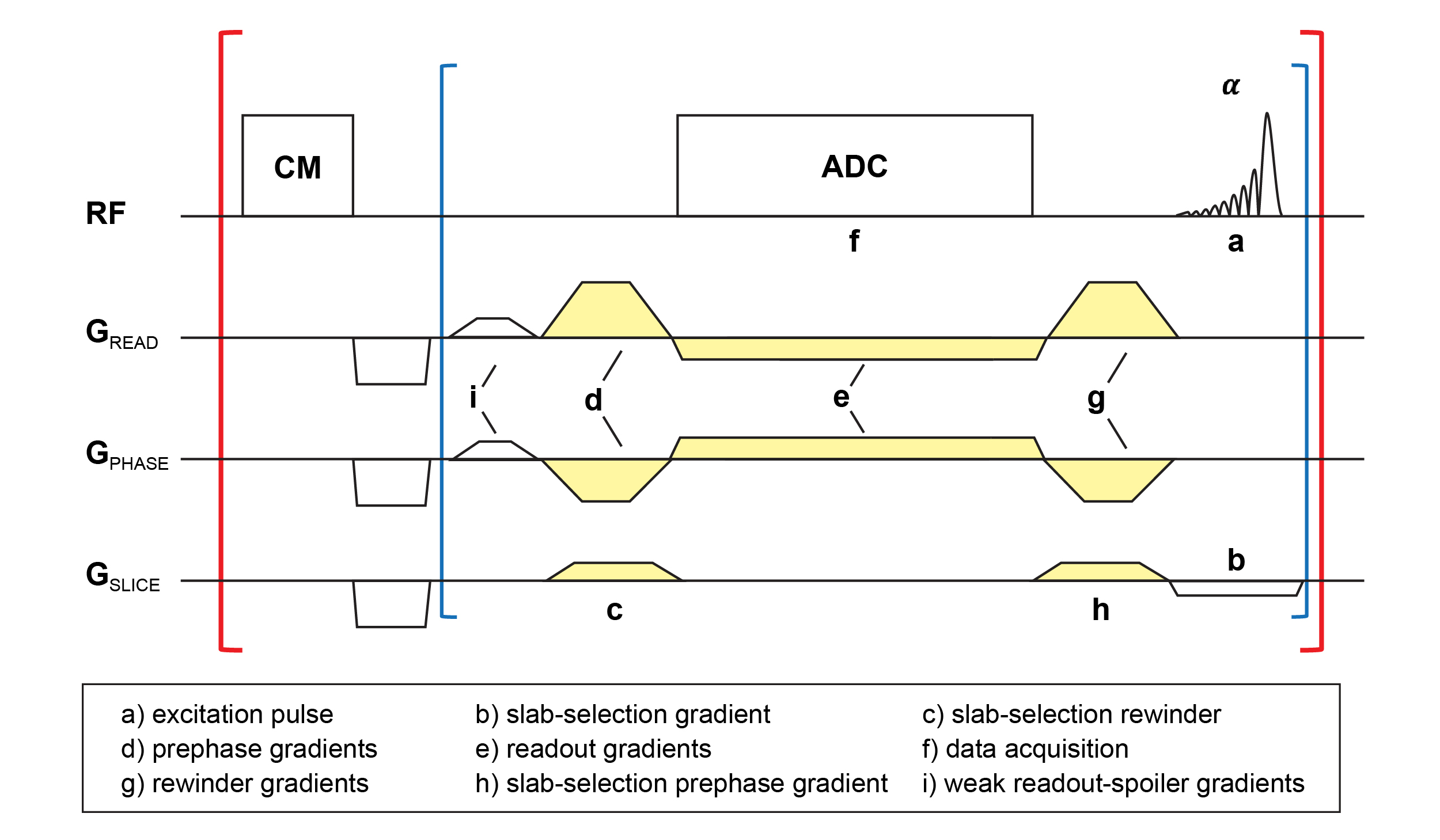
Sequence DesignA stack-of-stars echo-uT1RESS sequence (Figure 1) with golden-angle ordering has been implemented following the general scheme for radial 3D GRE acquisition4. Prior to acquiring the stack of partitions for one radial angle, a non-selective contrast-modifying (CM) saturation-recovery module is executed to induce T1 weighting. The sequence is acquired with a 3D PSIF (reversed FISP) readout to collect the spin-echo signal, which results in dark blood signal. At the start of each TR, a weak gradient spoiler (Figure 1.i) is applied in constant frequency-encoding direction throughout the entire acquisition, which creates a controllable amount of flow-induced dephasing. An option for slab-selective binomial water excitation was implemented as alternative to regular fat saturation.
Experiments
IRB-approved acquisitions were performed using clinical 3T systems (MAGNETOM Skyrafit, Siemens Healthineers). 11 subjects were imaged for clinical indications including brain metastases, peripheral artery disease, cancer (prostate, rectal), and lymphadenopathy. Typical scan parameters included: axial orientation, isotropic resolution (1.0 or 1.17 mm), 96 partitions, 512 radial views, BW 690 Hz/px, TA 4:58 min. Comparisons were made to MP-RAGE, VIBE, SPACE, and/or HASTE.
Dynamic Imaging
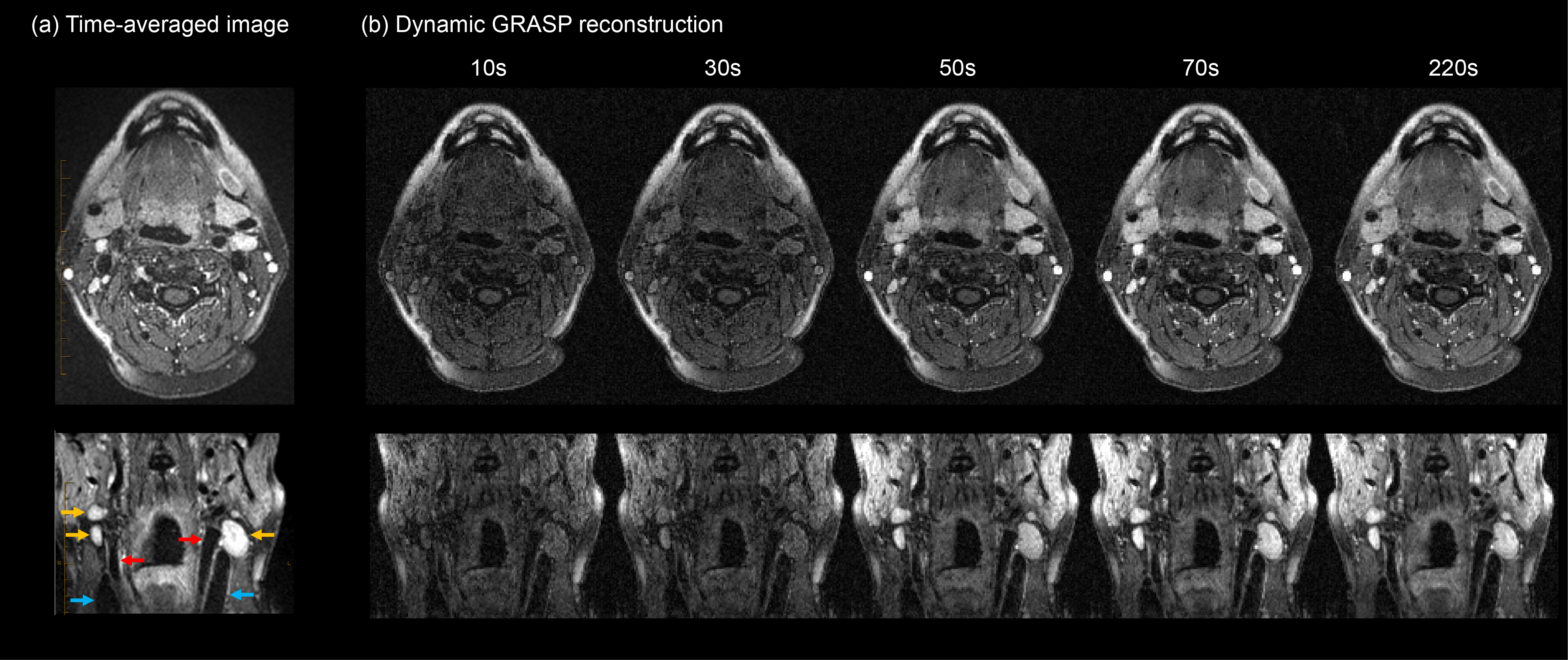
For dynamic acquisition, the sequence was started 20 seconds before injection of 0.1 mmol/kg of gadobutrol. Time-averaged images, reconstructed from all radial views, were calculated at the scanner to provide immediate feedback. Dynamic images were reconstructed offline from the same dataset using the GRASP algorithm5, combining 5 or 13 radial views into each frame for a temporal resolution of 4 or 10 seconds/frame.
Results
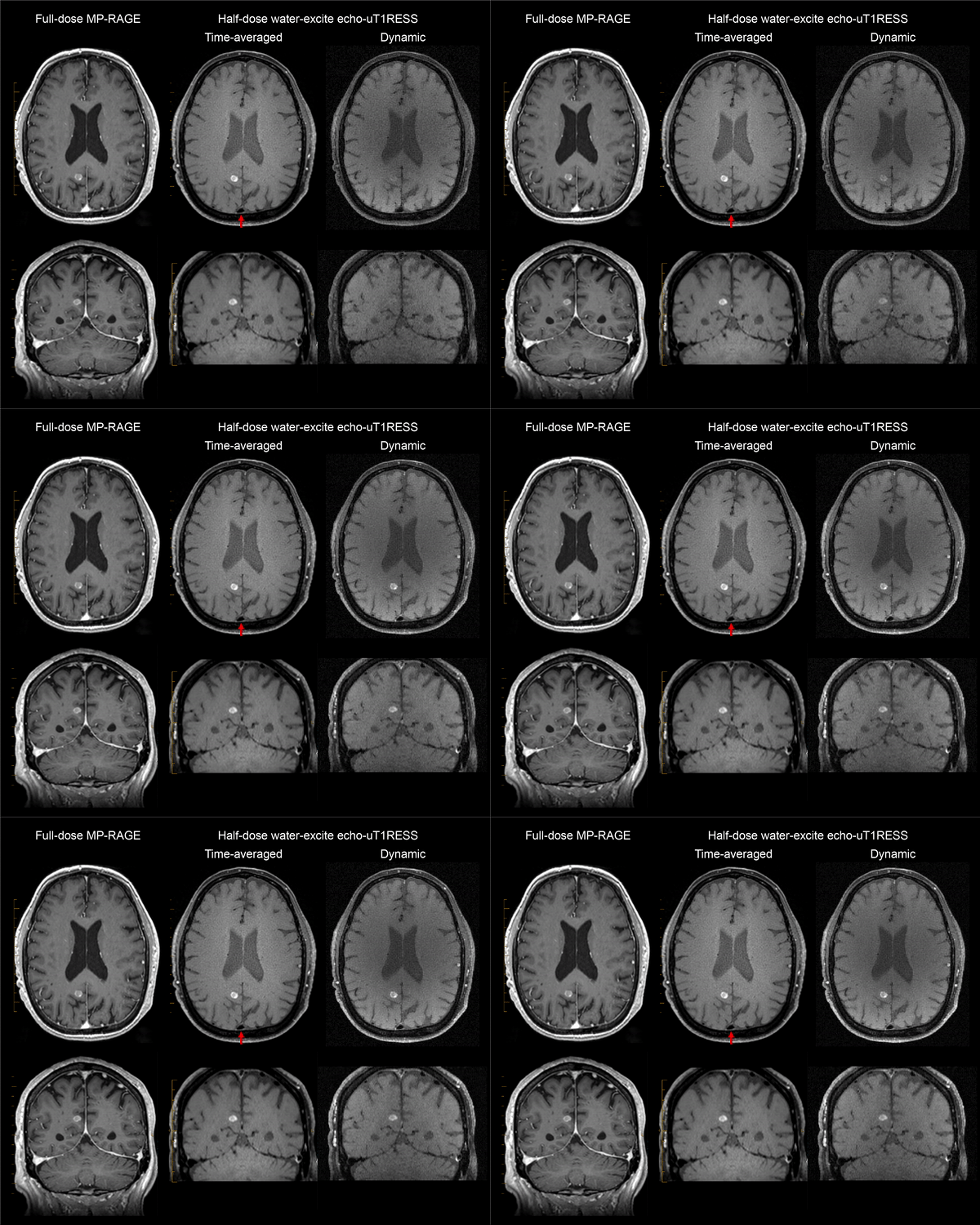
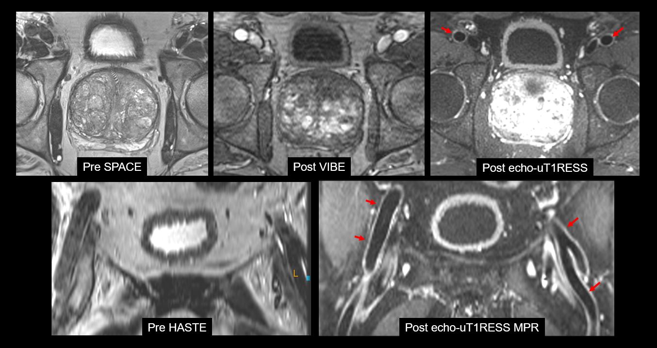
Figure 2 compares images acquired with MP-RAGE and echo-uT1RESS for a patient with brain metastases. Echo-uT1RESS provides higher lesion conspicuity using a half dose of gadobutrol compared to the full-dose scan acquired with MP-RAGE. Blood vessels remain dark in time-averaged and dynamic images. The temporal enhancement can be appreciated in the dynamic reconstruction.Figure 3 shows images of a patient examined due to elevated PSA value. The femoral arteries appear dark in the post-contrast echo-uT1RESS image compared to VIBE. Moreover, no motion artifacts are seen with radial stack-of-stars acquisition, compared to blurring or aliasing seen in the Cartesian VIBE and HASTE images.
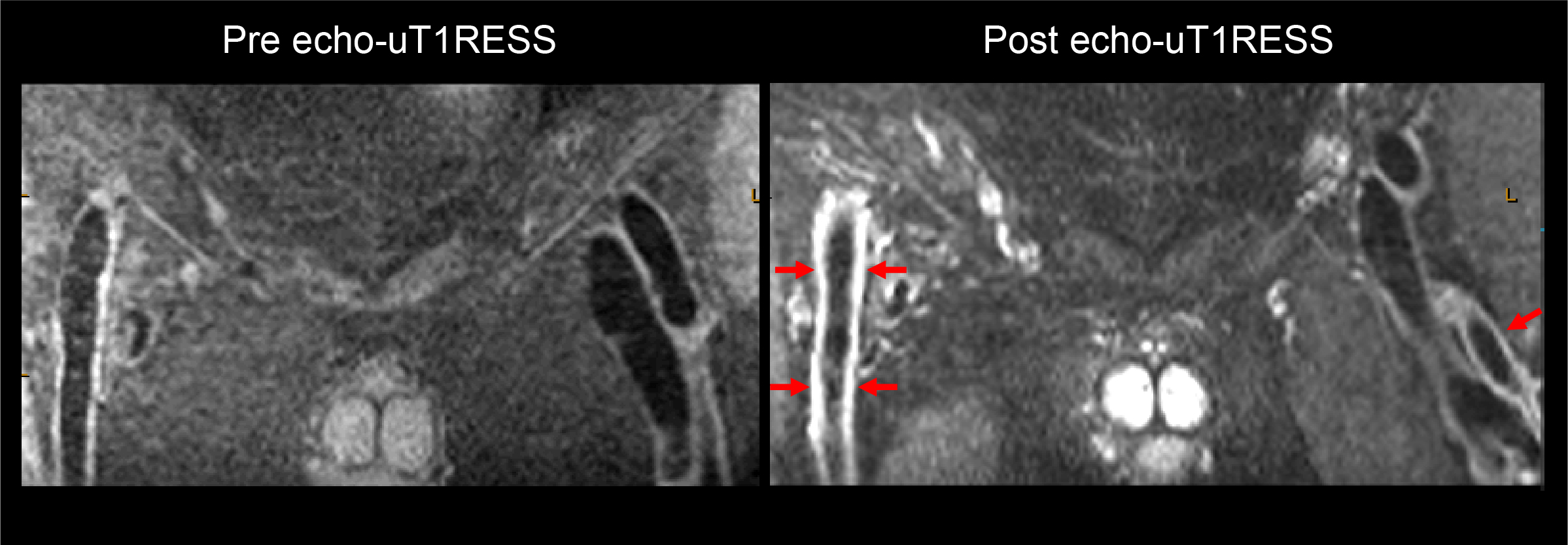
Figure 4 depicts the wall enhancement seen in the femoral arteries after contrast administration. The intraluminal blood appears dark in pre- and post-contrast echo-uT1RESS images.
Figure 5 shows time-averaged and dynamic images of a patient with cervical lymphadenopathy. The enhancing lymph nodes (yellow arrows) can be easily recognized compared to the dark internal carotid arteries (red arrows) and jugular veins (blue arrows). Gradual enhancement of the lymph nodes is observed in the dynamic frames while blood vessels remain dark.
Discussion
This work presents a novel pulse sequence for volumetric dark-blood imaging. First evaluations of the stack-of-stars echo-uT1RESS sequence in a small patient cohort revealed good-to-excellent image quality. The high motion robustness of radial sampling mitigates artifacts normally induced by respiration or bowel peristalsis and, thus, enables free-breathing scans with isotropic resolution. In addition, the sequence can be combined with the GRASP algorithm for generating a dark-blood DCE-MRI series with high temporal resolution. Similar to the previously described uT1RESS sequence with FISP readout2,3, echo-uT1RESS provides higher lesion-to-background contrast than MP-RAGE, allowing to reduce the amount of gadobutrol administration at a comparable level of lesion conspicuity. Moreover, the dark-blood contrast of echo-uT1RESS helps to identify small lesions near blood vessels, which may pose an advantage over the original uT1RESS sequence.Planned next steps include optimizing and shortening the total scan time by applying GRAPPA acceleration along the kz direction, and evaluating the clinical performance and reliability systematically in a larger patient cohort.
Acknowledgements
This work was supported by NIH R01 CA263091 and R21 CA273280. It was performed under the rubric of the Center for Advanced Imaging Innovation and Research (CAI2R, www.cai2r.net), an NIBIB National Center for Biomedical Imaging and Bioengineering (NIH P41 EB017183).References
1. Kato Y, Higano S, Tamura H, Mugikura S, Umetsu A, Murata T, Takahashi S. Usefulness of contrast-enhanced T1-weighted sampling perfection with application-optimized contrasts by using different flip angle evolutions in detection of small brain metastasis at 3T MR imaging: comparison with magnetization-prepared rapid acquisition of gradient echo imaging. American Journal of Neuroradiology. 2009, 30 (5): 923-929.
2. Edelman R, Leloudas N, Pang J, Bailes J, Merrell R, Koktzoglou I. Twofold improved tumor-to-brain contrast using a novel T1 relaxation-enhanced steady-state (T1RESS) MRI technique. Science advances. 2020 Oct 28;6(44):eabd1635.
3. Edelman RR, Koktzoglou I, Leloudas N, Pang J. T1 Relaxation-Enhanced Steady-State (T1RESS): An Improved Three-Dimensional Method for Contrast-Enhanced Imaging of Brain Tumors.
4. Block KT, Chandarana H, Milla S, Bruno M, Mulholland T, Fatterpekar G, et al. Towards Routine Clinical Use of Radial Stack-of-Stars 3D Gradient-Echo Sequences for Reducing Motion Sensitivity. Vol. 18, Journal of the Korean Society of Magnetic Resonance in Medicine. 2014;p.87.
5. Feng L, Grimm R, Block KT, Chandarana H, Kim S, Xu J, Axel L, Sodickson DK, Otazo R. Golden-angle radial sparse parallel MRI: combination of compressed sensing, parallel imaging, and golden-angle radial sampling for fast and flexible dynamic volumetric MRI. Magn Reson Med. 2014 Sep;72(3):707-17.
Figures