2480
Neuroimaging Insights: Structural Changes and Classification in Ménière's Disease1Union Hospital, Tongji Medical College, Huazhong University of Science and Technology, Wuhan, China, 2Philips Healthcare, Wuhan, China
Synopsis
Keywords: Diagnosis/Prediction, Brain, neuroanatomical
Motivation: Emerging evidence suggests that Ménière's disease (MD) may extend beyond the confines of the inner ear, and involved the central nervous system.
Goal(s): To investigate the neuroanatomical alterations associated with MD and to develop a machine learning classification model to effectively discriminate between MD patients and HC.
Approach: A case-control morphometry study was performed to examine potential brain structural changes and delineate the diagnostic utility of these identified brain alterations.
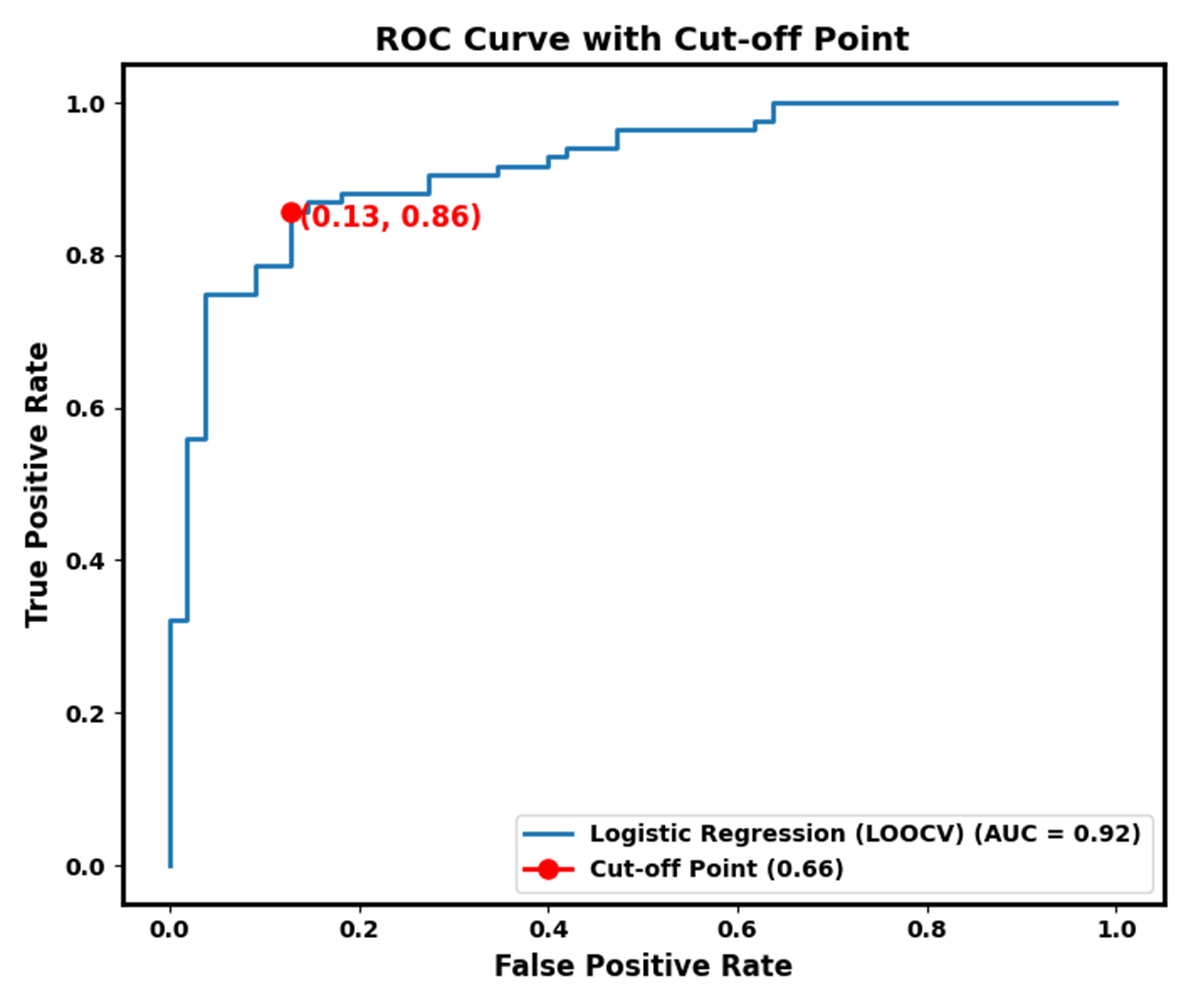
Results: Distinctive alterations in gray matter volume and cortical thickness were identified in regions implicated in emotional processing and sensory integration. The classification model showcased a discriminative power with an impressive AUC value(0.92).
Impact: MD patients showed distinctive morphometry alterations, and were leveraged as potential biomarkers, facilitating the discrimination between MD and HC.These findings provide critical insights into the intricate neuroanatomical alterations in MD and highlight the diagnostic potential of advanced neuroimaging techniques.
Introduction
Ménière's disease(MD) is a distinctive inner ear disorder characterized by spontaneous episodic vertigo, fluctuating sensorineural hearing loss, tinnitus, and other aural symptoms. While the pathophysiology of Meniere's disease has traditionally been attributed to disturbances in the inner ear's fluid dynamics, however, emerging evidence suggests potential involvement of the central nervous system1,2, prompting investigations beyond the peripheral manifestations. In this study, we aimed to address this gap in the literature by employing both Voxel-Based Morphometry(VBM) and Surface-Based Morphometry(SBM) analyses to comprehensively examine potential brain structural changes in MD patients and delineate the diagnostic utility of these identified brain alterations.By intertwining structural brain alterations, clinical parameters, and diagnostic potential, it is hoped that our study not only extends the current understanding of MD but also establishes a novel diagnostic framework for integrating neuroimaging findings with clinical metrics.Methods
A case-control study of 55 MD and 50 healthy control(HC) participants were collected. All participants underwent MRI scanning using a 3.0 Tesla MRI scanner(Philips, Ingenia CX) equipped with a 32-channel head coil. The acquisition of anatomical images utilized a high-resolution T1-weighted 3-dimensional fast gradient echo sequence [“Turbo Field Echo,” Repetition Time (TR) = 11.2 s, Echo Time (TE) = 5.1 s, Flip Angle (FA) = 8o, slice thickness = 0.7 mm, slices number = 258]. Intratympanic injection of a contrast agent (Gd-DTPA-dimeglumine solution; MultiHance, Braccosine, Shanghai, China) was administered, and delayed 3D-FLAIR MRI was employed to evaluate the extent of endolymphatic hydrops(EH)3.The vestibular hydrops ratio (VHR), which reflects the proportion of the endolymphatic space area relative to the total lymphatic space within the vestibule, was defined by the following formula: VHR = (number of negative pixels for the endolymph in the region of interest (ROI) / total number of pixels in the ROI) 4. The processing and analysis of the anatomical images were conducted utilizing the CAT12 toolbox5. In order to assess the discriminatory potential of these varied brain regions in distinguishing MD patients from HCs, a machine-learning classification model was developed.The Logistic Regression model was utilized for both feature selection and classification tasks. To select informative features, the SelectFromModel method with L2 regularization was employed, optimizing the feature set by using a threshold set to the median of feature importances. Leave-One-Out Cross Validation (LOOCV) was employed for robust model evaluation.
Results
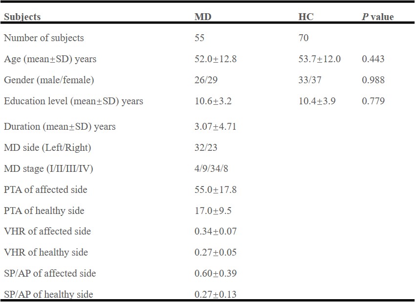
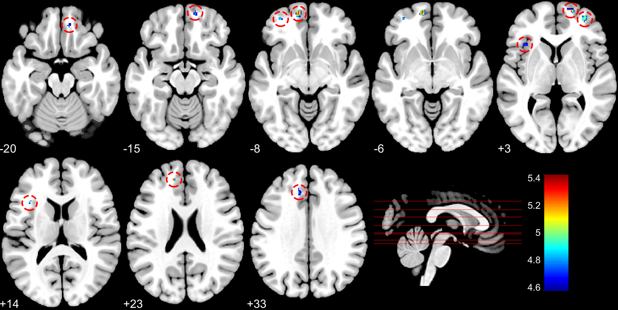
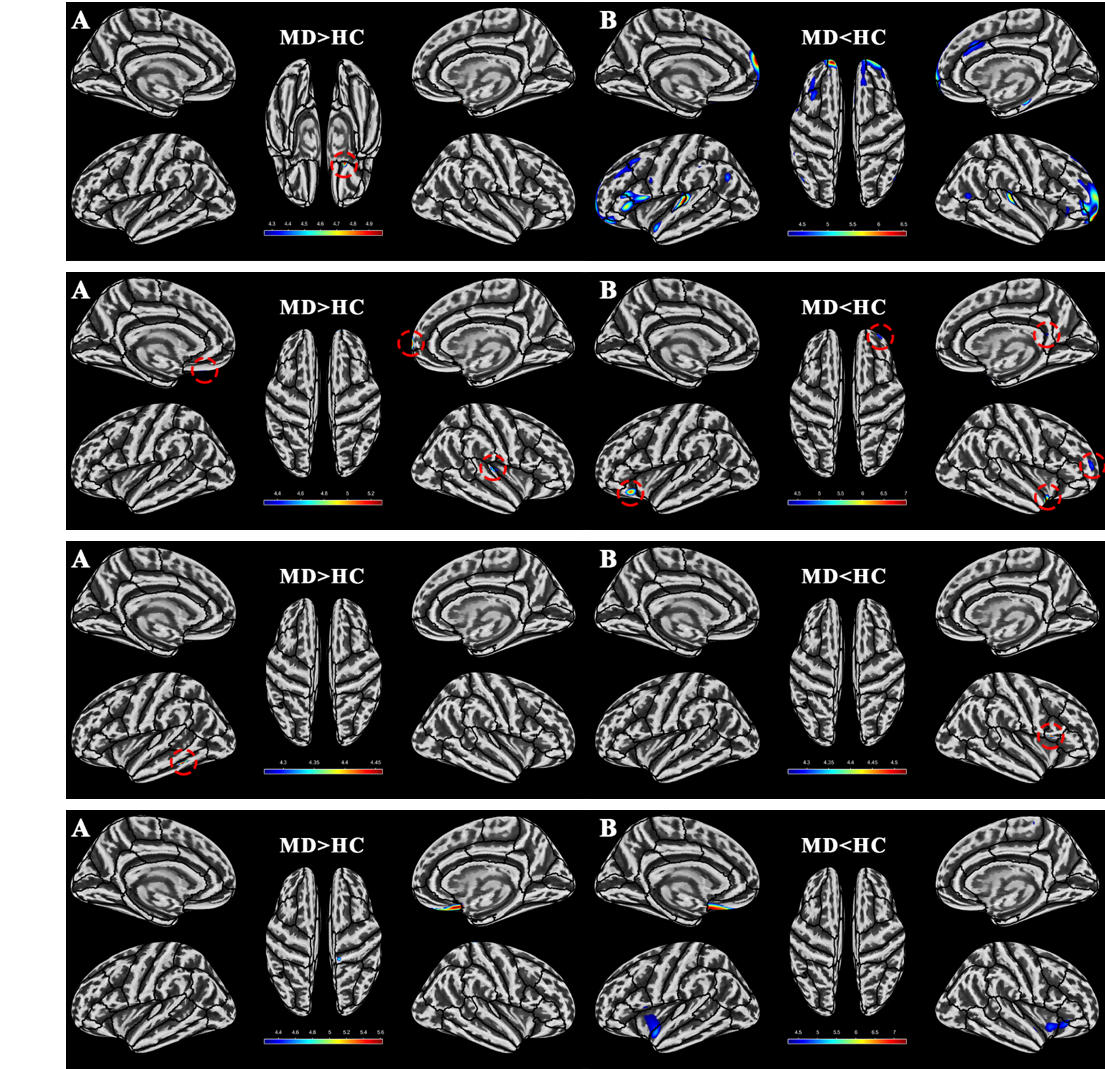
The demographic characteristics of the participants are presented in Figure 1. VBM analysis unveiled extensive alterations in brain gray matter volume among individuals with MD. As illustrated Figure 2, compared to the HC group, MD patients exhibited reduced gray matter volume in the bilateral superior frontal gyrus(dorsolateral, medial, medial orbital), bilateral middle frontal gyrus, left insula, left inferior frontal gyrus(triangular part), left middle cingulate and paracingulate gyri and right gyrus rectus(p < 0.05, FWE correction).SBM analysis revealed distinct patterns of cortical thickness, cortical complexity, gyrification index, and sulcal depth alterations among MD patients(Figure 3). Distinctive patterns of alterations of these four parameters of MD patients were observed in brain regions associated with emotional processing and sensory integration.
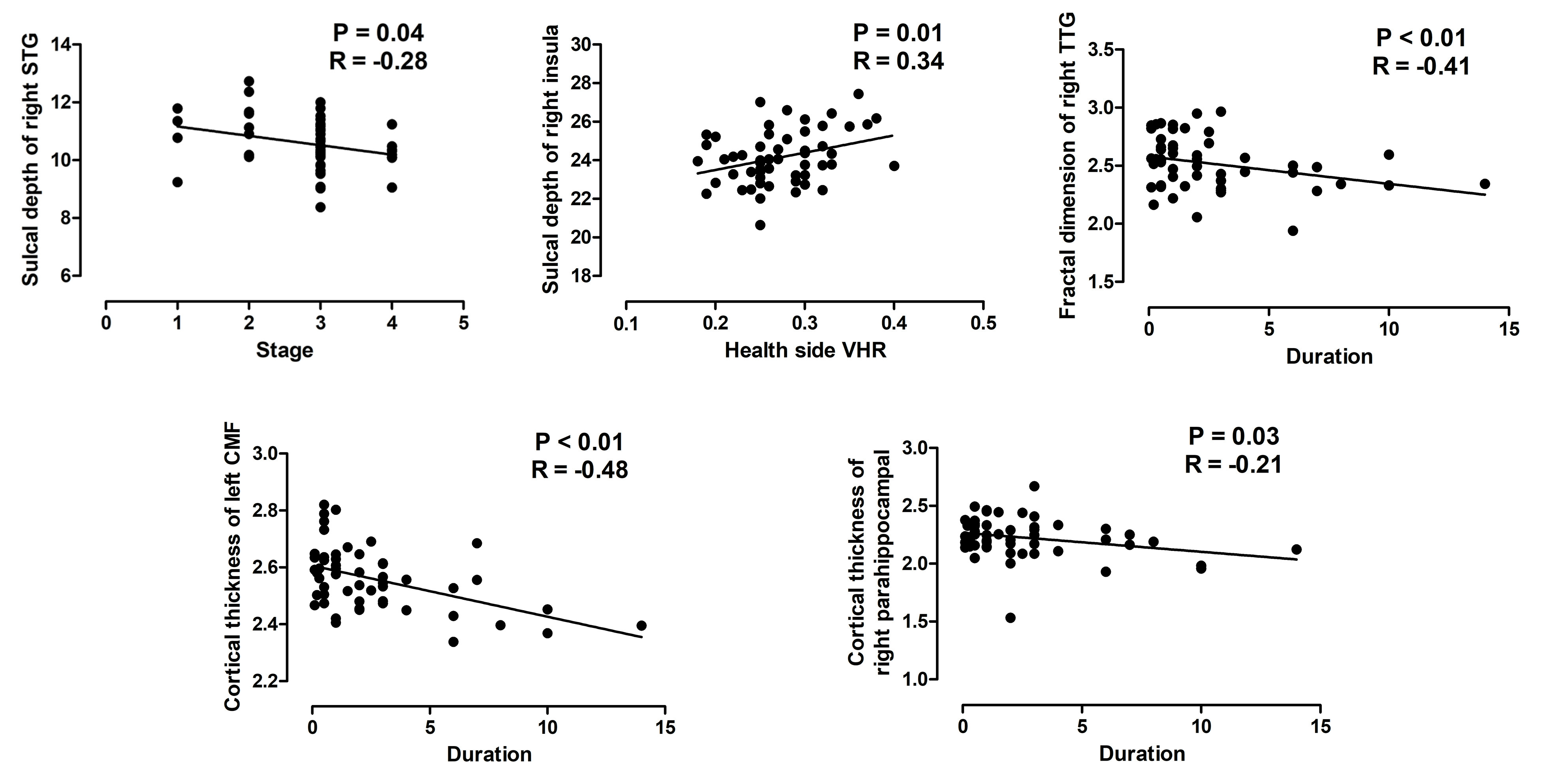
As portrayed in Figure 4, significant correlations emerged within the MD patient group, revealing associations between specific neuroanatomical metrics and distinct aspects of the disease.
The LOOCV results exhibit promising classification accuracy, achieving an overall accuracy of 84%. The model demonstrated a precision of 0.81 for MD and 0.86 for HC, indicating the ability to correctly classify individuals with MD and HC. Additionally, the recall was 0.78 for MD and 0.88 for HC, highlighting the model's capability to accurately identify MD and HC cases. The F1-scores for MD and HC were 0.80 and 0.87, respectively, suggesting a balanced performance in terms of precision and recall. Visual inspection of the ROC curve reveals a well-discriminating classifier(Figure 5). The AUC value indicates a strong performance with an AUC of 0.92. The cut-off point on the ROC curve, marked by a red circle, corresponds to a sensitivity of 0.86 and specificity of 0.87.
Discussion and Conclusion
In conclusion, our study provides a comprehensive exploration of neuroanatomical alterations in MD patients using VBM and SBM techniques. The observed changes in brain gray matter volume, cortical thickness, fractal dimension, gyrification index, and sulcal depth collectively contribute to our understanding of the complex neural underpinnings of MD. By employing a comprehensive array of neuroimaging features and a machine-learning approach, we achieve high diagnostic accuracy of MD. Our findings contribute to the growing body of knowledge regarding the neurobiological underpinnings of MD, opening new avenues for further research and clinical applications in the field of auditory and vestibular disorders.Acknowledgements
We thank all participants in this study.
References
1, Jian, H., Wang, S., Li, X., Zhao, H., Liu, S., Lyu, Y., . . . Zhang, D. (2023). Effect of Late-Stage Meniere's Disease and Vestibular Functional Impairment on Hippocampal Atrophy. Laryngoscope. https://doi.org/10.1002/lary.30816
2, Seo, Y. J., Kim, J., & Kim, S. H. (2016). The change of hippocampal volume and its relevance with inner ear function in Meniere's disease patients. Auris Nasus Larynx, 43(6), 620-625. https://doi.org/10.1016/j.anl.2016.01.006
3, Leng, Y., Fan, W., Liu, Y., Xia, K., Zhou, R., Liu, J., . . . Liu, B. (2023). Comparison between audio-vestibular findings and contrast-enhanced MRI of inner ear in patients with unilateral Meniere's disease. Front Neurosci, 17, 1128942. https://doi.org/10.3389/fnins.2023.1128942
4, Nakashima, T., Naganawa, S., Pyykko, I., Gibson, W. P., Sone, M., Nakata, S., & Teranishi, M. (2009). Grading of endolymphatic hydrops using magnetic resonance imaging. Acta Otolaryngol Suppl(560), 5-8. https://doi.org/10.1080/00016480902729827
5, Gaser C, Dahnke R, Kurth K, Luders E, Alzheimer"s Disease Neuroimaging Initiative. (2022). A Computational Anatomy Toolbox for the Analysis of Structural MRI Data. bioRxiv. https://doi.org/10.1101/2022.06.11.495736
Figures