2479
Automatic Quantitative Identification of Disproportionately Enlarged Subarachnoid-Space Hydrocephalus in iNPH Using Deep Learning Models1Neurosurgery, Nagoya City University Graduate School of Medical Science, Nagoya, Japan, 2Interfaculty Initiative in Information Studies/Institute of Industrial Science, The University of Tokyo, Tokyo, Japan, 3Medical System Research & Development Center, FUJIFILM Corporation, Tokyo, Japan, 4Faculty of System Design, Tokyo Metropolitan University, Tokyo, Japan, 5Mechanical Science and Bioengineering, Graduate School of Engineering Science, Osaka University, Osaka, Japan, 6Behavioral Neurology and Cognitive Neuroscience, Tohoku University Graduate School of Medicine, Sendai, Japan, 7Neurology and Clinical Neuroscience, Yamagata University School of Medicine, Yamagata, Japan, 8Radiology, Shiga University of Medical Science, Otsu, Japan
Synopsis
Keywords: Diagnosis/Prediction, Machine Learning/Artificial Intelligence, 3D MRI
Motivation: Automated detection for disproportionately enlarged subarachnoid-space hydrocephalus (DESH) using 3D MRIs.
Goal(s): We developed robust deep learning models for accurate DESH detection by automatically segmenting regions.
Approach: Utilized 3D U-Net for segmentation and multimodal convolutional neural network for classification. Achieved high accuracy, with mean Dice scores ranging 0.60 – 0.84 and softmax probability scores exceeding 0.95. All of the area under the curves exceeded 0.97.
Results: Successfully developed the highly accurate deep learning models in automatically segmentation of ventricles and regional subarachnoid spaces and in the detecting DESH, ventricular dilatation, tightened sulci in the high convexities, and Sylvian fissure dilatation.
Impact: Combining a 3D U-Net model and a multi-modal convolutional neural network model, disproportionately enlarged subarachnoid-space hydrocephalus (DESH) for idiopathic normal pressure hydrocephalus (iNPH) was automatically detected with automatically segmented regions from 3D T1- and T2-weighted MRIs.
METHODS: The study involved 160 participants, consisting of 27 iNPH patients and 133 healthy individuals. All participants underwent three-dimensional (3D) T1-weighted and T2-weighted MRIs. The deep learning models for this study consisted of two tasks: semantic segmentation task of cerebrospinal fluid (CSF) spaces and image classification task to assess DESH and other relevant features. For the semantic segmentation task, we employed a 3D U-Net model to automatically segment the total ventricles, total subarachnoid spaces, Sylvian fissure and basal cistern, and high-convexity part of the subarachnoid space. High-convexity part of the subarachnoid space was defined as the subarachnoid space located above the body of the lateral ventricles, with the anterior end on the coronal plane perpendicular to the anterior commissure–posterior commissure line passing through the front edge of the genu of corpus callosum, the posterior end in the bilateral posterior parts of the callosomarginal sulci, and the lateral end at 3 cm from the midline on the coronal plane perpendicular to the anterior commissure–posterior commissure line passing through the midpoint between anterior commissure and posterior commissure.3 This segmentation process aimed to provide a precise anatomical breakdown of these structures, which is crucial for the subsequent diagnostic step. The second task involved image classification, where the presence of DESH, ventricular dilatation, tightened sulci in the high convexities, and Sylvian fissure dilatation was assessed using a multi-modal convolutional neural network model. A total of 110 T1-weighted and 130 T2-weighted MRIs were used for training, while 30 T1- and 30 T2-weighted MRIs for internal validation. The remaining 20 T1- and 20 T2-weighted MRIs were used for external validation.
RESULTS: Automatic region extraction from 3D T1- and T2-weighted MRIs showed high accuracy for various brain regions. The mean Dice scores, which measure the overlap between manually and automatically segmented regions, were notably high for the total ventricles (0.85 and 0.83), Sylvian fissure and basal cistern (0.70 and 0.69), and high-convexity part of the subarachnoid space (0.68 and 0.60) in T1- and T2-weighted MRIs, respectively. In the image classification task, the models displayed impressive reliability in assessing DESH, ventricular dilatation, tightened sulci in the high convexities, and Sylvian fissure dilatation. All mean softmax probability scores exceeded 0.95, indicating the models' high confidence in their predictions. Furthermore, the areas under the receiver-operating characteristic curves (AUC) for the DESH, Venthi, and Sylhi indices, calculated based on the segmented regions for detecting DESH objectively, exceeded 0.97. These AUC scores demonstrate the models' ability to distinguish iNPH patients from healthy controls with a high degree of accuracy.
DISCUSSION: Our developed deep learning models have provided an objective and automatic approach to detect DESH, a critical feature of iNPH. By accurately segmenting CSF spaces and classifying DESH features, the models eliminate the subjective evaluation. The decision support tool can be expected to give patients with iNPH a better chance of receiving appropriate treatment earlier, to reduce ambiguity in the interpretation of DESH. The implementation of this technology in clinical practice has the potential to reduce the burden on healthcare professionals and improve the precision of iNPH diagnosis. By reducing the subjectivity in assessments, it can help ensure that individuals with iNPH receive timely and accurate treatment, potentially improving their quality of life and clinical outcomes. In addition, quantitative measurements and indices ensure objectivity and allow for easier interpretation of classification results, especially in cases where the clinical diagnosis is not clear.
CONCLUSION: We developed a novel diagnostic support tool for iNPH based on the combined use of a 3D U-Net model for semantic segmentation and a multi-modal convolutional neural network for image classification. Through these models, DESH in iNPH can be automatically detected and assessed with a high degree of accuracy. This advancement holds the potential to revolutionize the diagnosis of iNPH by providing a more objective and consistent method for evaluating DESH and other related features.
Acknowledgements
We would like to thank the radiology staff of the Shiga University of Medical Science, particularly Shinnosuke Hiratsuka, Masahiro Yoshimura, Asuka Nishihara, Kohei Ohashi, and Mika Adachi. We are grateful to the FUJIFILM Corporation for using the latest version of the SYNAPSE 3D workstation.
This research was supported by research grants from the Japan Society for the Promotion of Science, KAKENHI (Grant number: 21K09098, 22H03020, and 22H00190); from the FUJIFILM Corporation; from the G-7 Scholarship Foundation; from the Taiju Life Social Welfare Foundation; from the Osaka Gas Group Welfare Foundation; and from the Ministry of Education, Culture, Sports, Science and Technology as “Program for Promoting Researches on the Supercomputer Fugaku” (Development of human digital twins for cerebral circulation using Fugaku, JPMXP1020230118). The funders had no influence or involvement in the writing of this article. The funders had no effect or involvement in the writing of this article. The funders had no role in the study design, data collection and analysis, decision to publish, or preparation of the manuscript.
References
1. Nakajima M, Yamada S, Miyajima M, et al. Guidelines for management of idiopathic normal pressure hydrocephalus (Third edition): Endorsed by the Japanese society of normal pressure hydrocephalus. Neurol Med Chir (Tokyo) 2021
2. Hashimoto M, Ishikawa M, Mori E, et al. Diagnosis of idiopathic normal pressure hydrocephalus is supported by MRI-based scheme: a prospective cohort study. Cerebrospinal Fluid Research 2010;7
3. Yamada S, Ito H, Matsumasa H, et al. Tightened Sulci in the High Convexities as a Noteworthy Feature of Idiopathic Normal Pressure Hydrocephalus. World Neurosurg 2023;176:e427-e37
Figures
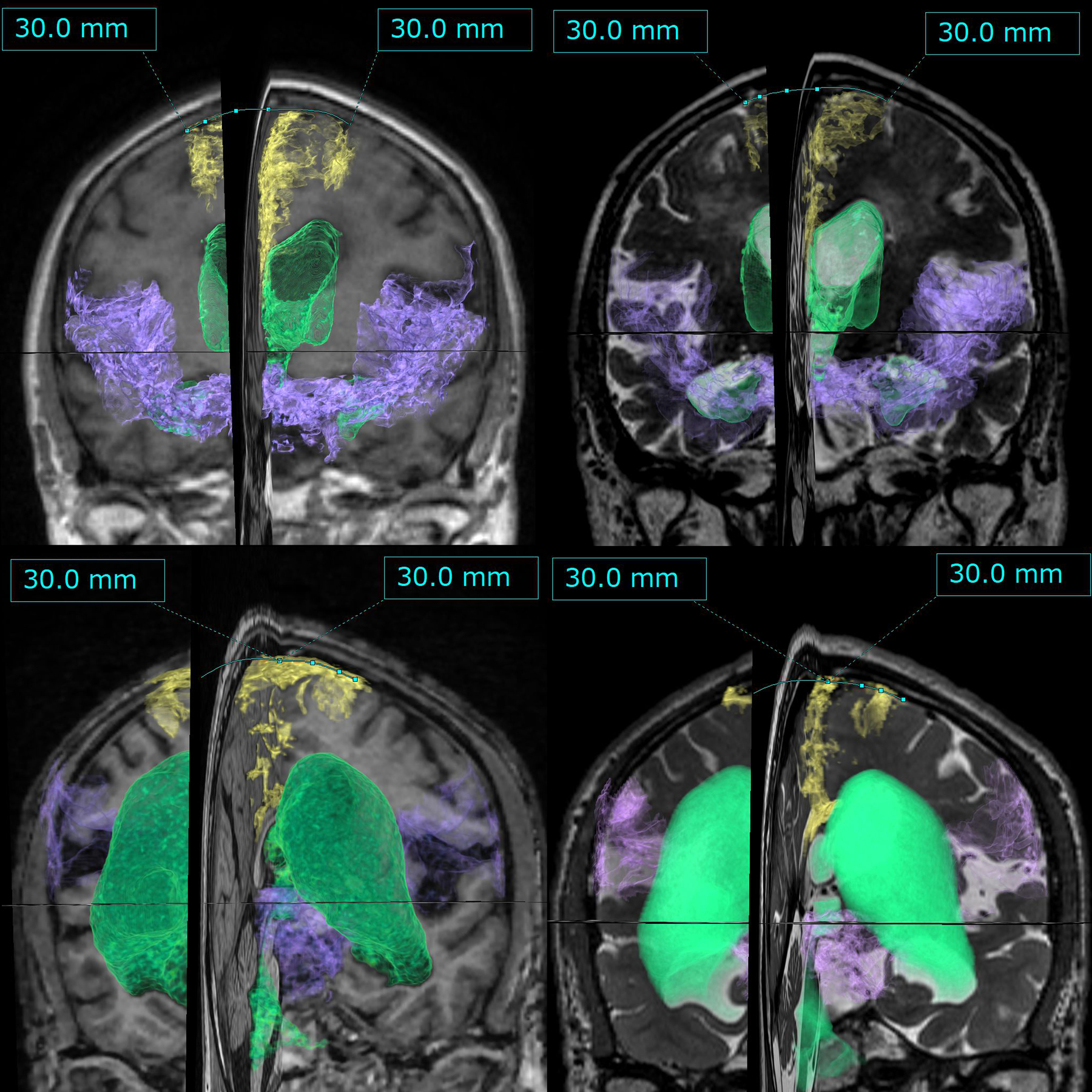
Segmentation of three volumes of interest from 3D T1-weighted and T2-weighted MRI in iNPH
The green indicates the total ventricle, the yellow indicates the high-convexity part of the subarachnoid space, and the purple indicates the Sylvian fissure and basal cistern. The upper left is from anterior to 3D T1-weighted MRI, the upper right is from anterior to T2-weighted MRI, the lower left is from posterior to T1-weighted MRI, and the lower right is from posterior to T2-weighted MRI.
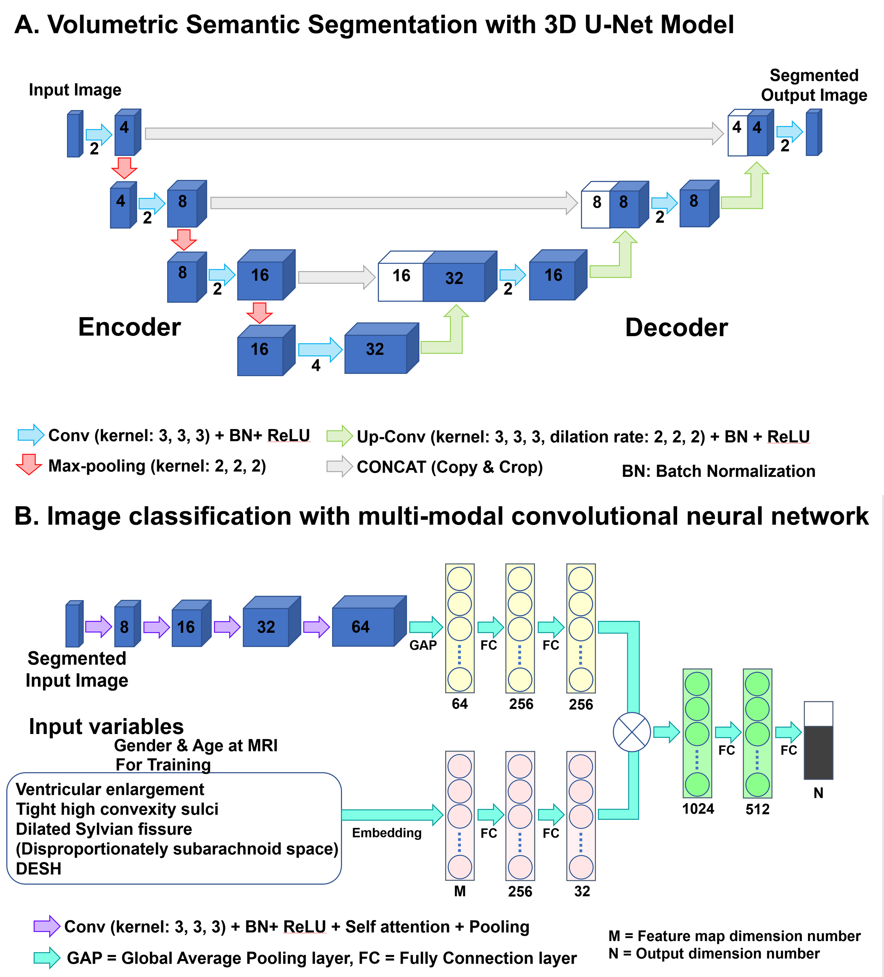
Two combined deep learning models; A. 3D U-Net model with four layers for volumetric semantic segmentation task and B: Multi-modal convolutional neural network for image classification task
Each blue box corresponds to a multi-channel feature map. The number of channels is denoted on front of the box. White boxes indicate copied feature maps. The image intensities of input images were normalized to [0, 1] by their maximum and minimize values.
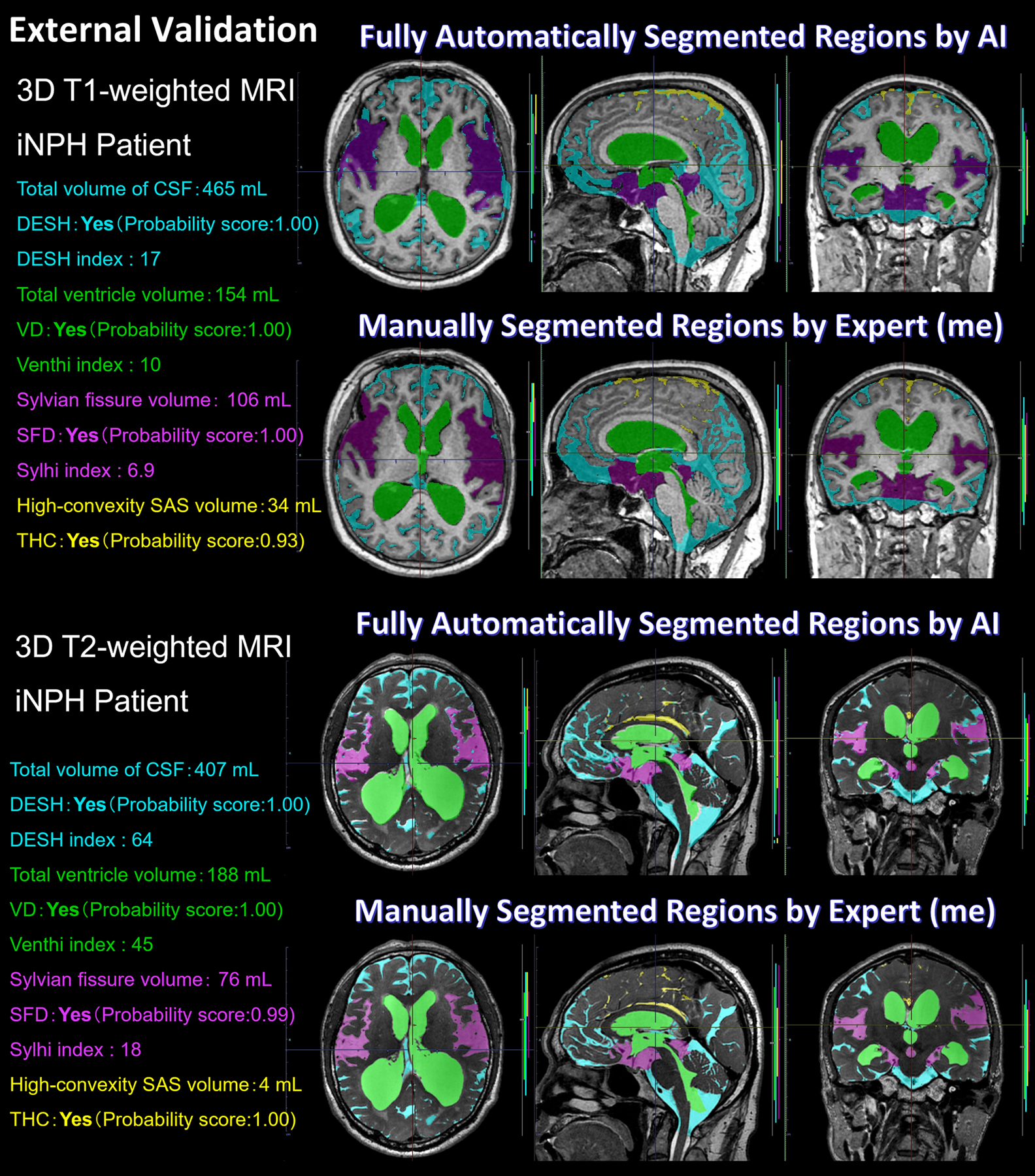
External validation by comparing manually and automatically segmented regions from 3D T1-weighted and T2-weighted images
Green indicates the total ventricles, purple indicates Sylvian fissure and basal cistern, yellow indicates high-convexity part of the subarachnoid space, and marine blue indicates other subarachnoid spaces.
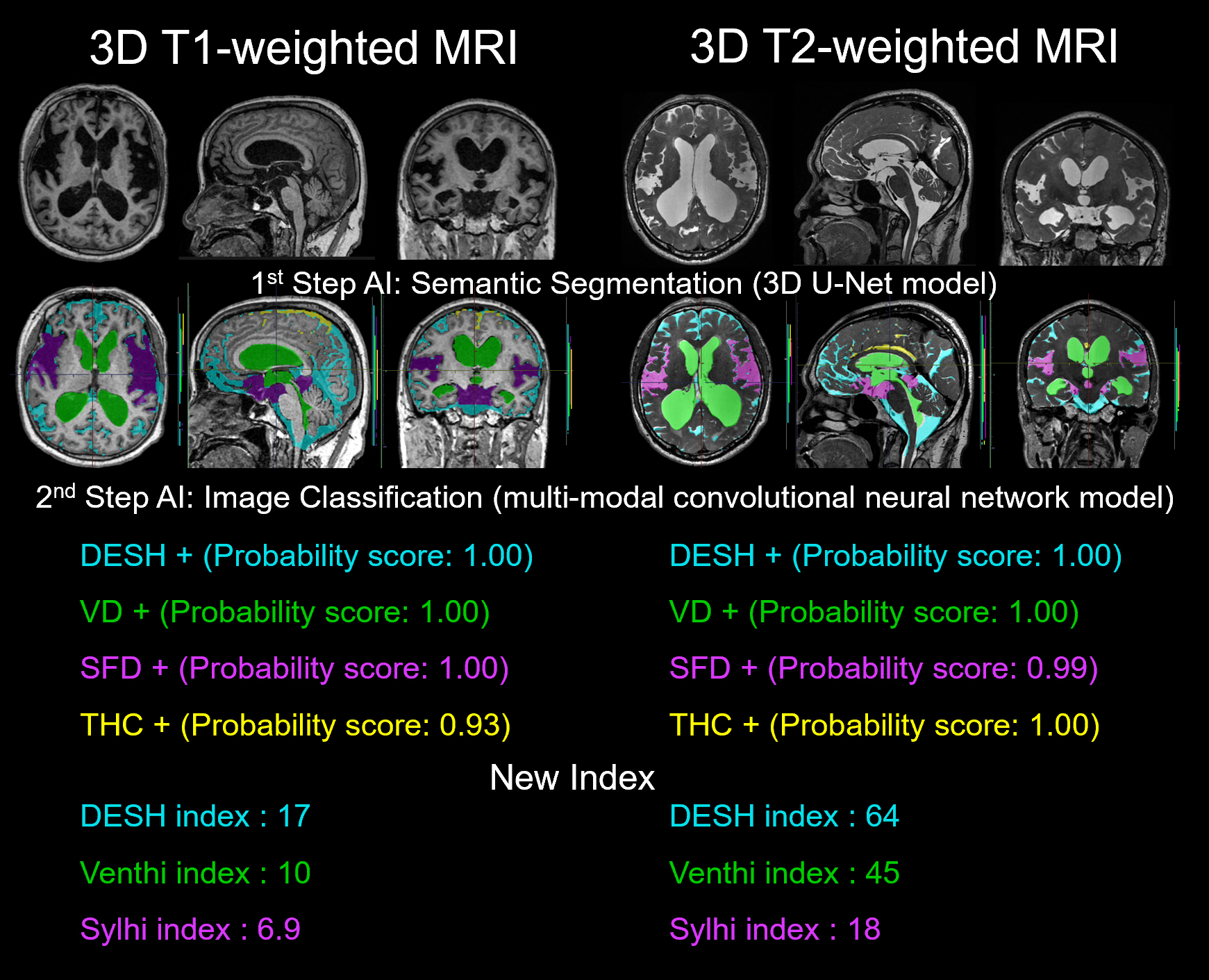
Flow of deep learning models
In the first step, the volumetric semantic segmentation task employed a 3D U-Net with four layers, consisting of 3D convolution with a batch normalization layer, ReLU activation layer, max pooling layer, and 3D up-convolution layer. In the second step, the image classification task employed a multimodal convolutional neural network. As supplemental indices for DESH diagnosis, the DESH index, Venthi index, and Sylhi index were created.