2477
Study of the preprocessing impact on the Deep Learning automatic segmentation of Choroid Plexus in Multiple Sclerosis1Department of Information Engineering, University of Padova, Padova, Italy, 2Department of Diagnostic and Public Health, University of Verona, Verona, Italy, 3Department of Neurosciences, Biomedicine and Movement Sciences, University of Verona, Verona, Italy, 4Department of Information Engineering, Padova Neuroscience Center, University of Padova, Padova, Italy, 5Neuroradiology Section, Diagnostic Pathology Department, University of Verona, Verona, Italy
Synopsis
Keywords: Data Processing, Machine Learning/Artificial Intelligence, Multiple Sclerosis
Motivation: The Choroid Plexus (ChP) is a vascular structure involved in brain regulatory functions. The relation between ChP Volume and brain disorders raises the interest on this structure and the need for an accurate segmentation, questioning whether to introduce a preprocessing step.
Goal(s): This work studies the preprocessing impact on the ChP segmentation with Deep Neural Networks (DNN) ensemble.
Approach: Three different preprocessing steps (brain extraction, N4 intensity correction, combination of both) were applied to 128 T1-w MRI images before DNN training. These approaches performances were compared to that without preprocessing.
Results: The preprocessing step does not improve DNN performance for the ChP segmentation.
Impact: The preprocessing steps of brain extraction and N4 intensity normalization correction on T1-w MRI images do not have an impact on Deep Neural Networks performance during the automatic segmentation of Choroid Plexus on Multiple Sclerosis patients.
Introduction
The Choroid Plexus (ChP) is a vascular structure located inside the brain ventricles. It is part of the glymphatic system1. ChP main roles are the production of the CSF and the regulation of brain homeostasis and clearance2. Recently, it has been hypothesized a relationship between the ChP Volume (ChPV) alteration and neurodegenerative disorders like Multiple Sclerosis (MS)3. Hence, there is a growing need for accurate delineation of ChP using automated methods avoiding the time-consuming manual segmentation on T1-w MRI image, the gold-standard approach. Moreover, recent works4,5 have studied the impact of preprocessing steps when performing medical imaging segmentation tasks with Deep Neural Networks (DNN) approaches, but results are non-uniform. The aim of this work is to evaluate the influence of three preprocessing steps on T1-w MRI images when performing the automatic segmentation of ChP with DNN in a cohort of MS patients.Methods
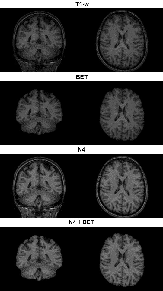
The original dataset was composed of 128 subjects and was supplied by the University Hospital of Verona - Multiple Sclerosis Center. Both Healthy Controls (HC, 24) and MS patients (104) composed the images collection. Two 3T MRI scanners were used for the acquisition of 3D T1-w MPRAGE images (FA 8°, 1x1x1 mm): Philips Acheiva-TX for 67 subjects (HC/MS 40.9 ± 9.9 years) and a Philips Elition-S for 61 subjects (MS 36.7 ± 10.1 years). The manual segmentation performed by two neuroradiologists was used as Ground Truth reference (GT).Three datasets were generated starting from the untouched one (Figure 1). The first dataset (BET) was a collection of the brain-extracted images. The brain mask was obtained by the intersection of the brain extraction masks derived from ROBEX6 and DeepBrain7. The second dataset (N4) was built applying the Advanced Normalization Toolbox8 (v.2.4.3) to the T1-w images to obtain a nonparametric nonuniform normalization with the N4BiasFieldCorrection method9 (standard parameters, shrink-factor=4, convergence to 50 iterations, cubic spline fitting). The last dataset (N4+BET) was obtained applying the brain extraction procedure to the N4 dataset.
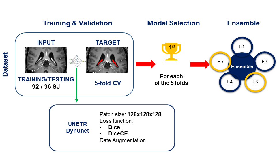
For the available datasets, images were randomly divided, balancing scanner/HC/MS, into training and testing sets (92/36 subjects). A five-fold cross-validation training procedure was implemented in MONAI10 (v.1.0.1) (Figure 2). We tested four combinations of DNN architectures (DynUnet, MONAI nnU-Net11; UNETR12) and loss functions (GeneralizedDice, combination of Dice and Cross-Entropy) with 128x128x128 patch-size and data augmentation transforms. We used the Adam-Weighted optimizer with fixed learning rate (1e-04), weight decay (1e-05), max iterations (2e04) and batch size (1). To improve the reliability of the final segmentation, the ensemble by major voting of the best-fold models was performed.
The analyzed performance metrics were Dice Coefficient, 95% Hausdorff Distance (95% HD), (Absolute) Percentage Volume Difference (ΔVol%), Pearson’s Volume Correlation Coefficient (r).
$$PercentageVolumeDifference = 100*\frac{(Volume_{Prediction} - Volume_{GT})}{Volume_{GT}}$$
One-way ANOVA and post-hoc t-test were performed on Dice, 95% HD and Absolute Volume.
Results
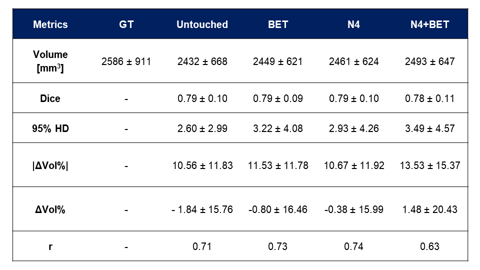
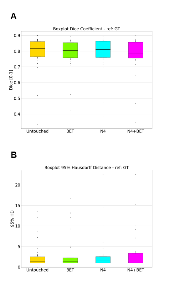
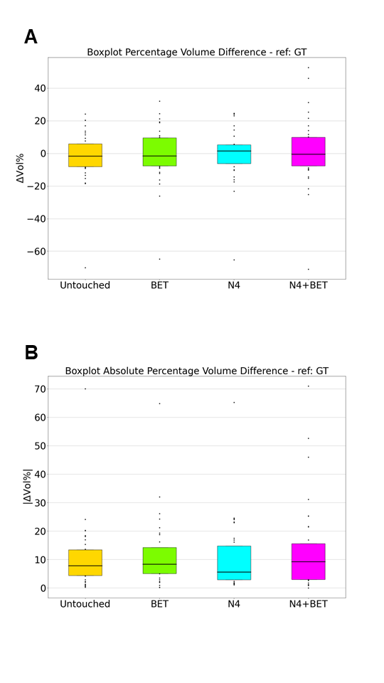
Table 1 shows the results of the comparison between the predicted segmentations of the four tested datasets and the GT. One-way ANOVA, as post-hoc t-tests, on Dice, 95% HD and Absolute Volume do not find differences between the groups. Untouched, BET and N4 show equal mean Dice Coefficient (0.79). Non preprocessed images bring to the best 95% HD (2.60±2.99) and |ΔVol%| (10.56±11.83%) metrics, while in relative terms N4 has higher performance (ΔVol%: -0.38±15.99%) and volume correlation with GT (0.74), while the application of both approaches (N4+BET) makes performance drop (r: 0.63, |ΔVol%|: 13.53±15.37%). Figure 3 displays the Dice Coefficient and the 95% HD of the compared approaches, while Figure 4 shows the Volume Analysis (|ΔVol%|, ΔVol%): no statistically significant differences have been detected.Discussion
Applying a preprocessing step on T1-w MRI images is supposed to mitigate variability of input images or reduce the space of feature to be learnt, equalizing datasets derived from different scanners. Results have highlighted the preprocessing impact when performing ChP automatic segmentation is negligible. Moreover, there are no statistically significant differences when applying or not the preprocessing steps. Applying intensity normalization brings to slightly higher performance in terms of ChPV estimate, while performance drops when combined with brain extraction as described in a recent study13. Moreover, the number of outliers is higher when images are preprocessed compared to the untouched ones, suggesting the loss of relevant information to accurately complete the segmentation.Conclusion
Preprocessing has a negligible impact on the estimates of the ChPV obtained with state-of-the-art segmentation DNNs. Therefore, DNN based automatic segmentation of ChP does not require preprocessing steps of brain extraction and intensity correction on T1-w MRI images derived from a cohort of MS subjects.Acknowledgements
No acknowledgement found.References
- Jessen NA, Munk ASF, Lundgaard I, Nedergaard M. The Glymphatic System: A Beginner’s Guide. Neurochem Res. 2015;40(12):2583-2599.
- Damkier HH, Brown PD, Praetorius J. Cerebrospinal fluid secretion by the choroid plexus. Physiol Rev. 2013;93(4):1847-1892.
- Ricigliano VAG, Morena E, Colombi A, et al. Choroid Plexus Enlargement in Inflammatory Multiple Sclerosis: 3.0-T MRI and Translocator Protein PET Evaluation. Radiology. 2021;301(1):166-177.
- De Raad KB, Van Garderen KA, Smits M, et al. The Effect of Preprocessing on Convolutional Neural Networks for Medical Image Segmentation. IEEE ISBI. 2021; 655-658.
- Kondrateva E, Druzhinina P, Dalechina A, et al. Negligible effect of brain MRI data preprocessing for tumor segmentation. Published online April 11, 2022.
- Iglesias JE, Liu C-Y, Thompson PM, Tu Z. Robust Brain Extraction Across Datasets and Comparison With Publicly Available Methods. IEEE Trans Med Imaging. 2011;30(9):1617-1634.
- DeepBrain: https://github.com/iitzco/deepbrain
- Avants BB, Tustison NJ, Song G, et al. A reproducible evaluation of ANTs similarity metric performance in brain image registration. NeuroImage. 2011;54:2033–2044.
- Tustison NJ, Avants BB, Cook PA, et al., N4ITK: Improved N3 Bias Correction, IEEE Trans Med Imaging. 2010;29(6):1310-1320.
- Consortium MONAI. MONAI: Medical Open Network for AI. https://zenodo.org/record/7245821. Published online October 24, 2022.
- Isensee F, Jaeger PF, Kohl SAA, Petersen J, Maier-Hein KH. nnU-Net: a self-configuring method for deep learning-based biomedical image segmentation. Nat Methods. 2021;18(2):203-211.
- Hatamizadeh A, Tang Y, Nath V, et al. UNETR: Transformers for 3D Medical Image Segmentation. Published online March 18, 2021.
- Pacheco BM, Cassia GS, Silva D. Towards fully automated deep-learning-based brain tumor segmentation: is brain extraction still necessary?. Biomed Signal Process Control. 2023;82:104514.
Figures