2474
Exploring the ocular glymphatic system: The association of MRI-visible perivascular spaces with intraocular pressure and tear total-tau1Department of Radiology & Nuclear Medicine, Maastricht University Medical Center, Maastricht, Netherlands, 2School for Mental Health and Neuroscience, Maastricht University, Maastricht, Netherlands, 3Department of Psychiatry & Neuropsychology, Maastricht University, Maastricht, Netherlands, 4University Eye Clinic, Maastricht University Medical Center+, Maastricht, Netherlands, 5Department of Intensive Care, Maastricht University Medical Center+, Maastricht, Netherlands, 6Cardiovascular Research Institute Maastricht, Maastricht University, Maastricht, Netherlands, 7Department of Electrical Engineering, Eindhoven University of Technology, Eindhoven, Netherlands
Synopsis
Keywords: Neurofluids, Neurofluids, Perivascular spaces, Waste clearance, Ocular glymphatics, Tau, Eye
Motivation: Preclinical research suggests an ocular glymphatic system similar to the cerebral system, driven by intraocular pressure (IOP). However, human studies are scarce.
Goal(s): To explore the eye-brain connection by investigating tear total-tau as a potential early marker of cerebral glymphatics and consider the link between IOP (driver of ocular glymphatics) and impaired cerebral waste clearance.
Approach: MRI-visible PVS were scored on 7T images and related to IOP and tear total-tau.
Results: Higher tear total-tau and lower IOP were associated with more PVS, implying a connection to impaired cerebral waste clearance and aligning with the potential presence of a human ocular glymphatic system.
Impact: Our exploratory results suggest that higher tear-tau and a reduced driving force of ocular waste clearance are connected to impaired cerebral waste clearance. Thereby, this study bridges the gap between the potential human ocular glymphatic system and cerebral waste clearance.
Introduction
Impaired cerebral waste clearance is a key feature of aging and various neurodegenerative conditions,1 including Alzheimer's disease (AD).2 Waste removal from the brain is facilitated by a network of perivascular spaces (PVS).3 Preclinical research suggests the existence of a similar system in the eyes: the ocular glymphatic system (Fig.1).4,5 The intraocular pressure (IOP) is hypothesized to drive this system, draining waste products from the eye through the optic nerve,6,7 towards the perivenous spaces8 of the cerebral waste clearance system.4,9 However, there is a scarcity of human studies investigating this mechanism.10In humans, quantifying MRI-visible PVS is an established non-invasive method to assess cerebral waste clearance function.11 Ultra-high field 7T MRI allows accurate and early stage quantification of PVS enlargement.12 Combining 7T MRI-visible PVS quantification with a measure for the driver of the ocular glymphatic system (i.e., IOP), provides a unique opportunity to explore the connection between the ocular and cerebral clearance system.
Furthermore, this eye-brain connection may also offer a means to gather insights about brain pathology. Tear fluid analysis was shown to be feasible to determine total-tau concentration (a marker for AD pathology),13 which was suggested as a proxy for cerebral tau presence and potentially indicatory of reduced cerebral waste clearance.
This study aimed to gain more insights into the eye-brain connection by examining the relation between PVS and tear total-tau in healthy elderly and explore the connection between IOP and PVS, which could support the potential presence of an ocular glymphatic system in humans.
Methods
MRI acquisition: Thirty cognitively healthy elderly subjects underwent 7T MRI (Siemens Healthineers, Erlangen, Germany) using a 32-element channel phased-array head coil, including whole-brain T2-weighted and T1-weighted scans (Fig.2).Ocular measures: Tear fluid was collected from both eyes using Schirmer’s strips while recording the length of tear wetting on the strip (tear-wetting length) and analysed for total-tau using the S-PLEX assay.13
In 23 subjects, the average IOP per eye was calculated over three subsequent measurements using the Auto Kerato-Refracto-Tonometer TRK-1P.14
Perivascular space scoring: PVS were visually rated on the T2-weighted images in the basal ganglia (BG) and centrum semiovale (CSO), two regions known for PVS occurrence.11 Each hemisphere was scored separately using a visual rating scale: 0 (<10), 1 (10-25), 2 (25-40), or 3 (>40), in the slice with the highest PVS count.11 Two independent raters performed consensus scoring while blinded to clinical data.
Anatomical brain size: White matter (WM) and BG volumes were automatically segmented per hemisphere from the T1-weighted images using Freesurfer (version 6.0.5)(Fig.2).15
Statistics: Partial Spearman’s correlation coefficients were computed between the PVS scores and both tear total-tau and IOP, while adjusting for age, sex, and tear-wetting length.
To account for potential atrophy effects, significant associations were further adjusted for the respective hemispheric WM or BG volumes.
Results
Subject characteristics and descriptive statistics are summarized in Table 1.Elevated tear total-tau levels were found to be significantly correlated to a higher CSO PVS score in both hemispheres (Fig.3). These associations remained significant after further accounting for WM volumes (Tab.2).
Lower right eye's IOP was significantly correlated with higher right hemispheric CSO PVS scores (Fig.3), and a similar significant association was found between the left eye's IOP and left hemispheric CSO PVS scores (Tab.2). These associations remained significant after additional correction for WM volumes.
No significant associations were found with the BG PVS scores (Tab.2).
Discussion and conclusion
This study found that higher levels of tear total-tau were related to more PVS in the CSO in both hemispheres, suggesting that elevated tear-tau might signify the accumulation of cerebral waste products due to impaired waste clearance.This relationship was specific to the CSO, where PVS enlargement is associated with pathological protein deposition (e.g., amyloid angiopathy),11 as opposed to the BG, where PVS are likely more influenced by vascular changes (e.g., arterial stiffness).11,16
Additionally, lower IOP was associated with more PVS in the ipsilateral CSO, aligning with the idea of the presence of an ocular glymphatic system in humans. Lower IOP may reduce the flow of waste-containing fluids through the optic nerve towards the cerebral waste clearance system (Fig.1).6
Notably, this relation was found ipsilaterally, but not contralaterally. This may be explained by the straight posterior pressure that is exerted on the fluid surrounding the optic nerve into the ipsilateral hemisphere, regardless of the optic nerve’s crossing at the chiasm.
While alternative indirect pathophysiological explanations for the IOP-PVS connection should be considered, our exploratory results suggest that a reduction in the pressure driving ocular waste clearance relates to impaired cerebral waste clearance, bridging the gap between these two systems.
Acknowledgements
No acknowledgement found.References
1. Rasmussen MK, Mestre H, Nedergaard M. The glymphatic pathway in neurological disorders. The Lancet Neurology. 2018;17(11):1016-1024.
2. Tarasoff-Conway JM, Carare RO, Osorio RS, et al. Clearance systems in the brain—implications for Alzheimer disease. Nature Reviews Neurology. 2015;11(8):457-470.
3. Jessen NA, Munk ASF, Lundgaard I, Nedergaard M. The glymphatic system: a beginner’s guide. Neurochemical research. 2015;40(12):2583-2599.
4. Wang X, Lou N, Eberhardt A, et al. An ocular glymphatic clearance system removes β-amyloid from the rodent eye. Science translational medicine. 2020;12(536):eaaw3210.
5. Mathieu E, Gupta N, Ahari A, Zhou X, Hanna J, Yücel YH. Evidence for cerebrospinal fluid entry into the optic nerve via a glymphatic pathway. Investigative ophthalmology & visual science. 2017;58(11):4784-4791.
6. Denniston A, Keane P. Paravascular Pathways in the Eye: Is There an 'Ocular Glymphatic System'? Investigative ophthalmology & visual science. 2015;56:3955-3956.
7. Wostyn P, Van Dam D, Audenaert K, Killer HE, De Deyn PP, De Groot V. A new glaucoma hypothesis: a role of glymphatic system dysfunction. Fluids and Barriers of the CNS. 2015;12:1-6.
8. Uddin N, Rutar M. Ocular lymphatic and glymphatic systems: implications for retinal health and disease. International Journal of Molecular Sciences. 2022;23(17):10139.
9. Xu Y, Cheng L, Yuan L, Yi Q, Xiao L, Chen H. Progress on Brain and Ocular Lymphatic System. BioMed Research International. 2022;2022.
10. Jacobsen HH, Ringstad G, Jørstad ØK, Moe MC, Sandell T, Eide PK. The human visual pathway communicates directly with the subarachnoid space. Investigative ophthalmology & visual science. 2019;60(7):2773-2780.
11. Wardlaw JM, Benveniste H, Nedergaard M, et al. Perivascular spaces in the brain: anatomy, physiology and pathology. Nature Reviews Neurology. 2020;16(3):137-153.
12. Yu L, Hu X, Li H, Zhao Y. Perivascular spaces, glymphatic system and MR. Front Neurol. 2022;13:844938.
13. Gijs M, Ramakers IH, Visser PJ, et al. Association of tear fluid amyloid and tau levels with disease severity and neurodegeneration. Scientific Reports. 2021;11(1):22675.
14. Forbes M, Pico G, Jr., Grolman B. A Noncontact Applanation Tonometer: Description and Clinical Evaluation. Archives of Ophthalmology. 1974;91(2):134-140.
15. Fischl B. FreeSurfer. Neuroimage. 2012;62(2):774-781.
16. van den Kerkhof M, van der Thiel MM, van Oostenbrugge RJ, et al. Impaired damping of cerebral blood flow velocity pulsatility is associated with the number of perivascular spaces as measured with 7T MRI. Journal of Cerebral Blood Flow & Metabolism. 2023:0271678X231153374.
Figures
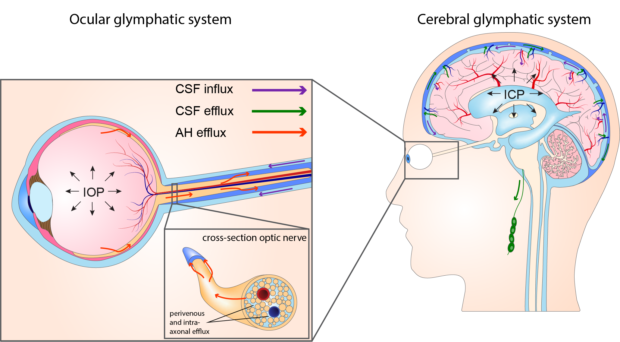
Fig.1. Theorized connection between the ocular (left) and cerebral (right) glymphatic systems. The ocular glymphatic system is thought to use IOP to drive waste products through the optic nerve into the subarachnoid space, towards the cerebral glymphatic system.
The cerebral glymphatic system transports CSF into perivascular spaces, transporting soluble waste products from the interstitial fluid through perivenous spaces towards the meningeal lymph nodes.
Abbreviations: IOP = intraocular pressure, ICP = intracerebral pressure, CSF = cerebral spinal fluid, AH = aqueous humor
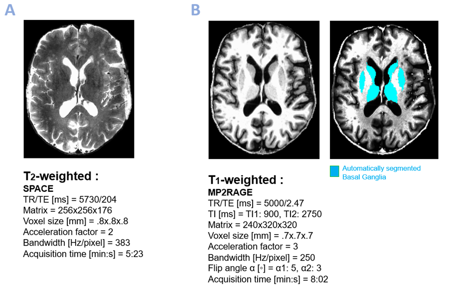
Abbreviations: TR = repetition time, TE = echo time, TI = inversion time
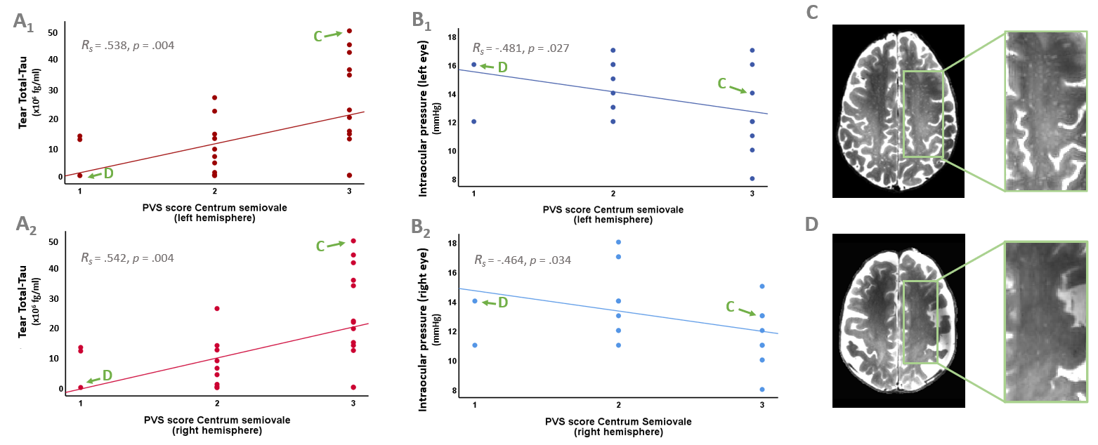
Fig.3. Scatterplots show that higher tear total-tau levels
are associated with higher Centrum semiovale (CSO) PVS scores in both left (A1) and
right (A2) hemispheres, and that lower intraocular pressure (IOP) in both the left (B1) and right (B2) eye is linked to higher ipsilateral
CSO PVS scores.
Arrows highlight exemplary subjects (C) with
a high CSO PVS score and tear total-tau, and low IOP
(77y, M) and (D) with a low CSO PVS score and tear total-tau, and high IOP (60y, F).
Least-square regression lines are added for visualization.
Abbreviations: PVS = perivascular space, Rs = Spearman rho
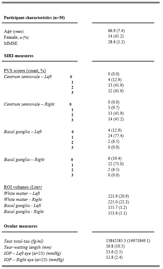
Abbreviations: IOP = intraocular pressure, PVS = perivascular space, ROI = region of interest, MMSE = Mini Mental State Examination.
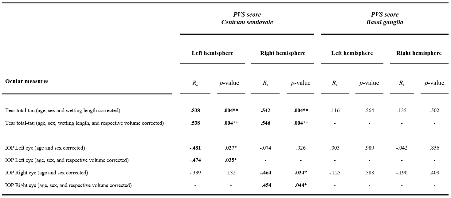
Tab.2. Partial Spearman rho correlations of MRI-visible PVS scores in the Centrum semiovale and Basal ganglia with tear total-tau and IOP. As noted within brackets, correlations were adjusted for age, sex, and tear-wetting length. To address potential atrophy effects, significant associations were further adjusted for the volume of the region where PVS were counted (e.g., BG and white matter), while non-significant associations were not further adjusted (-).
**p < .01, *p < .05.
Abbreviations: IOP = intraocular pressure, PVS = perivascular space, Rs = Spearman rho.