2473
Magnetic resonance imaging-based evaluation of parasagittal dura in young children with autism spectrum disorder1Diagnostic Imaging and Neuroradiology Unit, Scientific Institute IRCCS Eugenio Medea, Bosisio Parini, Italy, 2Neuroimaging Unit, Scientific Institute IRCCS Eugenio Medea, Bosisio Parini, Italy, 3Child Psychopathology Unit, Scientific Institute IRCCS Eugenio Medea, Bosisio Parini, Italy
Synopsis
Keywords: Neurofluids, Quantitative Imaging, Autism Spectrum Disorder
Motivation: To date, the assessment of cerebral parasagittal dura (PSD), a tissue lining the superior sagittal sinus, remains unexplored in young children with autism spectrum disorder (ASD).
Goal(s): Our goal was to quantify the volume of PSD using 3D-T2 Fluid Attenuated Inversion Recovery (3D-FLAIR) in young children with ASD and investigate correlations between PSD volume and both morphological and clinical variables.
Approach: We employed a customized U-net for the automatic segmentation of PSD.
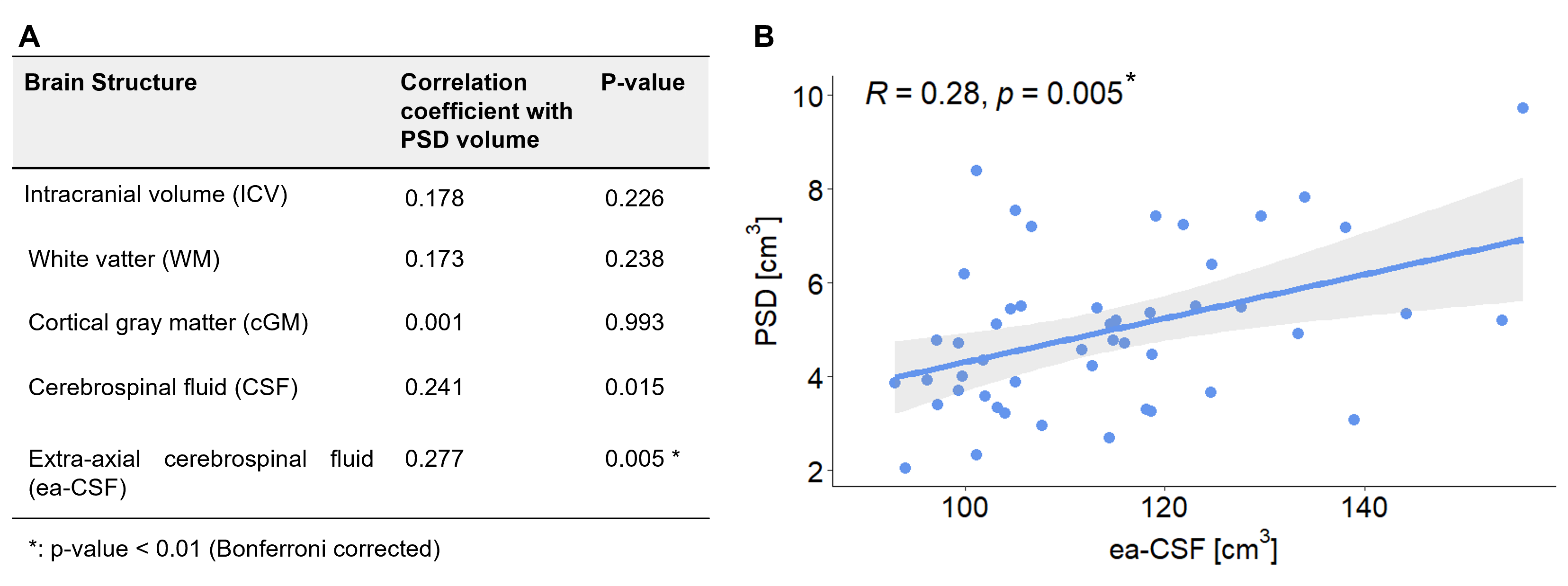
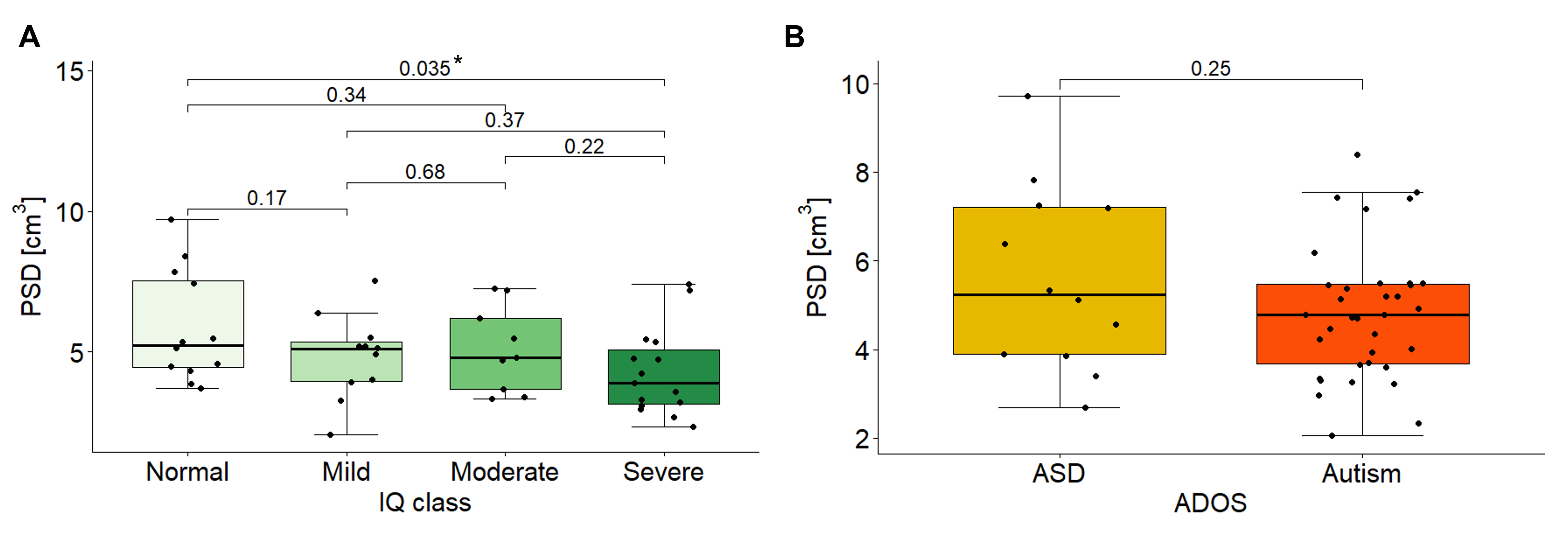
Results: The study revealed a significant positive correlation between PSD volume and extra-axial cerebrospinal fluid volume, and a significant negative correlation with the degree of developmental delay in children with ASD.
Impact: Our findings indicate that PSD volume may play a key role in neurodevelopment by affecting cerebrospinal fluid dynamics. This highlights the need for further research to understand alterations in the dynamics of neurofluids in the developing brain and in ASD.
Introduction
Autism Spectrum Disorder (ASD) is a multifaceted neurodevelopmental condition with a rising global prevalence estimated at 27.6 per 1000 children1. The etiology of ASD remains unknown, with genetic and environmental factors representing highly variable contributors to the ASD phenotype. There is recent evidence that derangements in the immune system are present in children with ASD. Such alterations can trigger a neuroinflammatory cascade leading to significant consequences for normal developmental processes2,3.There are no magnetic resonance imaging (MRI) based biomarkers to address neuroinflammation in ASD.Cerebral parasagittal dura (PSD), a tissue lining the walls of the superior sagittal sinus, plays essential roles in the drainage of cerebrospinal fluid (CSF), in the elimination of metabolic waste and in facilitating brain immunosurveillance4,5. Recent research indicates that in individuals with ASD, the subarachnoid space is enlarged, with increased CSF volume.6. While PSD volume has been investigated in adults using Magnetic Resonance Imaging (MRI)7,8,9,10, such investigation remains largely unexplored in children, especially in ASD.
Our research aims to characterize PSD within a cohort of male children diagnosed with ASD. To achieve this, we used automatic segmentation based U-Net algorithm to extract PSD volume and assess its associations with other brain structure volumes (intracranial volume (ICV), white matter (WM), cortical gray matter (cGM), CSF and extra-axial CSF (ea-CSF)) and clinical variables (Autism Diagnostic Observation Scale - second edition (ADOS-2) and developmental delay scale(IQ class)).
Methods
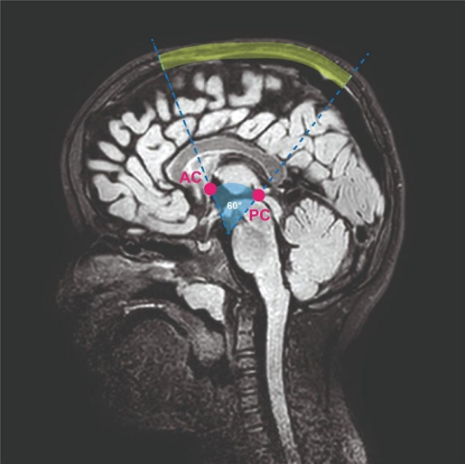
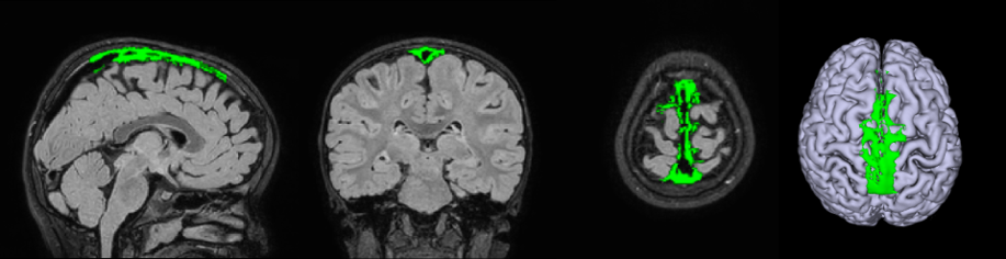
The study involved a cohort of 48 male children (age: 4.5 ± 1.5 years) diagnosed with ASD. MRI data were acquired using a Philips 3T scanner and a 32 channel head coil. The MRI protocol included 3D-T1 weighted (3D-T1w) (TR=8,3 ms, TE=3,9 ms, 1mm3 isotropic) and 3D-T2 Fluid Attenuated Inversion Recovery scans (3D-FLAIR) (TR=4800 ms, TE=289 ms, TI=1650 ms, 1mm3 isotropic). 3D-T1 images were processed through an ad-hoc pipeline developed in-house in order to extract brain structures volumes (ICV, WM, cGM and CSF). The ea-CSF was derived from the CSF mask by manually removing the ventricle and the components below the anterior commissure - posterior commissure line. PSD volume extraction was performed on 3D-FLAIR images, as previously employed by Ringstad et al.4. The segmentation of PSD volume was achieved using a customized Convolutional Neural Network (CNN), specifically the U-net11. The resulting masks underwent manual inspection and correction if necessary, overseen by a neuroradiologist. Notably, the PSD segmentation was limited to its central region to enhance reproducibility, as the anterior and posterior regions often proved either absent or challenging to differentiate from adjacent tissues.(Figure 1, Figure 2)Statistical analyses were conducted, with correlations assessed using Pearson or Kendall correlation tests. ANOVA test was applied to evaluate differences between groups in categorical variables. To further elucidate the obtained results, a multivariate regression analysis was performed and the best model was selected based on the Akaike Information Criteria (AIC) .
Results
Considering developing brains in our cohort of young children with autism, a significant age-related increase in ICV (p-value=0.03 , R=0.22) and WM (p-value=0.004, R=0.29) volume was found, while no age-related correlations were found with PSD, GM, CSF and ea-CSF volumes. PSD volume was positively correlated with ea-CSF volume (p-value = 0.005, Kendall correlation coefficient R= 0.28) and negatively correlated with IQ (p-value = 0.028 and F-value = 5.134, ANOVA). In particular, a significant difference in PSD volume was found between children with normal and severe IQ (p-value = 0.035). No correlations were found between PSD volume and other brain structure volumes or ASD severity. (Figure 3, figure 4) After multivariate regression analysis, PSD volume was best explained by a combination of ea-CSF volume and IQ. The correlations between PSD and the model independent variables were also tested including the others as confounding factors, confirming a significant relationship between the PSD and ea-CSF volume (p-value = 0.003) and between the PSD volume and children with normal and severe IQ (p-value = 0.045).Discussion
We found a robust positive correlation between PSD and ea-CSF volumes and a negative correlation between PSD volume and the severity of developmental delay. The present findings hold significant importance in light of the growing understanding of the role of PSD in brain waste clearance, CSF absorption and neuroinflammation. Severe developmental delay may be a consequence of an underdeveloped PSD which is inefficient in draining CSF, contributing to the accumulation of toxic material and promoting low-grade neuroinflammatory processes known to be associated with ASD.Conclusion
The study suggests that PSD volume may influence the neurodevelopmental process in children with ASD by altering overall dynamics of neurofluids.Acknowledgements
NA, LL and GF are supported partially from 5xMille and RC2023 funds. Fundings from RC2023 partially support DP.References
[1] MAENNER, Matthew J., et al. Prevalence and characteristics of autism spectrum disorder among children aged 8 years—Autism and Developmental Disabilities Monitoring Network, 11 sites, United States, 2020. MMWR Surveillance Summaries, 2023, 72.2: 1.
[2] TOSCANO, Chrystiane VA, et al. Neuroinflammation in autism spectrum disorders: Exercise as a “pharmacological” tool. Neuroscience & Biobehavioral Reviews, 2021, 129: 63-74.
[3] HUGHES, H. K.; MORENO, R. J.; ASHWOOD, P. Innate immune dysfunction and neuroinflammation in autism spectrum disorder (ASD). Brain, behavior, and immunity, 2022.
[4] RINGSTAD, G.; EIDE, P. K. Cerebrospinal fluid tracer efflux to parasagittal dura in humans. Nat Commun 11 (1): 354. 2020.
[5] RUSTENHOVEN, Justin, et al. Functional characterization of the dural sinuses as a neuroimmune interface. Cell, 2021, 184.4: 1000-1016. e27.
[6] SHEN, Mark D., et al. Extra-axial cerebrospinal fluid in high-risk and normal-risk children with autism aged 2–4 years: a case-control study. The Lancet Psychiatry, 2018, 5.11: 895-904.
[7] MELIN, Erik, et al. Human parasagittal dura is a potential neuroimmune interface. Communications Biology, 2023, 6.1: 260.
[8] SONG, Alexander K., et al. Parasagittal dural space hypertrophy and amyloid-β deposition in Alzheimer’s disease. Brain Communications, 2023, 5.3: fcad128.
[9] HETT, Kilian, et al. Parasagittal dural space and cerebrospinal fluid (CSF) flow across the lifespan in healthy adults. Fluids and Barriers of the CNS, 2022, 19.1: 1-13.
[10] PARK, Mina, et al. Aging is positively associated with peri-sinus lymphatic space volume: assessment using 3T black-blood MRI. Journal of Clinical Medicine, 2020, 9.10: 3353.
[11] RONNEBERGER, Olaf; FISCHER, Philipp; BROX, Thomas. U-net: Convolutional networks for biomedical image segmentation. In: Medical Image Computing and Computer-Assisted Intervention–MICCAI 2015: 18th International Conference, Munich, Germany, October 5-9, 2015, Proceedings, Part III 18. Springer International Publishing, 2015. p. 234-241.
Figures