2470
Perivascular and parenchymal fluid characteristics differentially contribute to cognition in typically developing children and adolescents1University of Southern California, Los Angeles, CA, United States
Synopsis
Keywords: Neurofluids, Neurofluids, Child development; diffusion MRI; perivascular spaces
Motivation: Perivascular spaces (PVS) play a critical role in fluid transfer and waste clearance in the brain, but few studies have explored how alterations to perivascular fluid flow may impact brain maturation and behavior
Goal(s): This study aims to characterize age-related alterations to perivascular and parenchymal fluid flow characteristics in typically developing children and assess their contribution to cognition.
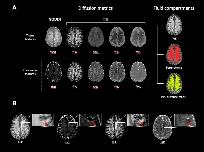
Approach: We employ multi-compartment diffusion models to quantify free water diffusion characteristics within automatically defined perivascular spaces using enhanced PVS contrasts.
Results: Our findings show free water diffusion characteristics within the PVS and surrounding parenchyma are associated with age and cognitive scores.
Impact: Our findings suggest alterations to perivascular space function may occur as early as childhood. Variations in perivascular and parenchymal fluid properties may be predictive of cognitive outcomes in adolescents, thus underscoring the importance of waste clearance functionality on brain health.
Introduction
Perivascular spaces (PVS) are fluid-filled cavities that surround penetrating blood vessels and provide a low resistance pathway for fluid transfer to facilitate efficient waste clearance from the brain (1, 2). We have previously shown that PVS become progressively larger and more numerous across the lifespan (3) which may confer altered waste clearance during aging. Diffusion MRI (dMRI) is a non-invasive neuroimaging method that is sensitive to displacement patterns of diffusing water molecules and can be used to evaluate fluid flow dynamics within and around PVS (4). PVS enlargement is associated with altered fluid flow dynamics as measured with free water dMRI in normal aging (5) as well as in patients with AD neuropathology (6), which may be indicative of flow distributions and waste clearance dysfunction. However, little is known regarding the functional implications of PVS enlargement during child development. The goal of this study is to characterize the age-related changes in PVS fluid flow dynamics in developing children and adolescents between 8 and 21 years of age using multi-compartment dMRI models and assess the functional implications of such changes with cognitive scores.Methods
High resolution T1w (voxel size: .8 mm isotropic; FOV: 256x240x166 mm; TR = 2500 ms, TI = 1000 ms, TE = 1.8/3.6/5.4/7.2 ms, FA = 8 degrees), T2w (voxel size: .8 mm isotropic; FOV: 224x224 mm; TR/TE: 3200/565 ms) and multi-shell dMRI scans (b=1500 s/mm2, 3000 s/mm2, 92-93 diffusion-encoding directions per shell, 1.5 mm isotropic voxel, TR=3.23 s) from 655 typically developing children and adolescents were acquired through the Lifespan Human Connectome Project in Development. PVS were automatically segmented from enhanced PVS contrasts (EPC) derived from T1w and T2w images using the protocol described in (7) (Figure 1). The NODDI metrics intracellular volume fraction (ficvf) and isotropic volume fraction (fiso) were calculated using the spherical mean technique and tissue tensor imaging free water diffusivity measures fRD, fMD and fFA were calculated using a bitensor model with an Aligned Zeppelin distribution implemented with QIT (8). Diffusion maps were the co-registered to the EPC using a rigid body transform. To assess diffusion metrics as a function of the distance from the PVS, 9 concentric rings were automatically generated from level sets of the PVS boundary with 1 mm spacing between them (Figure 1) and were bounded by the white matter tissue mask to exclude gray matter and ventricles from analyses. Cognitive performance was assessed using the NIH Toolbox composite scores for total cognition. The relationships between age, cognition, and diffusion characteristics were assessed with general linear models and linear mixed effects models.Results
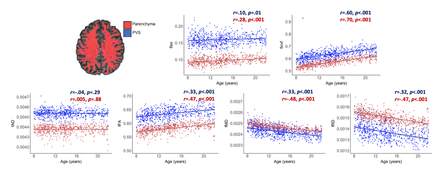
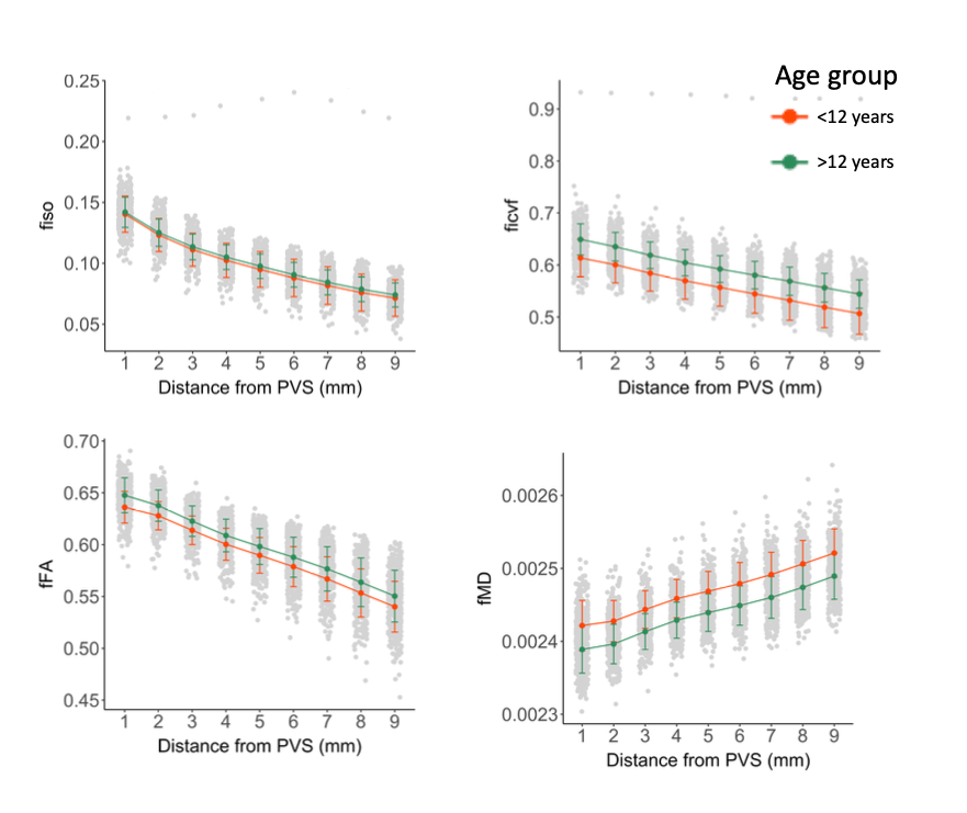
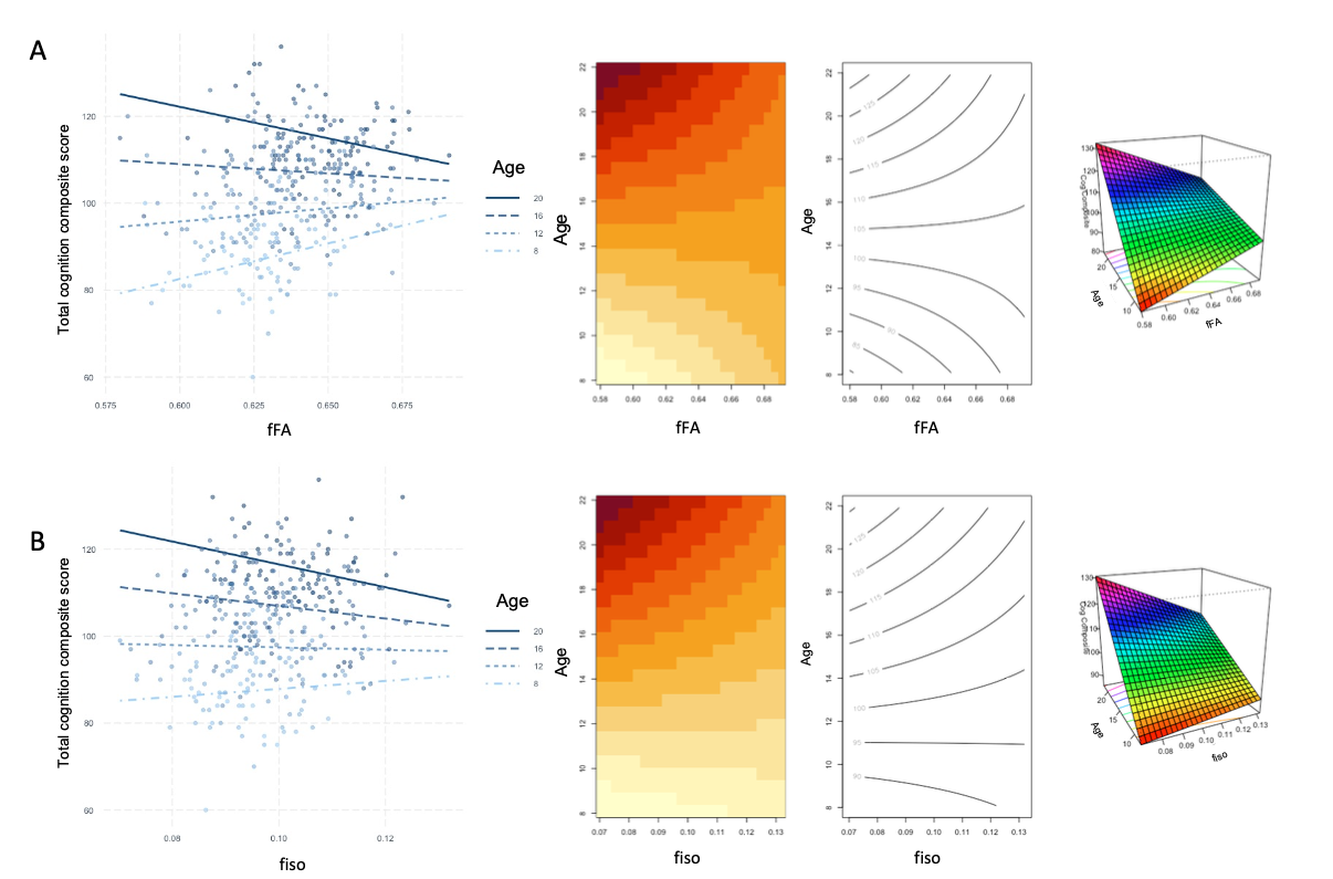
Age was significantly associated with free water diffusion features in the PVS and surrounding parenchyma (Figure 2). A significant interaction between perivascular and parenchymal FISO and age was observed, where age-related FISO alterations increased faster in the PVS compared to the parenchyma (B=.003, t=-3.13, p=.0013). fFA, ficvf and fiso decreased, while fMD increased monotonically with distance from the PVS in both young children and adolescents (Figure 2), indicative of decreased free water in parenchymal regions furthest from PVS. After adjusting for age, perivascular and parenchymal free water diffusivity measures did not significantly predict cognitive scores from the NIH toolbox. However, a significant interaction between fFA and age was observed in the PVS (p<.001), where fFA was associated with better cognitive performance in younger children and worse cognitive performance in older adolescents (Figure 4A). Additionally, a significant interaction between fiso and age was observed in the brain parenchyma (p=.01), where fiso was negatively correlated with total cognition composite scores in older subjects only (Figure 4B).Discussion
Our findings show free water diffusivity measures increases with age in children and adolescents within perivascular spaces and the surrounding parenchyma. While it has been shown that age-related PVS enlargement in older adults is accompanied by increased free water (5), our findings demonstrate this process begins much earlier in childhood, and may indicate changing waste clearance functionality in the growing brain. Our findings also show perivascular and parenchymal fluid characteristics are predictive of cognitive outcomes during child development, where increased free water may lead to preferential impairments to cognition in older adolescents. Our findings therefore suggest altered fluid characteristics in the maturing brain may disrupt cognitive development, potentially due to inefficient waste clearance.Acknowledgements
The image computing resources provided by the Laboratory of Neuro Imaging Resource (LONIR) at USC are supported in part by National Institutes of Health (grant number P41EB015922). Data collection and sharing for this project was funded by the Human Connectome Project. The research reported in this publication was supported by the National Institute of Mental Health (NIMH) of the NIH under the award number RF1MH123223, the NIA award numbers R01AG070825 and R41AG073024 and the USC ADRC 1P30AG066530–01.References
1. J. J. Iliff, et al., A paravascular pathway facilitates CSF flow through the brain parenchyma and the clearance of interstitial solutes, including amyloid β. Sci Transl Med 4 (2012).
2. M. Marín-Padilla, D. S. Knopman, Developmental aspects of the intracerebral microvasculature and perivascular spaces: Insights into brain response to late-life diseases. J Neuropathol Exp Neurol 70, 1060–1069 (2011).
3. K. M. Lynch, F. Sepehrband, A. W. Toga, J. Choupan, Brain perivascular space imaging across the human lifespan. Neuroimage 271 (2023).
4. T. Taoka, et al., Evaluation of glymphatic system activity with the diffusion MR technique: diffusion tensor image analysis along the perivascular space (DTI-ALPS) in Alzheimer’s disease cases. Jpn J Radiol 35, 172–178 (2017).
5. K.M. Lynch, R. Cabeen, R. Custer, F. Sibilia, A.W. Toga, J. Choupan, Age-related alterations to perivascular and parenchymal fluid flow characteristics with tissue tensor imaging (TTI) in International Society for Magnetic Resonance in Medicine, (2022).
6. K.M. Lynch, R. Custer, F. Sibilia, A.W. Toga, J. Choupan, Perivascular and parenchymal fluid flow characteristics and AD neuropathology in International Society for Magnetic Resonance in Medicine, (2023).
7. F. Sepehrband, et al., Image processing approaches to enhance perivascular space visibility and quantification using MRI. Sci Rep 9, 12351 (2019).
8. R. P. Cabeen, F. Sepehrband, A. W. Toga, Rapid and accurate NODDI parameter estimation with the spherical mean technique in Proc International Society for Magnetic Resonance in Medicine (ISMRM), (2019), p. 3363.
Figures