2466
Background R2* correction for χ-separation (chi-separation)1Department of Electrical and Computer Engineering, Seoul National University, Seoul, Korea, Republic of
Synopsis
Keywords: Susceptibility/QSM, Quantitative Susceptibility mapping, Susceptibility source separation, R2* mapping, Artifact correction, Iron imaging, Myelin imaging
Motivation: An advanced quantitative susceptibility mapping (QSM) technique, χ-separation, requires tissue field map and R2* to separate paramagnetic and diamagnetic components. As the background field is removed to acquire the tissue field map, the background R2* needs to be removed to accurately estimate paramagnetic and diamagnetic susceptibility maps.
Goal(s): Investigate the effect of background R2* correction on χ-separation.
Approach: The effect of background R2* correction on χ-separation was tested by varying the background R2* with multiple head orientations and k-space filtering.
Results: Background R2* correction successfully reduced overestimation in χ-separation maps in different head orientations and low-pass filtering levels.
Impact: Background R2* correction enables consistent χ-separation across different head orientations and k-space filtering.
Introduction
χ-separation (chi-separation) is an advanced quantitative susceptibility mapping (QSM) technique that jointly utilizes tissue phase and transverse relaxation rate maps to separate paramagnetic (χpara) and diamagnetic susceptibility (χdia) contributions in QSM1. Inaccurate tissue field or R2* (or R2) maps may result in erroneous χpara and χdia maps. When creating a tissue field map, background field from susceptibility difference (e.g. air-tissue interface) and incomplete shimming is removed by applying background field removal techniques2,3 to the phase of gradient-echo data. Background field variation also causes intravoxel dephasing, resulting in magnitude modulation of gradient-echo data and overestimation of R2*4. Therefore, this additional R2*, named as background R2*, should be corrected when conducting χ-separation to prevent potential bias in χpara and χdia maps. In this study, we demonstrated that background R2* correction can reduce artifactual overestimation of χpara and χdia. Moreover, we examined the effect of background R2* correction under 1) multiple head orientations and 2) varying k-space filtering, confirming that background R2* correction leads to consistent χ-separation maps across different head orientations and k-space filtering.Methods
A healthy volunteer was scanned using Siemens 3 T MRI with a 3D multi-echo gradient-echo sequence and a 2D multi-echo spin-echo sequence. The acquisition with the 3D multi-echo gradient-echo sequence was repeated with changing the head orientation.The effect of intravoxel field gradient on 3D multi-echo gradient-echo data was modelled using voxel spread function method5. In this model, the modulated signal from a voxel by linearly varying intravoxel field can be expressed as multiplication with a shifted sinc function in k-space, where the shift is proportional to the intravoxel field gradient. The voxel spread function can be calculated by inverse Fourier transform of this shifted sinc function. When k-space data is filtered with a windowing function, the effect of windowing can be accommodated by multiplying the shifted sinc function with the windowing function.
The schematic flowchart of background R2* correction for χ-separation is presented in Fig. 1. First, the field gradient was estimated from the numerical gradient of combined phase of multi-echo gradient-echo data. The voxel spread function was calculated from the field gradient map. Using this voxel spread function, the magnitude modulation by the field gradient was estimated, and utilized to generate a background-corrected R2* map. For χ-separation, a tissue field map was prepared by removing background field from the combine phase map6. The R2 map were derived from the multi-echo spin-echo data. Finally, χpara and χdia maps were acquired with χ-sepnet7 utilizing the corrected or uncorrected R2*, R2, and tissue field maps as inputs.
Results
In Fig. 2, the effect of background R2* correction on χpara and χdia maps were examined at the regions close to air cavities. While uncorrected χpara and χdia maps exhibited overestimation at regions marked with cyan lines, corrected χpara and χdia values were in normal ranges, as confirmed by the χpara, χdia, and R2* profile, which was plotted along cyan lines. Overall, the χpara and χdia bias from background R2* at regions close to nasal, frontal, and tympanic cavities were properly reduced in the corrected χpara and χdia maps.In Fig. 3, background R2* correction and χ-separation were conducted with multi-echo gradient-echo data acquired with three head orientations. When the head orientation was aligned with the direction of B0 field, the overestimation of χpara and χdia occurred bilaterally at the inferior part of frontal lobe. On contrary, when the B0 field was angled toward the patient’s right (or left), the overestimation predominantly occurred at the right (or left) part frontal lobe due to orientation dependence of background R2*. After background R2* correction, the orientational dependence of χpara and χdia was reduced, resulting in more consistent χpara and χdia maps across B0 directions.
Applying low-pass filtering in k-space results in larger effective voxel size, leading to increased intravoxel dephasing and background R2*. In Fig. 4, the same multi-echo gradient-echo data was reconstructed after Tukey-windowing with different low-pass level. The overestimation in uncorrected χpara and χdia maps intensified as increasing low-pass level. By applying background R2* correction, the overestimation in χpara and χdia was corrected, providing more consistent χpara and χdia maps across different low-pass levels.
Discussion
As MRI vendors provide reconstructed images at different low-pass levels, the background R2* differs vendor-to-vendor. Therefore, applying background R2* correction is crucial for consistent χ-separation maps across venders. As noted in Yablonskiy et al.5, the R2* correction based on voxel spread function may fail at tissue boundaries or voxels with highly nonlinear field gradient.Conclusion
Background R2* correction for χ-separation successfully reduced overestimation in χpara and χdia maps.Acknowledgements
This research was supported by the National Research Foundation of Korea (NRF-2019M3C7A1031994, NRF-2022R1A4A1030579), and INMC at Seoul National University.References
1. Shin, H.-G., et al., χ-separation: Magnetic susceptibility source separation toward iron and myelin mapping in the brain. NeuroImage 240, 118371 (2021).
2. Schweser, F., Deistung, A., Lehr, B. W. & Reichenbach, J. R., Quantitative imaging of intrinsic magnetic tissue properties using MRI signal phase: An approach to in vivo brain iron metabolism? NeuroImage 54, 2789-2807 (2011).
3. Liu, T., et al., A novel background field removal method for MRI using projection onto dipole fields (PDF). NMR in Biomedicine 24, 1129-1136 (2011).
4. Ordidge, R. J., Gorell, J. M., Deniau, J. C., Knight, R. A. & Helpern, J. A., Assessment of relative brain iron concentrations using T2-weighted and T2*-weighted MRI at 3 tesla. Magnetic Resonance in Medicine 32, 335-341 (1994).
5. Yablonskiy, D. A., Sukstanskii, A. L., Luo, J. & Wang, X., Voxel Spread Function Method for Correction of Magnetic Field Inhomogeneity Effects in Quantitative Gradient-Echo-Based MRI. Magnetic Resonance in Medicine 70, 1283-1292 (2013).
6. Wu, B., Li, W., Guidon, A. & Liu, C., Whole brain susceptibility mapping using compressed sensing. Magnetic Resonance in Medicine 67, 137-147 (2012).
7. Kim, M., et al., Chi-sepnet: Susceptibility source separation using deep neural network. Proceedings of International Society of Magnetic Resonance in Medicine 30, 2464 (2022).
Figures
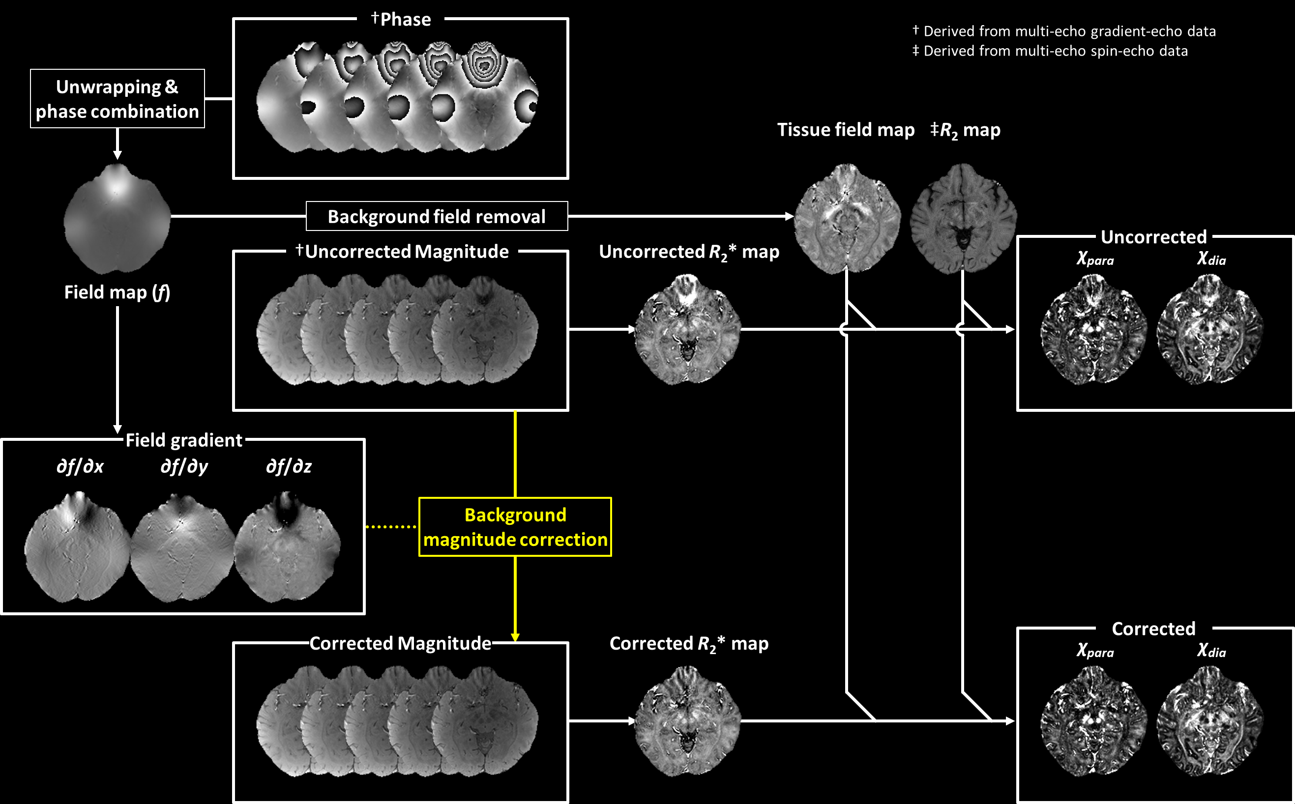
Fig. 1. Schematic flowchart illustrating the background R2* correction for χ-separation.
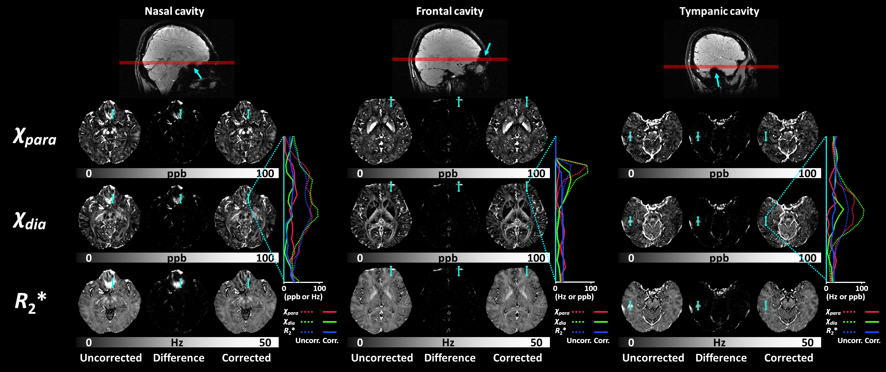
Fig. 2. The effect of background R2* correction on χpara and χdia maps at regions close to air cavities (cyan arrows). The χpara (red), χdia (green), and R2* (blue) profiles along cyan lines are plotted on the right of each set of imaging slices, demonstrating successful correction of overestimated χpara and χdia.
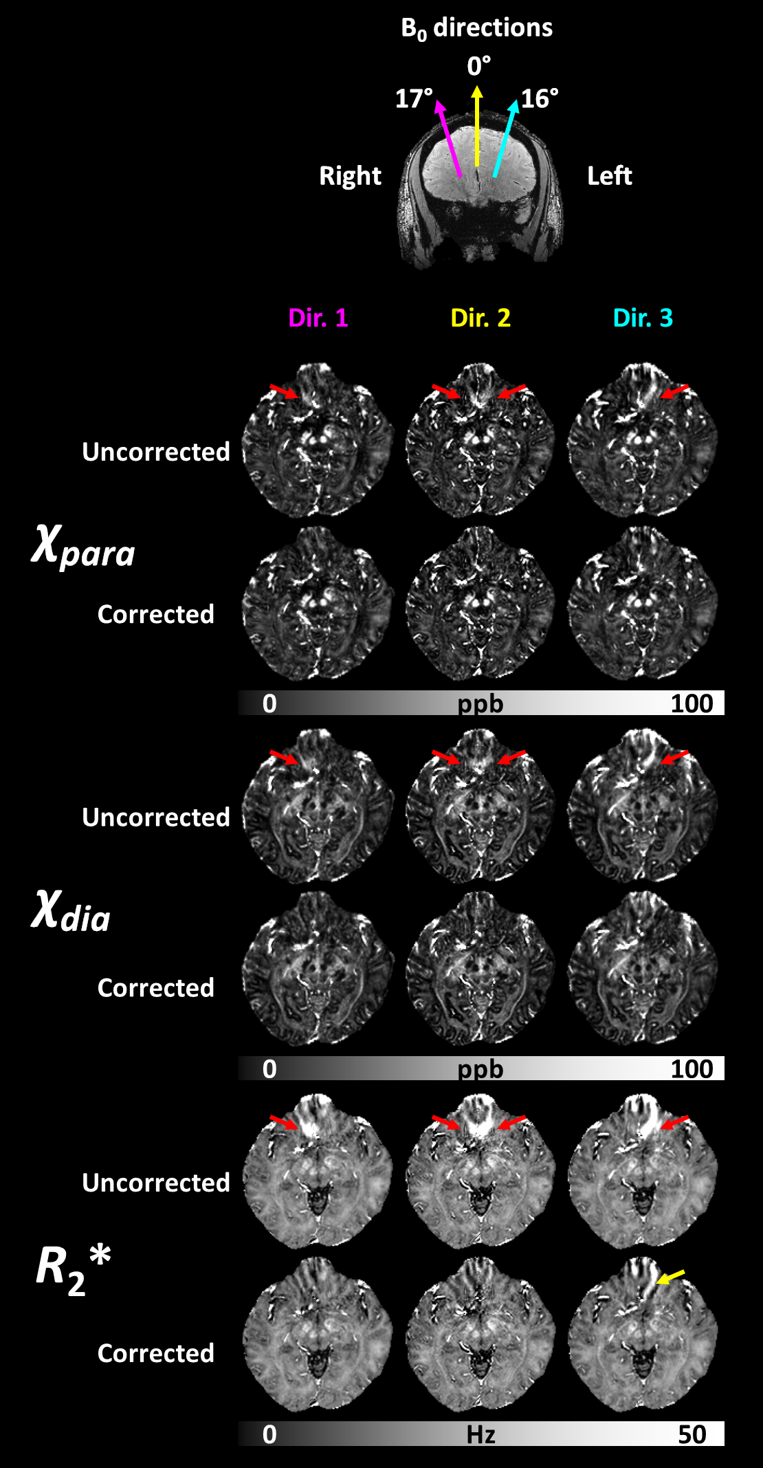
Fig. 3. Orientation dependence of background R2* and overestimation in χpara and χdia (red arrows). When B0 direction is angled toward right or left, the overestimation in χpara and χdia predominantly occurs at right or left. When B0 direction is aligned straight the overestimation occurs bilaterally. Background R2* correction effectively resolves this orientational dependency and provides consistent χpara and χdia maps across brain orientations. Background R2* correction may not properly work at tissue boundary or regions with highly nonlinear field gradient (yellow arrow).
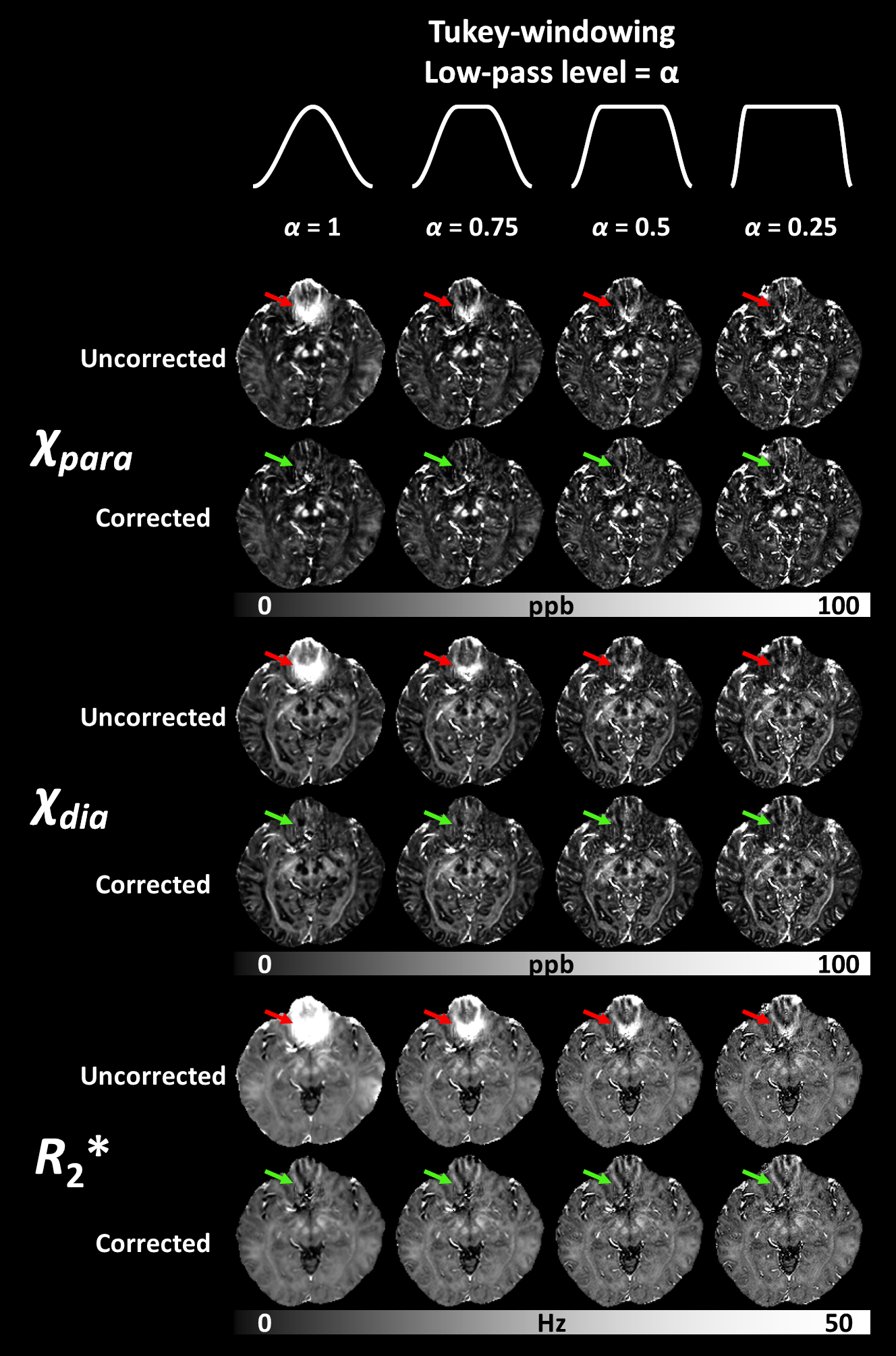
Fig. 4. Background R2* and overestimation in χpara and χdia (red arrows) at different levels of low-pass filtering in k-space. The background R2* intensifies when effective voxel size increases by higher level of low-pass filtering. The overestimation χpara and χdia successfully removed by background R2* correction (red arrows), providing consistent χpara and χdia maps across different low-pass levels.