2453
QSM Reconstruction of Arbitrary Dipole Orientations using an End-to-end Neural Network via Latent Feature Editing1School of Computer Science and Engineering, Central South University, Changsha, China, 2School of EECS, The University of Queensland, Brisbane, Australia, 3State Key Laboratory of Radiation, Medicine and Protection, Soochow University, Suzhou, China, 4University of Alberta, Edmonton, AB, Canada, 5University of Calgary, Calgary, AB, Canada
Synopsis
Keywords: Susceptibility/QSM, Quantitative Susceptibility mapping, Orientation-Adaptive, Latent Feature Editing, OA-iQSM
Motivation: The performances of most DL-QSM methods are limited to MRI phase data of pure-axial acquisition orientation.
Goal(s): In this work, we would like to propose a novel DL-based end-to-end neural network for QSM reconstruction from phase data of arbitrary dipole orientations.
Approach: A novel Latent Feature Editing (LFE) module to learn the encoding of acquisition orientation vectors and seamlessly integrate them into the latent features of deep networks to make them orientation-adaptive.
Results: Both simulated and in vivo experiments demonstrate that the proposed LFE module can result in desirable QSM images at arbitrary oblique head orientations.
Impact: This work introduces a new DL paradigm, allowing researchers to develop innovative QSM methods without requiring a complete overhaul of their existing architectures.
Introduction
QSM is a novel post-processing technique to extract the tissue susceptibility distribution from MRI phase data. Recently, deep neural networks [1-4] are emerging as alternatives to traditional QSM reconstruction methods. However, a significant challenge facing current deep learning (DL) QSM approaches is their limited adaptability to magnetic dipole field orientation variations during training and testing. Therefore, we propose a novel Latent Feature Editing (LFE) module to learn the encoding of the acquisition orientation vectors and integrate them into the latent features of deep neural networks. A novel single-step DLQSM method, namely Orientation-Adaptive iQSM (OA-iQSM), was constructed by incorporating the proposed LFE modules into our iQSM network [4], which was initially designed for phases of pure-axial orientations. Simulated and in vivo experiments were conducted to compare the proposed OA-iQSM with multiple state-of-the-art QSM methods.Method
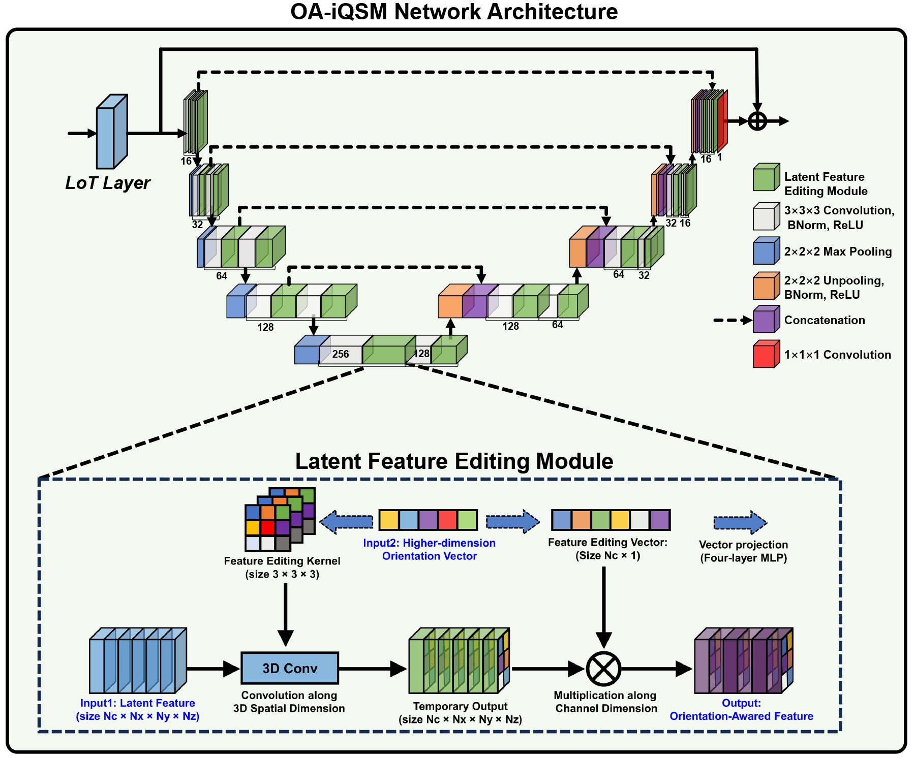
OA-iQSM NetworkAs shown in Fig. 1, the proposed OA-iQSM is constructed by combining the proposed LFE modules into our previously developed iQSM network backbone [4], which is composed of a self-tailored LoT Layer and a traditional U-net. The LFE modules are appended after every 3D convolutional layer in the U-net part to make full use of the orientation information. The LFE module learns the encoding of the acquisition orientation vectors using two MLPs and integrates them into the latent features of deep neural networks through 3D image convolution and multiplication. The U-net part contains 18 convolutional layers; 18 LFE modules; 4 max-pooling and transposed convolution layers; 22 ReLU units, and an output convolutional layer.
Training data preparation
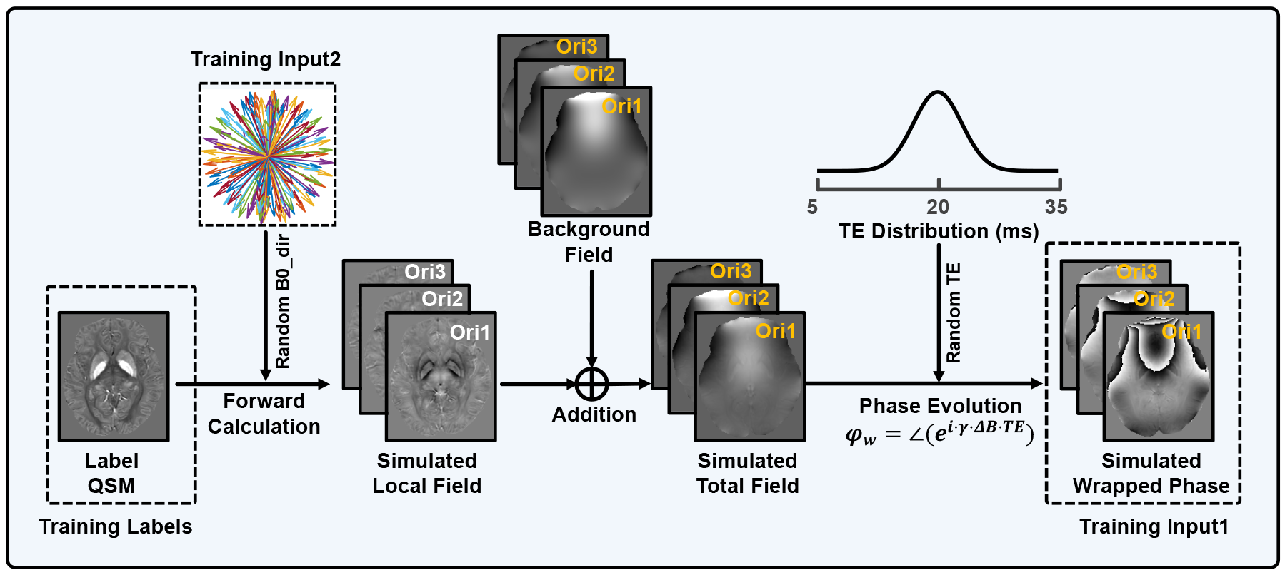
In this work, a self-supervised training scheme was introduced to simulate as many different dipole orientation vectors as possible for network training. A total of 14400 small susceptibility map patches (size: 643) were randomly cropped from 96 full-size brain volumes. Then, the training data were prepared using the pipeline shown in Fig. 2. OA-iQSM network was trained with simulated wrapped phase and dipole orientation vectors as inputs and original QSM data as labels.
Network training
All network parameters were initialized with normally distributed random numbers of 0 mean and 0.01 standard deviation. All networks were trained for 100 epochs (around 24 hours) on one Nvidia Tesla A6000 GPU using Adam optimizer. The batch size was 32, and the learning rate was 10-3, 10-4, and 10-5 for the first 40 epochs, 40-80 epochs, and the final 20 epochs. MSE was used as the loss function.
Validation on simulated and in vivo datasets
The proposed iQFM and iQSM were compared with several established methods, including multi-step iLSQR [5], LPCNN [1], AFTER-QSM [2], and our single-step iQSM method. All multi-step methods started with Laplacian Unwrapping and RESHARP background removal in this work.
Two healthy brain subjects were simulated from a COSMOS-QSM for an ablation study to quantitatively evaluate the effectiveness of the proposed LFE modules. Besides, two more simulated brains based on STI label and one in vivo brain data acquired at 3T were adopted to compare the proposed OA-iQSM with previous QSM methods.
Results
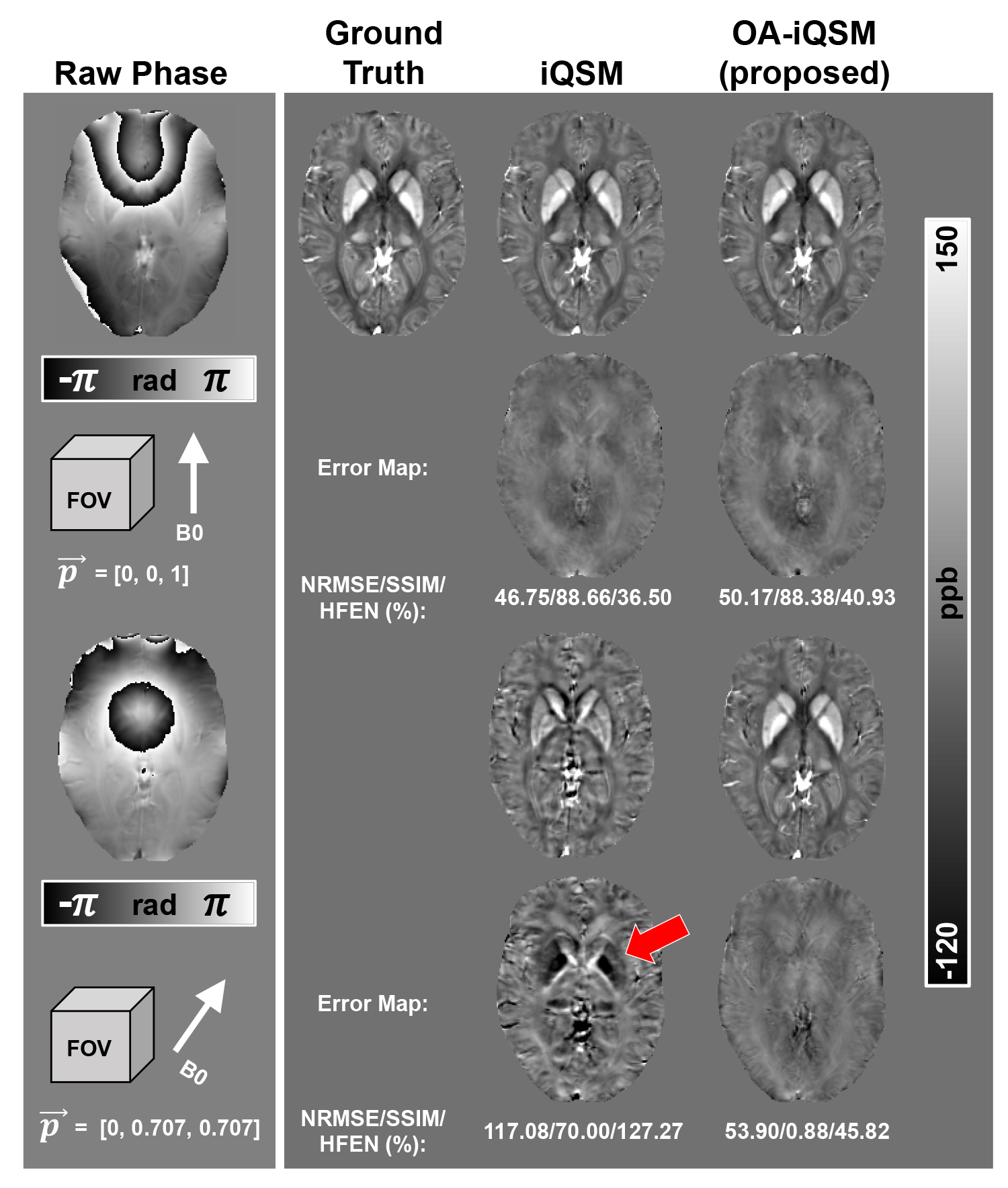
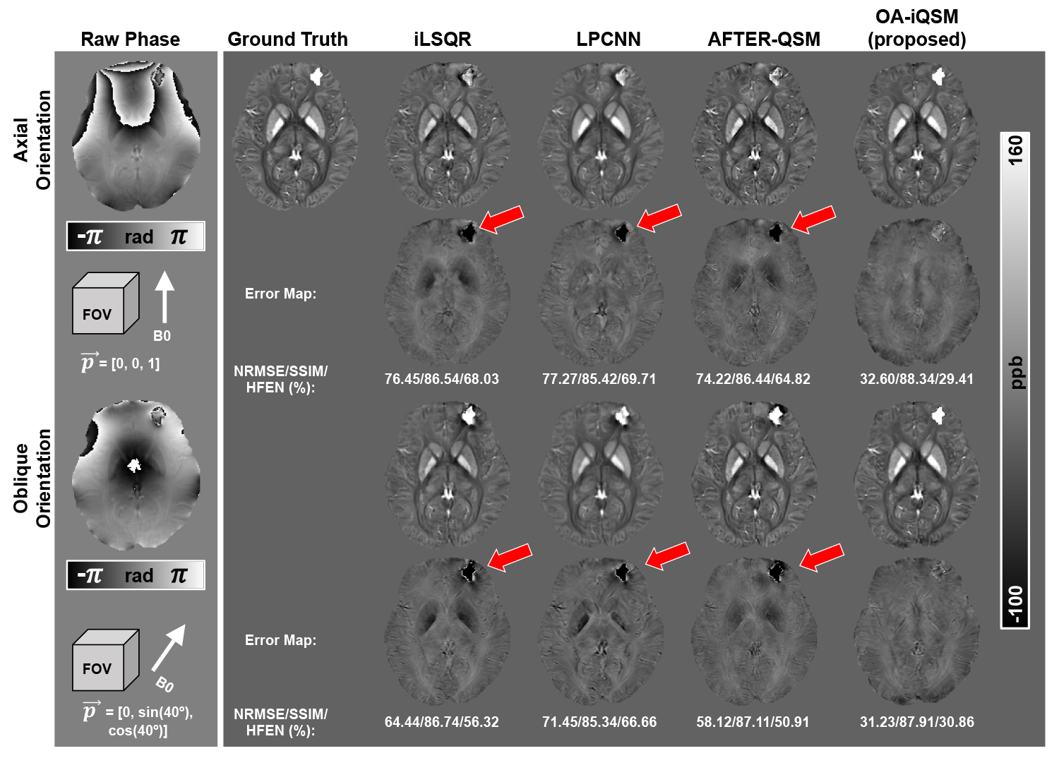
Figure 3 compares the proposed OA-iQSM with the original iQSM on two COSMOS-based simulated brain subjects to validate the effectiveness of the proposed LFE modules. OA-iQSM showed consistent reconstructions for phase data of both pure-axial and 45° oblique dipole orientations, while the original iQSM failed to present reasonable results for the latter case.Figure 4 compares the proposed OA-iQSM with various established QSM methods on two simulated phases from a χ33 QSM with a simulated hemorrhage lesion (0.8 ppm). The upper two rows compare different QSM methods on phase data of axial orientation, while the bottom two rows illustrate the results of the oblique case. According to the error maps and the numerical metrics, OA-iQSM showed the most consistent and best reconstruction results among all methods.
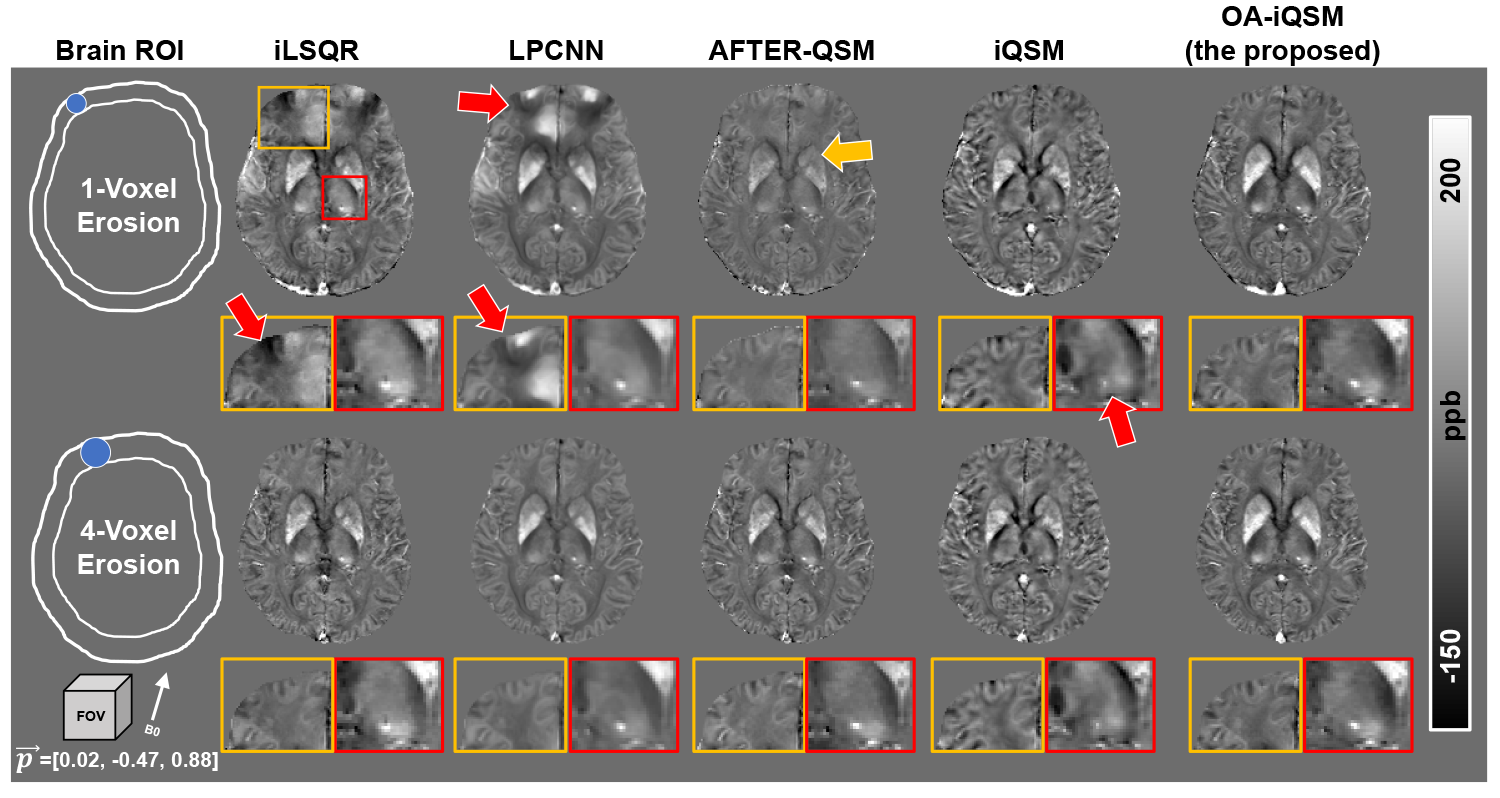
QSM results of various methods on the in vivo subject were compared in Fig. 5. The upper two row shows reconstructions of 1-voxel brain edge erosion, while the bottom two rows demonstrate results of 4-voxel brain mask erosion. The proposed OA-iQSM produced the most visually appealing results for 1-voxel mask erosion data, while iLSQR and LPCNN showed substantial artifacts, AFTER-QSM showed significant contrast loss compared with its 4-voxel erosion result, and iQSM failed to give correct reconstructions on the micro-bleeding lesion, as confirmed in the zoom-in region and the red arrow.
Discussion and Conclusion
This study proposed a novel OA-iQSM method for single-step QSM reconstruction from MRI phases of arbitrary acquisition orientations, thanks to the novel LFE design, which enables QSM researchers to develop novel orientation-adaptive QSM methods based on their existing network backbones.Acknowledgements
YG acknowledges support from the National Natural Science Foundation of China under Grant No. 62301616. HS acknowledges support from the Australian Research Council (DE210101297, DP230101628).
References
1. K. W. Lai, M. Aggarwal, P. van Zijl et al., “Learned Proximal Networks for Quantitative Susceptibility Mapping,” Med Image Comput Comput Assist Interv, vol. 12262, pp. 125-135, Oct, 2020.
2. Z. Xiong, Y. Gao, F. Liu et al., “Affine transformation edited and refined deep neural network for quantitative susceptibility mapping,” Neuroimage, vol. 267, pp. 119842, Feb 15, 2023.
3. Y. Gao, X. Y. Zhu, B. A. Moffat et al., “xQSM: quantitative susceptibility mapping with octave convolutional and noise-regularized neural networks,” NMR in Biomedicine, vol. 34, no. 3, Mar, 2021.
4. Y. Gao, Z. Xiong, A. Fazlollahi et al., “Instant tissue field and magnetic susceptibility mapping from MRI raw phase using Laplacian enhanced deep neural networks,” Neuroimage, vol. 259, pp. 119410, Oct 1, 2022.
5. W. Li, N. Wang, F. Yu et al., “A method for estimating and removing streaking artifacts in quantitative susceptibility mapping,” Neuroimage, vol. 108, pp. 111-122, Mar, 2015.
Figures