2452
Fast Multipole Method-Enhanced Boundary Element Modeling for Total Field Inversion in Quantitative Susceptibility Mapping1East China Normal University, Shanghai, China, 2Department of Radiology, Weill Medical College of Cornell University, New York, NY, United States, 3MR Scientific Marketing, Siemens Healthineers, Shanghai, China
Synopsis
Keywords: Susceptibility/QSM, Quantitative Susceptibility mapping, Total Field Inversion, Boundary Element method, Fast Multipole method
Motivation: The study aims to improve quantitative susceptibility mapping (QSM) precision at region of interest (ROI) boundaries where background field interference affects accuracy.
Goal(s): To introduce a novel boundary element method total field inversion (BEM-TFI) enhanced by the fast multipole method (FMM) for high-resolution QSM.
Approach: A comparative assessment of BEM-TFI technique was performed with traditional QSM methods, utilizing in-vivo data and simulated field maps to determine inversion quality and background field removal efficiency.
Results: Enhanced by FMM, the BEM-TFI method demonstrated significant improvements in accurately discerning tissue susceptibility from background noise, indicating a substantial advancement in the outcomes of QSM.
Impact: The BEM-TFI enhancement in QSM accuracy allows for more detailed characterization of the brain's cortex, potentially enriching neuroscientific research and elevating the quality of neuroimaging data.
Introduction
In neurological research, quantitative susceptibility mapping (QSM) has become a principal tool for evaluating magnetic susceptibility, particularly within deep brain nuclei.1, 2 The ability of QSM to discern alterations in cortical susceptibility is crucial for comprehensive whole-brain analyses, shedding light on the cortex's role in the pathophysiology of various neurological disorders, including Parkinson’s disease, Alzheimer’s disease, and Wilson’s disease.3-6 Accurate susceptibility mapping near the boundaries of regions of interest (ROI) is essential due to their susceptibility to background field interference.1, 7, 8 Traditional QSM approaches, reliant on sequential background removal and field inversion, often amplify errors in these boundary regions.8 Recent advancements have introduced single-step or total field inversion methods, aiming to streamline this process.9-13 However, these methods continue to confront challenges, particularly in accurately separating the background and local fields. To address these challenges, we propose a boundary element method total field inversion (BEM-TFI) method that integrates Dirichlet boundary conditions directly into the inversion process, bolstered by the computational efficiency of the fast multipole method (FMM),14, 15 thus achieving a more accurate mapping of tissue susceptibility.Theory
The cost function of our method is formulated as follow: $$\underset{\,\, \left[ \begin{array}{c} \boldsymbol{\chi }\\ \boldsymbol{f}_{\boldsymbol{\varGamma }}\\\end{array} \right]}{\boldsymbol{\chi }=argmin}\,\,\left\| \boldsymbol{m}\cdot e^{i\left( \boldsymbol{D\chi }\,+\,\boldsymbol{Lf}_{\boldsymbol{\varGamma }}\,\,-\,\,\boldsymbol{f}_{\boldsymbol{total}}\,\, \right)} \right\| _{2}^{2}+\lambda \left\| \boldsymbol{M}_G\boldsymbol{\nabla \chi } \right\| _1\,\, \ $$Where $$$\boldsymbol{\chi }$$$ is the local tissue susceptibility, m is a weighting matrix compensating for the nonuniform phase noise,16 fГ is the boundary value of background field, ftotal is the total field, MG is the edge mask,16 and L is the discretization matrix of boundary integral equation stored using FMM.Method
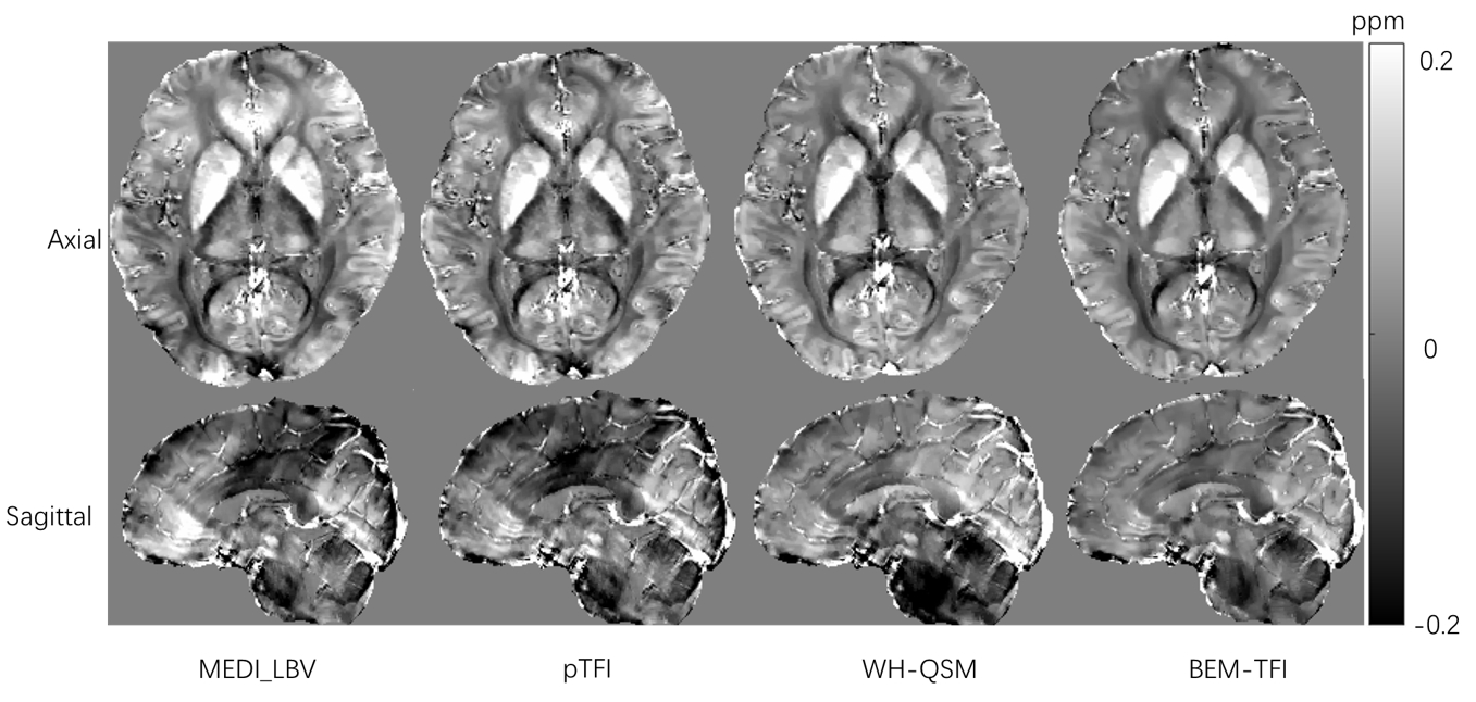
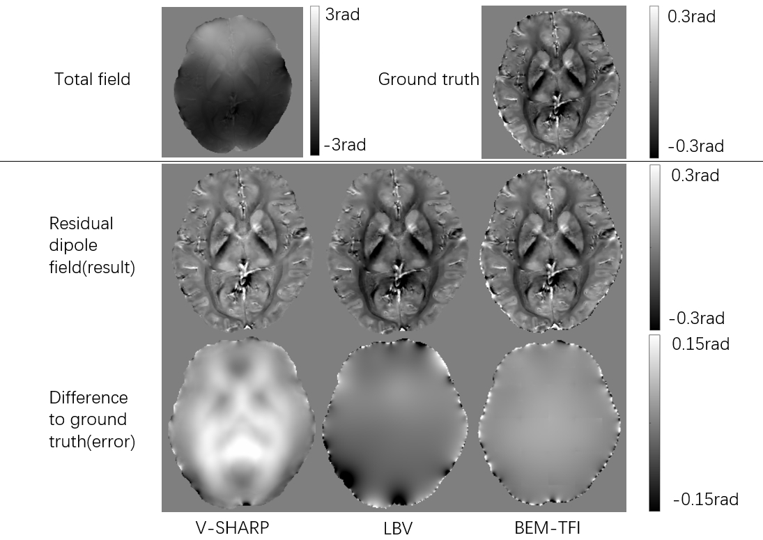
Two sets of experiments were performed to validate the efficacy of our proposed method. We firstly compared susceptibility maps reconstructed by our BEM-TFI technique with the established methods—nonlinear morphology enabled dipole inversion (MEDI) with Laplace boundary value (LBV) background removal,16, 17 preconditioned total field inversion (pTFI),10, 12 and weak-harmonic QSM18 on in-vivo data. The in vivo data were acquired with a 3T Siemens Prisma MRI scanner equipped with a 64-channel head coil. Data acquisition utilized a 3D gradient-recalled-echo (GRE) sequence with the following parameters: TR/ TE1/ΔTE = 38/6.4/5.2 ms, number of echoes = 6, flip angle =15°, monopolar readout, voxel size = 0.85× 0.85 × 0.85mm3, scan time = 7:32. The total field maps were obtained by nonlinear fitting of the multi-echo GRE data followed by graph cut based phase unwrapping.9,10 We then used a forward-simulated total field map based on the above in-vivo susceptibility maps to evaluate the performance our BEM-TFI technique in background field removal. The external susceptibility outside the region of interest was set at a constant value of 9 ppm. We compared the performance of our method to the LBV method and variable size sophisticated harmonic artifact reduction for phase data (V-SHARP) techniques.19Result
Fig. 1 shows the susceptibility maps of a healthy volunteer obtained by four QSM reconstruction method. Notably, our method demonstrated a more even susceptibility within the cortex and cerebellum, suggesting an improved delineation of these regions compared to the other methods.Fig. 2 shows the performances three QSM techniques in background field removal using a simulated total field map. V-SHARP is shown to have the limitation of erroneously filtering out essential low-frequency information from the local field signal. In comparison, while the LBV method generally reproduces residual dipole fields accurately, it introduces errors near the ROI boundaries. The BEM-TFI approach, however, consistently produces results that are in close agreement with the ground truth across the entire field.
Discussion
The integration of the FMM with BEM-TFI marks a notable progression in QSM, particularly enhancing the fidelity of susceptibility mapping in regions susceptible to background field interference. This enhancement is crucial, considering the precise quantification needed for studying neurodegenerative conditions. The superiority of our method is due to the use of an FMM-enhanced boundary element approach to model the background field. This enables our BEM-TFI technique to outperform other methods by avoiding the low-frequency errors commonly associated with background interference. Despite these advancements, our approach introduces new complexities, such as the possibility of FMM-related artifacts and the nuanced demands of surface mesh modeling, which necessitate further scrutiny. The adaptability of this approach to a variety of clinical scenarios also remains to be fully explored.Conclusion
The application of the FMM in the novel BEM-TFI technique has shown to effectively improve the quality of susceptibility maps, particularly in separating the background field influence from the true tissue susceptibility signal. This advancement signifies a promising step forward in the accuracy and utility of QSM for neurological diagnostics.Acknowledgements
No acknowledgement found.References
[1] Wang Y and Liu T. Quantitative susceptibility mapping (QSM): Decoding MRI data for a tissue magnetic biomarker. Magn Reson Med, 2015; 73(1): 82-101
[2] Deistung A, Schweser F and Reichenbach J R. Overview of quantitative susceptibility mapping. NMR Biomed, 2017; 30(4)
[3] Acosta-Cabronero J, Williams G B, Cardenas-Blanco A, Arnold R J, Lupson V and Nestor P J. In Vivo Quantitative Susceptibility Mapping (QSM) in Alzheimer's Disease. Plos One, 2013; 8(11)
[4] Acosta-Cabronero J, Cardenas-Blanco A, Betts M J, Butryn M, Valdes-Herrera J P, Galazky I and Nestor P J. The whole-brain pattern of magnetic susceptibility perturbations in Parkinson's disease. Brain, 2017; 140: 118-131
[5] Spotorno N, Acosta-Cabronero J, Stomrud E, Lampinen B, Strandberg O T, van Westen D and Hansson O. Relationship between cortical iron and tau aggregation in Alzheimer's disease. Brain, 2020; 143: 1341-1349
[6] Shribman S, Bocchetta M, Sudre C H, Acosta-Cabronero J, Burrows M, Cook P, Thomas D L, Gillett G T, Tsochatzis E A, Bandmann O, Rohrer J D and Warner T T. Neuroimaging correlates of brain injury in Wilson's disease: a multimodal, whole-brain MRI study. Brain, 2022; 145(1): 263-275
[7] Schweser F, Deistung A and Reichenbach J R. Foundations of MRI phase imaging and processing for Quantitative Susceptibility Mapping (QSM). Z Med Phys, 2016; 26(1): 6-34
[8] Schweser F, Robinson S D, de Rochefort L, Li W and Bredies K. An illustrated comparison of processing methods for phase MRI and QSM: removal of background field contributions from sources outside the region of interest. NMR Biomed, 2017; 30(4)
[9] Chatnuntawech I, McDaniel P, Cauley S F, Gagoski B A, Langkammer C, Martin A, Grant P E, Wald L L, Setsompop K, Adalsteinsson E and Bilgic B. Single-step quantitative susceptibility mapping with variational penalties. NMR Biomed, 2017; 30(4)
[10] Liu Z, Kee Y, Zhou D, Wang Y and Spincemaille P. Preconditioned total field inversion (TFI) method for quantitative susceptibility mapping. Magn Reson Med, 2017; 78(1): 303-315
[11] Acosta-Cabronero J, Milovic C, Mattern H, Tejos C, Speck O and Callaghan M F. A robust multi-scale approach to quantitative susceptibility mapping. Neuroimage, 2018; 183: 7-24
[12] Liu Z, Wen Y, Spincemaille P, Zhang S, Yao Y, Nguyen T D and Wang Y. Automated adaptive preconditioner for quantitative susceptibility mapping. Magn Reson Med, 2020; 83(1): 271-285
[13] Wen Y, Spincemaille P, Nguyen T, Cho J, Kovanlikaya I, Anderson J, Wu G, Yang B, Fung M, Li K, Kelley D, Benhamo N and Wang Y. Multiecho complex total field inversion method (mcTFI) for improved signal modeling in quantitative susceptibility mapping. Magn Reson Med, 2021; 86(4): 2165-2178
[14] Greengard L and Rokhlin V. A new version of the fast multipole method for the Laplace equation in three dimensions. Acta Numerica, 1997; 6: 229-269
[15] Nishimura N, Yoshida K and Kobayashi S. A fast multipole boundary integral equation method for crack problems in 3D. Engineering Analysis with Boundary Elements, 1999; 23(1): 97-105
[16] Liu J, Liu T, de Rochefort L, Ledoux J, Khalidov I, Chen W, Tsiouris A J, Wisnieff C, Spincemaille P, Prince M R and Wang Y. Morphology enabled dipole inversion for quantitative susceptibility mapping using structural consistency between the magnitude image and the susceptibility map. Neuroimage, 2012; 59(3): 2560-8
[17] Zhou D, Liu T, Spincemaille P and Wang Y. Background field removal by solving the Laplacian boundary value problem. NMR Biomed, 2014; 27(3): 312-9
[18] Milovic C, Bilgic B, Zhao B, Langkammer C, Tejos C and Acosta-Cabronero J. Weak-harmonic regularization for quantitative susceptibility mapping. Magn Reson Med, 2019; 81(2): 1399-1411
[19] Li W, Avram A V, Wu B, Xiao X and Liu C. Integrated Laplacian-based phase unwrapping and background phase removal for quantitative susceptibility mapping. NMR Biomed, 2014; 27(2): 219-27
Figures