2446
Submillimeter in vivo human brain diffusion MRI at 500 mT/m with concurrent field monitoring1Athinoula A. Martinos Center for Biomedical Imaging, Massachusetts General Hospital and Harvard Medical School, Charlestown, MA, United States, 2Institute of Medical Physics and Radiation Protection, Mittelhessen University of Applied Sciences, Giessen, Germany, 3Q Bio Inc, San Carlos, CA, United States, 4Department of Diagnostic and Interventional Radiology, University Hospital Marburg, Philipps University of Marburg, Marburg, Germany
Synopsis
Keywords: Diffusion Acquisition, Data Acquisition
Motivation: High-fidelity, artifact-free diffusion MRI (dMRI) in high-performance gradient systems requires more advanced encoding and reconstruction approaches than those widely used.
Goal(s): To achieve ghosting- and eddy current distortion-free in vivo submillimeter human brain dMRI with concurrent field monitoring.
Approach: A 16-ch clip on field probe system was integrated onto custom-built 72ch to map high-order field perturbations during the acquisition. SENSE-based reconstruction was informed with the monitored phase evolution as expressed in a 3rd-order spherical harmonic model.
Results: Concurrent field monitoring reconstruction reduces non-linear Nyquist ghosting and geometric distortions generated by high-order eddy currents from ultra-strong diffusion gradients.
Impact: We expect concurrent field monitoring to become essential in achieving high-quality image reconstruction as the adoption of high-performance gradients systems grows and image acquisition strategies evolve with more sophisticated image encodings.
Introduction
High-performance gradients are becoming increasingly prevalent in human MRI scanners as gradient technology and hardware advance.1-4 Maximizing the benefits afforded by high-performance gradient technology1-4 for diffusion MRI requires overcoming the technical challenges associated with strong diffusion gradients, including more pronounced high-order eddy currents and pervasive concomitant field effects. In this work, we first investigate the impact of eddy currents from the ultra-strong diffusion gradients (500 mT/m) of the Connectome 2.0 scanner1,5 on the typically long image readouts of high-spatial-resolution diffusion MRI acquisitions . We evidence the limitation of conventional ghosting-removal reconstruction methods to provide high-quality images. We then achieve eddy-current-induced geometric distortion and ghosting-free submillimeter in vivo human diffusion MRI at an unprecedentedly short echo time at Gmax = 500 mT/m with concurrent field monitoring integrated into a custom-built 72-ch in vivo coil.Methods
Acquisition protocol: A healthy volunteer (23F) underwent a diffusion MRI scan on the Connectome 2.0 scanner1,5 (MAGNETOM Connectom.X, Siemens Healthineers, Erlangen, Germany) with a custom-built 72-channel head coil.6 The coil was equipped with a 16-channel 19F clip-on field probe system (Skope, Inc., Zurich, Switzerland) for concurrent field monitoring.7Image acquisition parameters: Whole brain dMRI data was acquired with a 2D Pulsed-Field Gradient Spin Echo (PGSE) EPI sequence with axial slices, 0.9 mm in-plane resolution, slice thickness = 0.9 mm, TR/TE = 12,000/47 ms, phase encoding direction A>P, in-plane acceleration factor = 2, PF = 6/8, no SMS, BW = 1912 Hz, echo spacing = 0.57 ms. Readout time = 46 ms; Total acquisition time = 7.2 min.
Diffusion parameters: 30 diffusion encoding directions at b = 1, 200 s/mm2 (Gmax = 500 mT/m, $$$\Delta$$$=8.7 ms, $$$\delta$$$=2.9 ms) + 1 b0-image. A gradient-recalled echo (GRE) scan was performed after the diffusion scan to estimate the coil sensitivities.
Image reconstruction: Triggers and synchronization pulses were added to the beginning of the 2D-PGSE EPI sequence to make sure that the monitored field and the k-space data were in sync during the image readout. 8 A third-order spherical harmonics (SH) model was fitted to the 16 NMR probes’ signal phase with a linear least squares (LLS) algorithm.9 B0-eddy-current-compensation (B0-ECC) applied by the scanner was removed prior to reconstruction. Images were reconstructed within a SENSE reconstruction framework. The image encoding matrix was informed with the phase evolution, expressed in a 3rd-order SH expansion model.9,10 The ESPIRiT algorithm was used to estimate the coil sensitivities.11 The magnitude of the reconstructed complex images was taken for analysis and visualization.
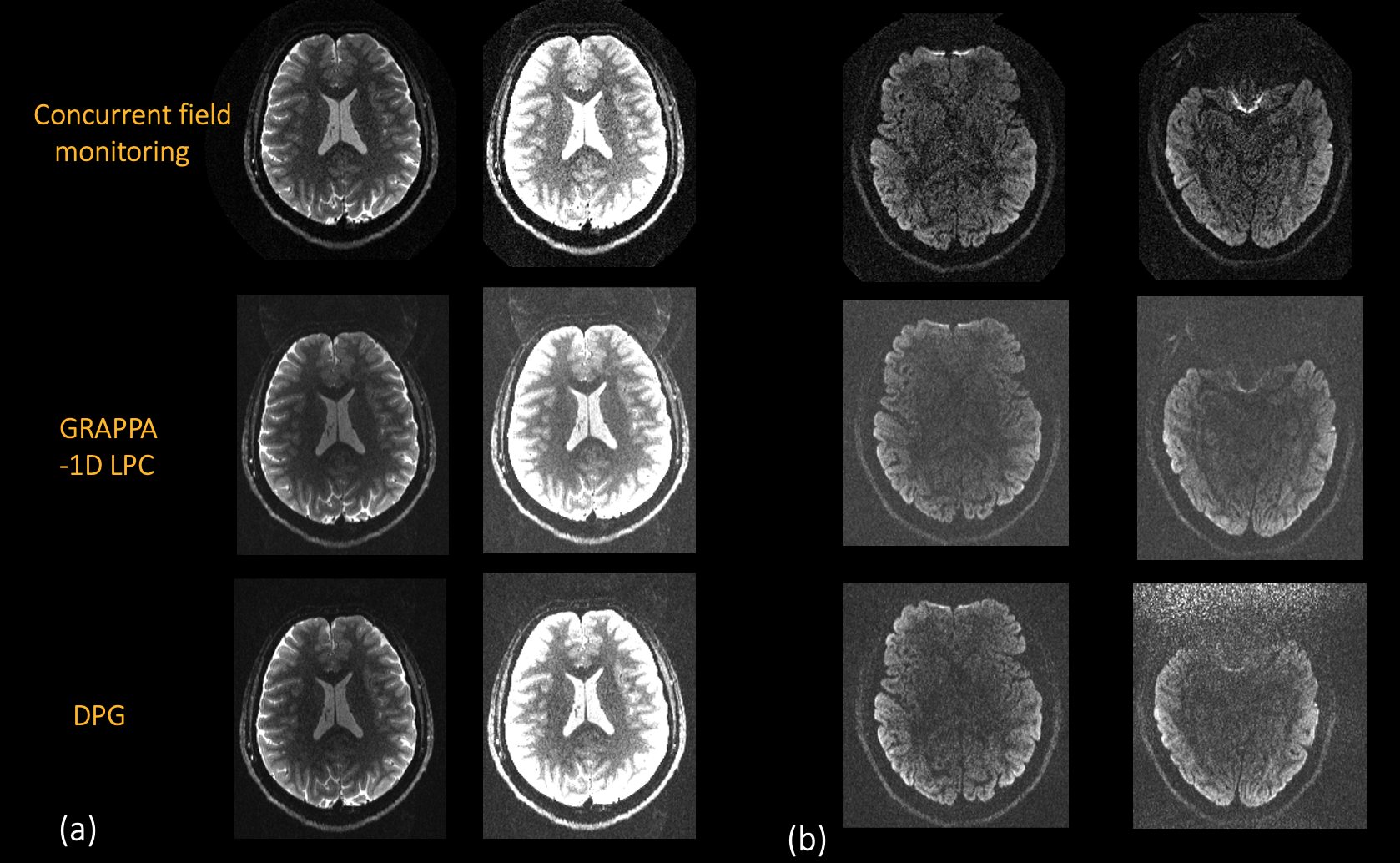
Validation: Concurrent field monitoring-based image reconstruction was compared with a) standard GRAPPA12 reconstruction with 1D navigators13 for Nyquist ghosting correction based on a linear phase model (1D LPC) and with b) Dual Polarity Grappa (DPG)14. On-line reconstruction was used. Reconstructed images with GRAPPA-1D LPC and DPG were corrected for eddy current-induced geometric distortion with the FSL post-processing tool ‘eddy’’15. No susceptibility distortion correction was used for GRAPPA-1D LPC, DPG, or reconstruction based on concurrent field monitoring.
Results
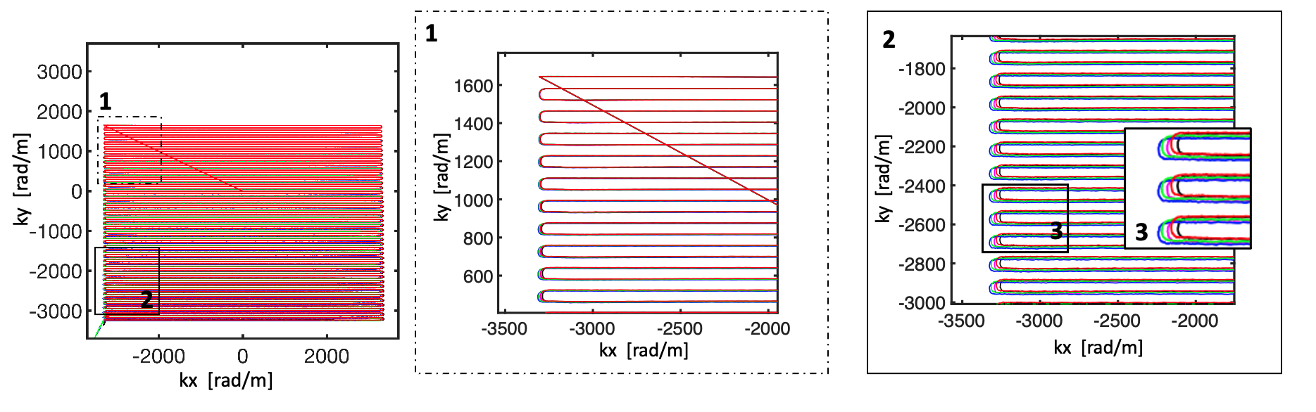
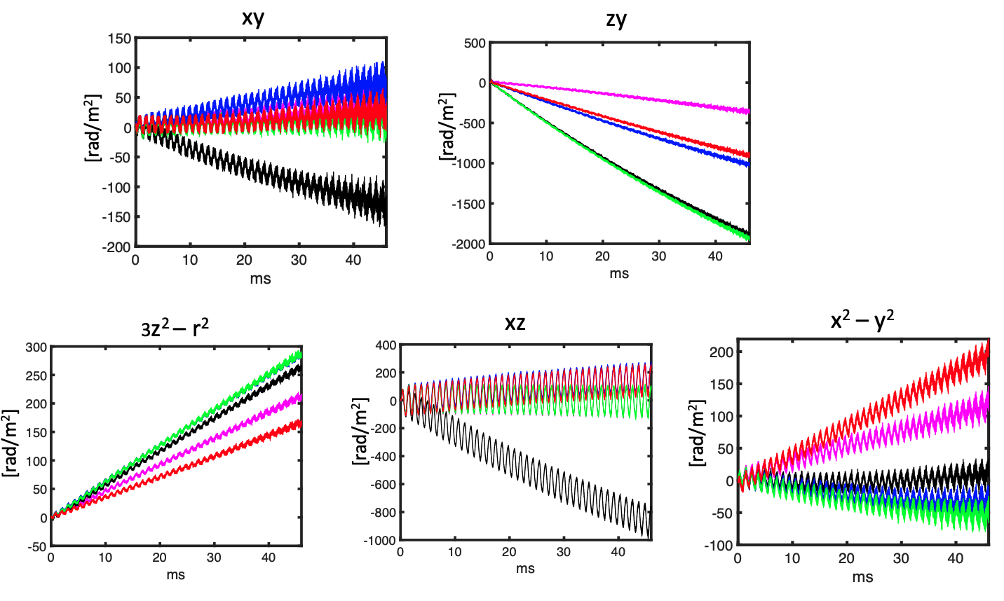
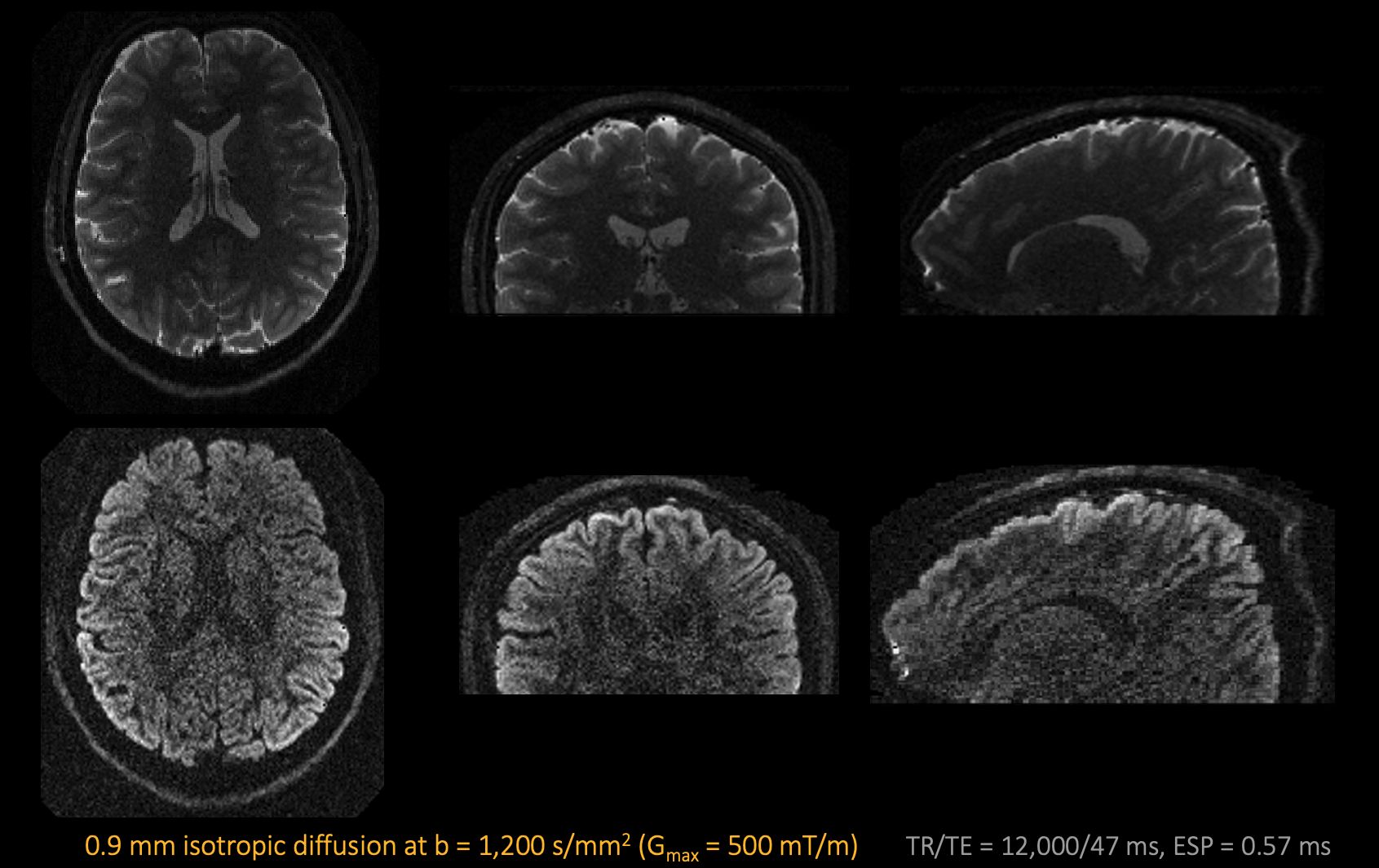
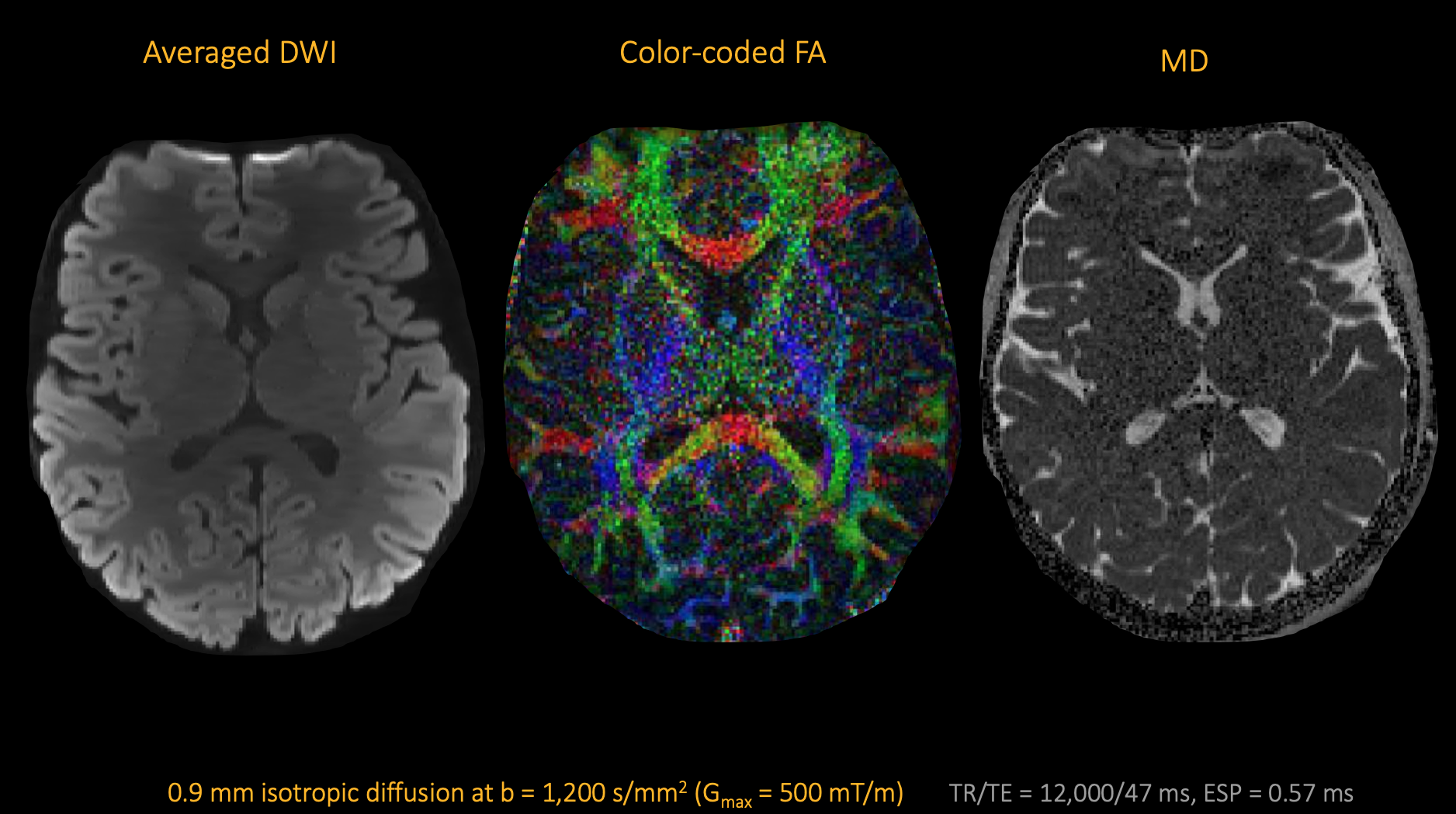
Eddy currents from the ultra-strong diffusion gradients perturb the image encoding, causing each DWI's k-space trajectory to deviate from the nominal k-space trajectory (Fig. 1). Higher order phase terms are also prominent with the asymmetric Connectome 2.0 head gradient coil (see those that are a function of z, in Fig. 2), and diffusion-direction dependent due to remaining long-decay eddy currents. Standard reconstruction methods like GRAPPA-1D LPC, widely used with conventional gradient strength regimes, cannot model such complex phase modulations. Axial, coronal, and sagittal views of a b0-image and a diffusion-weighted image reconstructed with concurrent field monitoring are shown in Fig. 3. A comparison with GRAPPA-1D LPC and DPG is also presented in Fig. 4. Ghosting artifacts remain visible with GRAPPA-1D LPC but are largely mitigated with DPG and slightly more with concurrent field monitoring-based reconstruction (Fig. 4a). Images reconstructed with concurrent field monitoring present better contrast and higher SNR than DPG results (Fig. 4b). Color-coded FA maps and mean diffusivity maps are shown in Fig. 5 alongside the directionally-averaged diffusion-weighted image. Note the high contrast on the averaged DWI map revealing finer details visible from the geometric distortion correction provided by concurrent field monitoring.Conclusion
High spatial-order magnetic fields can cause image artifacts during the readout of an in vivo submillimeter diffusion MRI acquisition of the human brain at 500 mT/m. These artifacts can only be partially fixed with standard reconstruction methods. We show here that concurrent field monitoring effectively reduces non-linear Nyquist ghosting and geometric distortions generated by eddy currents from ultra-high-strength diffusion gradients.Acknowledgements
The research reported in this abstract was supported by the National Institute of Biomedical Imaging and Bioengineering of the National Institutes of Health under award number U01EB026996 and P41EB030006. We thank Cameron Cushing, Christian Mirkes, and Paul Weavers from Skope, Inc. for their assistance and support.References
1Huang SY, Witzel T, Keil B, et al. Connectome 2.0: Developing the next-generation ultra-high gradient strength human MRI scanner for bridging studies of the micro-, meso- and macro-connectome. Neuroimage. 2021 Nov;243:118530.
2Setsompop K, Kimmlingen R, Eberlein E, et al. Pushing the limits of in vivo diffusion MRI for the Human Connectome Project. Neuroimage. 2013 Oct 15;80:220-33. doi: 10.1016/j.neuroimage.2013.05.078. Epub 2013 May 24. PMID: 23707579; PMCID: PMC3725309.
3Foo TKF, Tan ET, Vermilyea ME, et al. Highly efficient head-only magnetic field insert gradient coil for achieving simultaneous high gradient amplitude and slew rate at 3.0T (MAGNUS) for brain microstructure imaging. Magn Reson Med. 2020 Jun;83(6):2356-2369. doi: 10.1002/mrm.28087. Epub 2019 Nov 25. PMID: 31763726.
4Feinberg D, Dietz P, Liu, C et al. Design and Development of a Next-Generation 7T human brain scanner with high-performance gradient coil and dense RF arrays. Proc. Intl. Soc. Mag. Reson. Med. 29 (2021)
5Ramos-Llorden G, Dietz P, Davids M et al. Connectome 2.0: Performance evaluation and initial in vivo human brain diffusion MRI results. Submitted to ISMRM 2024.
6Mahmutovic M, Shrestha M, Ramos-Llordén G, et al. A 72-channel Head Coil with an Integrated 16-Channel Field Camera for the Connectome 2.0 Scanner. Submitted to ISMRM 2024.
7Wilm BJ, Nagy Z, Barmet C, et al. Diffusion MRI with concurrent magnetic field monitoring. Magn Reson Med. 2015 Oct;74(4):925-33. doi: 10.1002/mrm.25827. Epub 2015 Jul 17. PMID: 26183218.
8Barmet C, De Zanche N, Pruessmann KP. Spatiotemporal magnetic field monitoring for MR. Magn Reson Med. 2008 Jul;60(1):187-97. doi: 10.1002/mrm.21603. PMID: 18581361.
9Wilm BJ, Barmet C, Pavan M, Pruessmann KP. Higher order reconstruction for MRI in the presence of spatiotemporal field perturbations. Magn Reson Med. 2011 Jun;65(6):1690-701. doi: 10.1002/mrm.22767. Epub 2011 Apr 22. PMID: 21520269.
10Ramos-Llordén G, Park DJ, Kirsch JE, et al. Eddy current-induced artifact correction in high b-value ex vivo human brain diffusion MRI with dynamic field monitoring. Magn Reson Med. 2023 Sep 27. doi: 10.1002/mrm.29873. Epub ahead of print. PMID: 37753621.
11Uecker M, Lai P, Murphy MJ, et al. ESPIRiT--an eigenvalue approach to autocalibrating parallel MRI: where SENSE meets GRAPPA. Magn Reson Med. 2014 Mar;71(3):990-1001. doi: 10.1002/mrm.24751. PMID: 23649942; PMCID: PMC4142121.
12Griswold MA, Jakob PM, Heidemann RM et al. Generalized autocalibrating partially parallel acquisitions (GRAPPA). Magn Reson Med. 2002 Jun;47(6):1202-10. doi: 10.1002/mrm.10171. PMID: 12111967.
13Feiweier, T. Magnetic resonance method and apparatus to determine phase correction parameters. US Patent. 8,497,681. 2013
14Hoge WS, Polimeni JR. Dual-polarity GRAPPA for simultaneous reconstruction and ghost correction of echo planar imaging data. Magn Reson Med. 2016 Jul;76(1):32-44. doi: 10.1002/mrm.25839. Epub 2015 Jul 24. PMID: 26208304; PMCID: PMC4758917.
15Andersson JLR, Sotiropoulos SN. An integrated approach to correction for off-resonance effects and subject movement in diffusion MR imaging. Neuroimage. 2016 Jan 15;125:1063-1078. doi: 10.1016/j.neuroimage.2015.10.019. Epub 2015 Oct 20. PMID: 26481672; PMCID: PMC4692656.
Figures