2443
3D diffusion MRI at 7T with Universal Pulses for improved image uniformity1Department of Neurology and Neurosurgery, McGill University, Montreal, QC, Canada, 2McConnell Brain Imaging Centre, Montreal Neurological Institute, McGill University, Montreal, QC, Canada, 3German Center for Neurodegenerative Diseases (DZNE), Bonn, Germany, 4Siemens Healthcare Limited, Montreal, QC, Canada, 5Department of Physics and Astronomy, University of Bonn, Bonn, Germany, 6Department of Biomedical Engineering, McGill University, Montreal, QC, Canada
Synopsis
Keywords: Diffusion Acquisition, Brain, Universal Pulses
Motivation: 2D multi-slice diffusion-weighted MRI (dMRI) suffers from severe signal non-uniformity caused by B1+ inhomogeneity at ultra-high fields.
Goal(s): To design a 3D dMRI sequence with Universal parallel transmission radio-frequency Pulses (UPs) to improve image uniformity across the brain at 7T without lengthening workflow.
Approach: We propose a 3D spin-echo sequence with an inversion pulse before excitation to enhance SNR. 3D UPs were designed for inversion, excitation, and refocussing and implemented in a sequence with a stack-of-EPI readout.
Results: Diffusion-weighted image uniformity was significantly improved using the 3D sequence with UPs in comparison to 2D dMRI at 7T.
Impact: We propose a 3D dMRI sequence that includes UPs to improve image uniformity across the brain at 7T, including the temporal lobes and cerebellum. This improvement in image quality is critical for precision mapping of whole brain structural connectivity.
Introduction
Ultra-high field (UHF) imaging has been used to increase the SNR and resolution of diffusion MRI (dMRI) [1, 2]. However, 2D dMRI suffers from image non-uniformity, in particular signal loss in the temporal lobes and cerebellum, due to the enhanced B1+ field inhomogeneity at UHF. 3D sequences are more SNR efficient than 2D multi-slice sequences. Additionally, 3D Universal parallel transmission Pulses (UPs) [3] significantly improve B1+ uniformity to achieve uniform images at 7T without lengthening workflow. We propose a fast, multi-shot 3D dMRI sequence that includes UPs and has a similar scan time to a typical 2D DWI sequence with multi-band acceleration.Methods
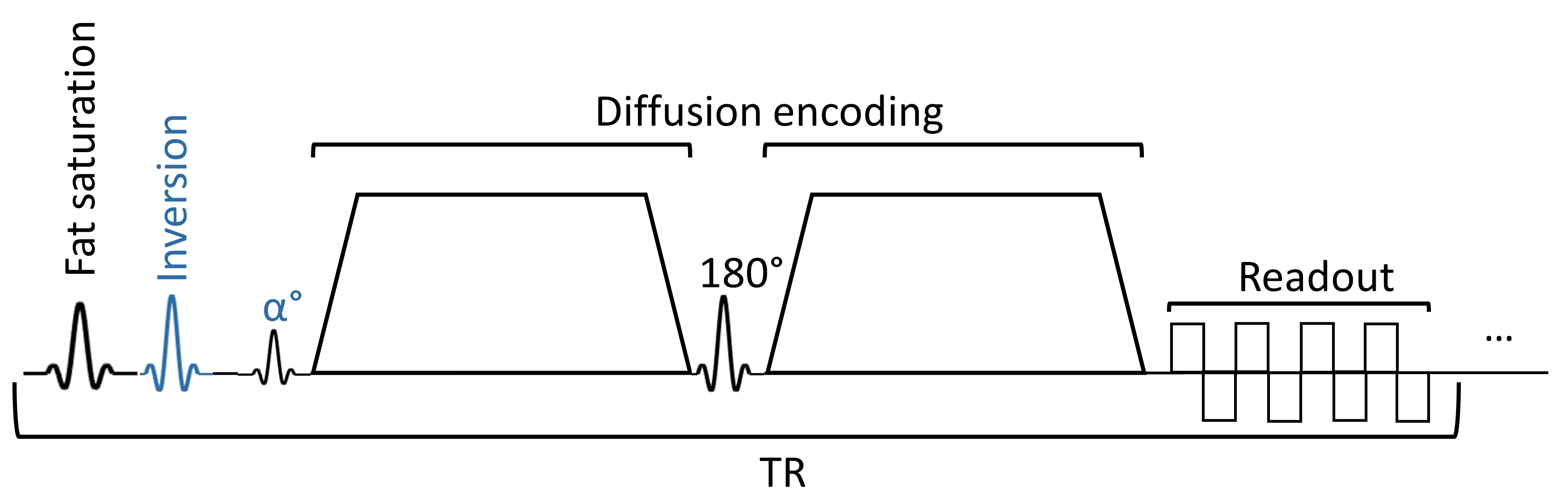
The proposed 3D dMRI sequence diagram is shown in Figure 1. The longitudinal magnetization is inverted before the excitation to enhance its recovery after the short TR and thus increase SNR. The excitation flip angle is reduced to the Ernst angle to maximize the available signal. 3D k-space is acquired in multiple shots such that the total scan time per volume is similar to the TR of a 2D DWI sequence. Bloch simulations were performed to compare the steady-state signal of the 3D sequence to a typical 2D spin-echo (SE), and to investigate the sensitivity of the 3D sequence to B1+ non-uniformity. The SE excitation pulse was maintained at 90° to achieve maximum signal. T1 and T2 relaxation times were set to 1400 and 47 ms, respectively, to model the white matter at 7T. 3D inversion, excitation, and refocusing UPs were designed and implemented in a sequence with a stack-of-EPIs readout trajectory. A GRAPE 180° pulse (8ms duration) was optimized using a dataset of 10 healthy subjects (scanned previously), for both the inversion and refocussing UPs [3]. The inversion pulse had a 90° phase difference from the excitation and refocusing pulses. After extracting the phase profile from Bloch simulations, a phase coherent vFA GRAPE excitation pulse (0.5ms duration) was designed [4, 5]. A human subject was scanned on a Siemens Terra 7T scanner with the 8-channel transmit and 32-channel receive Nova coil. Scan parameters were: field-of-view of 240x240x192 mm, matrix size of 200, 160 sagittal partitions, in-plane acceleration factor of 3, excitation flip angle of 29°, and TE/TR of 49/190 ms. A 2D DWI protocol with similar parameters and a multi-band factor of 2 with circularly polarized (CP) RF pulses was acquired for comparison. No diffusion encoding was performed as part of this feasibility study that focusses on image uniformity of 3D dMRI at 7T.Results
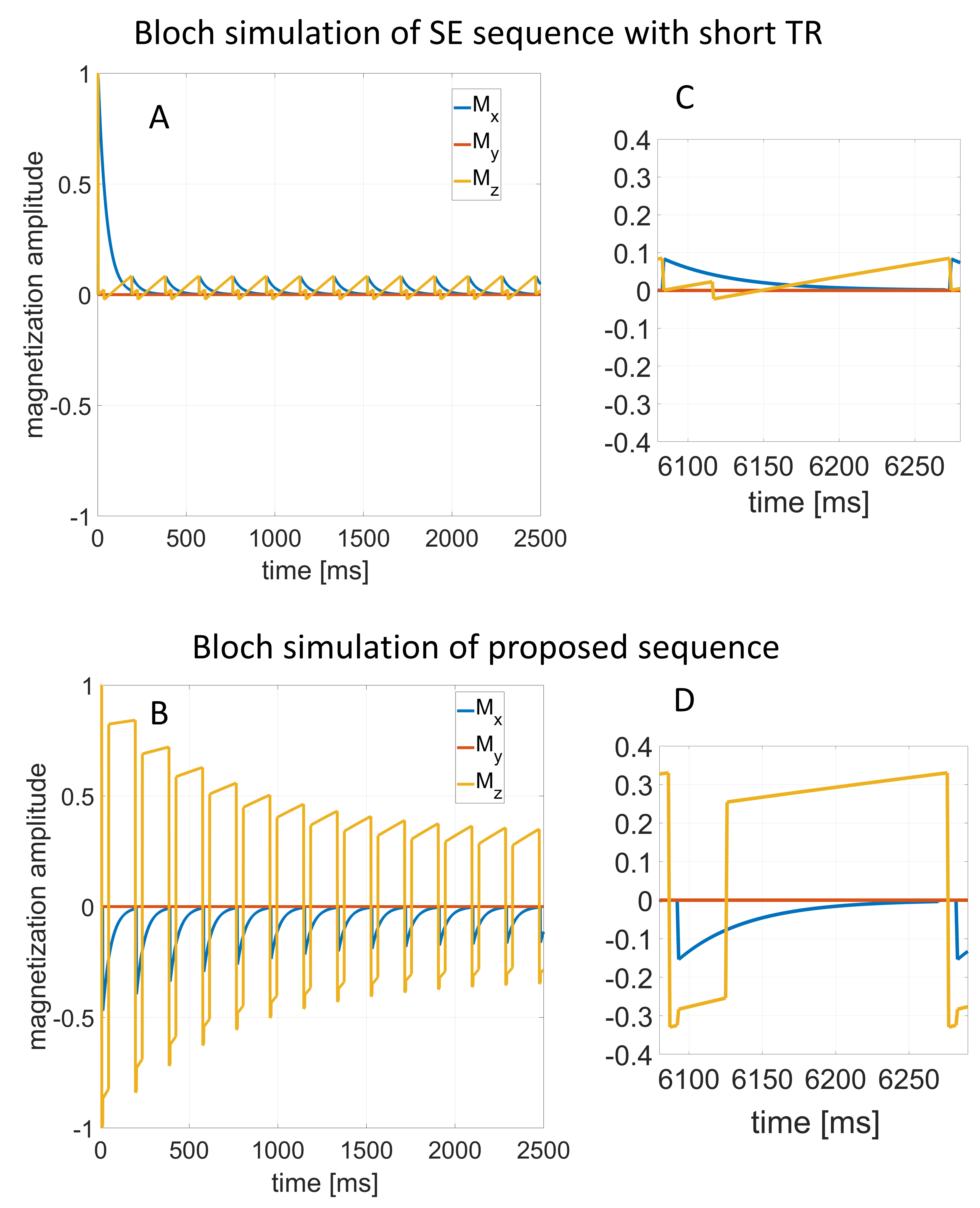
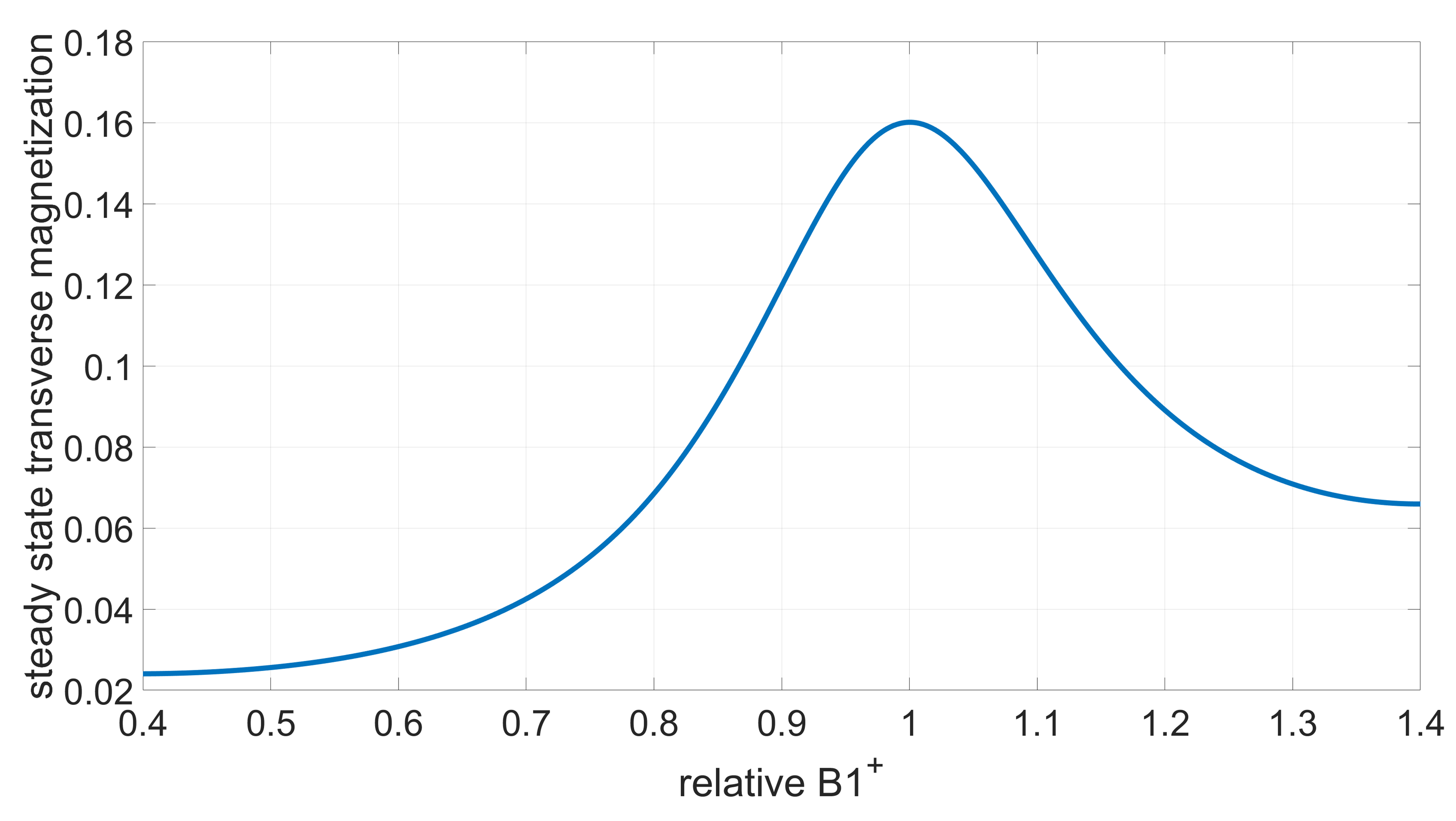
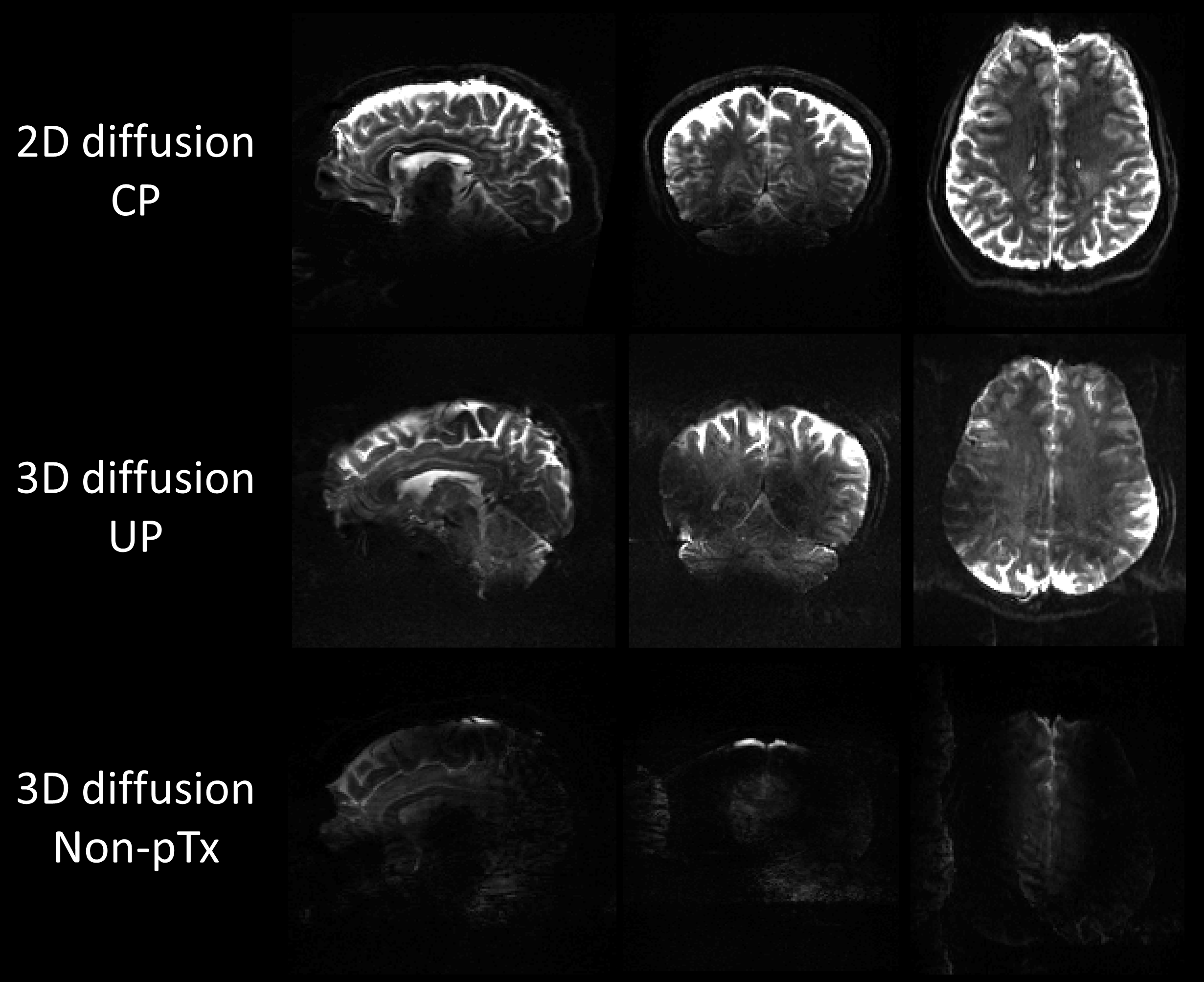
Bloch simulations of the SE and proposed 3D sequence are shown in Figure 2. The short TR causes a significant reduction in the steady state magnetization for the SE sequence. Adding an inversion pulse before the excitation and reducing the excitation flip angle to the Ernst angle doubles the available transverse magnetization. The impact of B1+ inhomogeneity on the 3D SE signal is illustrated in Figure 3. A 17% B1+ error reduces the SNR by 50%. These simulation results highlight the importance of using parallel transmission to achieve uniform RF pulses across the 3D field-of-view. Figure 4 shows scans of a subject using a typical 2D SE sequence and the 3D sequence with and without UPs, clearly showing that parallel transmission is critical for 3D DWI at 7T. The images acquired with the 3D sequence also have a stronger T1-weighting than the 2D SE sequence due to the shorter TR.Discussion
Adding an inversion pulse before the excitation doubles the available transverse magnetization, leading to an SNR increase. However, it also increases the sequence’s sensitivity to B1+ non-uniformity. UPs restored the signal in the cerebellum and temporal lobes, and image uniformity can be further improved by improving the UPs or using subject-tailored pulses. Multi-shot approaches in diffusion imaging are sensitive to phase differences between shots, which were not corrected in the 3D diffusion scan and cause image artifacts as seen in Figure 4. These phase inconsistencies can also cause signal loss in some areas, similar to B1+ inhomogeneity. We will implement the Trajectory Using Radially Batched Internal Navigator Echoes (TURBINE) readout trajectory [6] in combination with image-based phase correction [7] to correct these motion artifacts in future work.Conclusion
We designed a 3D dMRI sequence with enhanced SNR and B1+ uniformity. Feasibility was demonstrated using Bloch simulations and human scans. Future work will focus on refining the RF pulse designs to further improve uniformity while respecting SAR constraints. This new 3D sequence design will enable high-resolution whole brain diffusion imaging at 7T, which is critical for precision structural connectivity mapping.Acknowledgements
Authors would like to thank Nicolas Boulant at NeuroSpin for their tools used for pTx pulse implementation, and David Costa, Ronaldo Lopez, and Soheil Mollamohseni Quchani (McConnell Brain Imaging Centre) for helping with human scans. This work was supported by: the Montreal Neurological Institute, the Brain Canada Foundation, Healthy Brains for Healthy Lives, Fonds de Recherche du Québec - Santé, Fonds de Recherche du Québec - Nature et technologie, and the Natural Sciences and Engineering Research Council of Canada.References
[1] Feizollah, S., & Tardif, C. L. (2023). High-resolution diffusion-weighted imaging at 7 Tesla: single-shot readout trajectories and their impact on signal-to-noise ratio, spatial resolution and accuracy. NeuroImage, 274, 120159.
[2] Ma, R., Akçakaya, M., Moeller, S., Auerbach, E., Uğurbil, K., & Van de Moortele, P. F. (2020). A field-monitoring-based approach for correcting eddy-current-induced artifacts of up to the 2nd spatial order in human-connectome-project-style multiband diffusion MRI experiment at 7T: A pilot study. Neuroimage, 216, 116861.
[3] Gras, V., Vignaud, A., Amadon, A., Le Bihan, D., & Boulant, N. (2017). Universal pulses: a new concept for calibration‐free parallel transmission. Magnetic resonance in medicine, 77(2), 635-643.
[4] Khaneja N., Reiss T, Kehlet C., Schulte-Herbrüggen T., Glaser S. J. (2005). Optimal control of coupled spin dynamics: design of NMR pulse sequences by gradient ascent algorithms. Journal of Magnetic Resonance, 172(2), 296-305.
[5] Gras V., Mauconduit F., Vignaud A., Amadon A., Stöcker T., Boulant N. (2018). Design of universal parallel‐transmit refocusing kT‐point pulses and application to 3D T2‐weighted imaging at 7T. Magnetic resonance in medicine, 80(1), 53-65.
[6] Graedel, N. N., McNab, J. A., Chiew, M., & Miller, K. L. (2017). Motion correction for functional MRI with three‐dimensional hybrid radial‐C artesian EPI. Magnetic resonance in medicine, 78(2), 527-540.
[7] Pipe, J. G., Farthing, V. G., & Forbes, K. P. (2002). Multishot diffusion-weighted FSE using PROPELLER MRI. Magnetic Resonance in Medicine: An Official Journal of the International Society for Magnetic Resonance in Medicine, 47(1), 42-52.
Figures