2442
Three-dimensional phase-based diffusion imaging using RF phase-modulated gradient echo imaging with Stack-of-Stars1Department of Radiology, University of Wisconsin-Madison, Madison, WI, United States, 2Department of Medical Physics, University of Wisconsin-Madison, Madison, WI, United States, 3Department of Biomedical Engineering, University of Wisconsin-Madison, Madison, WI, United States, 4Department of Medicine, University of Wisconsin-Madison, Madison, WI, United States, 5Department of Emergency, University of Wisconsin-Madison, Madison, WI, United States
Synopsis
Keywords: Diffusion Acquisition, Diffusion/other diffusion imaging techniques
Motivation: There is increased interest in high-resolution diffusion-weighted imaging (DWI); however, conventional DWI methods have limitations such as image distortion and motion sensitivity.
Goal(s): To demonstrate the feasibility of high-resolution DWI using phase-based diffusion with radial Stack-of-Stars acquisition (PBD-SoS).
Approach: To evaluate PBD-SoS, quantitative ADC measurements were performed with a phantom and compared with conventional methods. In vivo imaging was performed to demonstrate the clinical feasibility of PBD-SoS.
Results: The results showed ADC values of the phantom measured using PBD-SoS were consistent (R2=0.99) with conventional DWI, and there were fewer motion artifacts compared to conventional methods in vivo.
Impact: PBD-SoS enables high-resolution DWI that is robust to motion, which can be particularly beneficial for body imaging where motion artifacts are common. While current PBD-SoS scans may take longer, future implementation of acceleration techniques could overcome this limitation.
Introduction
There is increasing interest in high-resolution diffusion-weighted imaging (DWI), particularly for applications such as the detection and monitoring of small lesions1,2.Several high-resolution DWI have been proposed, including those based on multi-shot echo-planar imaging (EPI)3,4 and dual-echo steady-state (DESS)5,6. Although these methods have demonstrated high-resolution and susceptibility-robust imaging7, they still suffer from long acquisition time and motion sensitivity.
We recently proposed a phase-based diffusion (PBD) imaging that utilizes radiofrequency (RF) phase-modulated gradient-echo sequences to address these challenges8. Although the PBD provides a high-resolution DWI, motion sensitivity remains a challenge.
In this study, we developed a PBD using a stack-of-stars (SoS) sampling (PBD-SoS) to enable high-resolution DWI with reduced sensitivity to motion.
Theory
Signal Representation:T2 and diffusion can be encoded into the MR signal by using GRE with incrementing the transmit RF phase quadratically8. Small RF phase increments, such as 1-3° preserve transverse magnetization, and encodes both T2 and diffusion weighting into signal phase.
Figure 1a illustrates the plots of the real and imaginary parts of the signal as functions of T2 and diffusion, using a large gradient moment. The real part of the signal is mainly affected by diffusion, while the imaginary part is a FID-dominant signal. Consequently, diffusion can be encoded into the signal phase, which is less sensitive to T1 and flip angle as demonstrated in the previous study8. Figure 1b presents plots of the signal phase as a function of T2 and the diffusion coefficient. These plots indicate that the phase increases in conjunction with increases in both T2 and ADC.
Image Acquisition:
Image acquisition was implemented using an RF phase-modulated GRE with SoS to reduce motion sensitivity. The gradient moment is maintained consistently across all the spoke angles to maintain steady-state of the magnetization.
A four-pass acquisition scheme was adopted (Figure 2a). The first two passes use a small gradient moment, and the last two passes use a larger moment to encode T2 and diffusion, respectively. Opposite polarity of RF phase increments of ±θ° are used in each set of two passes to remove background phase components (Figure 2b). The phase can be obtained by subtracting between the two passes. ADC and T2 maps are reconstructed using a lookup table-based method previously introduced8.
Methods
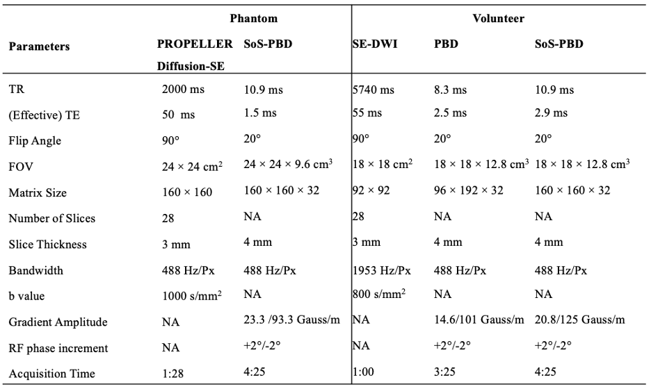
Phantom and in vivo experiments were performed to demonstrate the feasibility of the SoS-PBD. The parameters used in the experiments are listed in Table 1. Acquisitions were performed on a clinical 3.0T MRI system (Signa Premier, GE Healthcare, Waukesha, WI, USA).Phantom Study:
To compare the quantitative values, phantom experiments were conducted using the proposed method and conventional DWI. A phantom comprising 16 vials with varying amounts of PVP (10-50%) and MnCl2 (0.01-0.08mM) was used to encode ADC and T2, respectively. A PROPELLER Fast Spin Echo (FSE) imaging was chosen as the conventional method to avoid susceptibility-related distortion. ADC and T2 values of the acquired maps for the central slice were measured. Bland-Altman analysis was performed to evaluate systematic bias and variability.
In vivo study: To demonstrate in vivo feasibility, the prostate of a healthy male (46y.o.) volunteer was acquired using EPI, PBD, and PBD-SoS. PBD was acquired using a Cartesian trajectory.
Results
Phantom Study:As shown in Figure 3, the phantom data indicates that ADC (LoA =-130-+36mmm2/s, mean difference=-45mmm2/s) values measured using PBD-SoS are consistent (R2=0.99) with those obtained using conventional DWI, with a slight bias. T2 values obtained from PBD-SoS and conventional methods are generally in agreement (R2=0.97), although the PBD-SoS measurements tend to slightly overestimate high T2 values and underestimate low T2 values.
In vivo study:
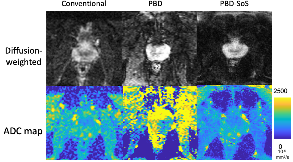
In vivo imaging demonstrated the feasibility of high-resolution imaging using PBD-SoS compared to conventional DWI (Figure 4). Modest streak artifacts were present in the ADC map acquired using PBD-SoS. In contrast, severe artifacts were visible in the ADC map acquired using PBD, leading to a non-uniform distribution of the ADC.
Discussion
In this work, we have developed a novel quantitative DW method that utilizes RF phase-modulated GRE and SoS sampling. Both phantom and in vivo studies indicated that PBD-SoS enables high-resolution imaging without motion artifacts.Several studies have reported motion-robust DESS-DWI using non-Cartesian trajectories and a balanced gradient moment for high-resolution imaging9,10. However, DESS-based methods inherently have a longer TR due to two consecutive readouts , which could potentially extend the acquisition time. Although the proposed PBD still has a relatively long acquisition time, it should be amenable to acceleration and optimization of the acquisition scheme.
In conclusion, we have successfully developed and demonstrated the feasibility of PBD-SoS for simultaneous high-resolution, motion-robust quantitative ADC mapping.
Acknowledgements
Dr. Reeder is the John. H Juhl Endowed Chair of Radiology.References
1. Medved M, Soylu-Boy FN, Karademir I, Sethi I, Yousuf A, Karczmar GS, Oto A. High-resolution diffusion-weighted imaging of the prostate. American Journal of Roentgenology 2014;203(1):85-90.
2. Sharif-Afshar A-R, Nguyen C, Feng TS, Payor L, Fan Z, Saouaf R, Li D, Kim HL. Prospective pilot trial to evaluate a high resolution diffusion-weighted MRI in prostate cancer patients. EBioMedicine 2016;7:80-84.
3. Chen N-k, Guidon A, Chang H-C, Song AW. A robust multi-shot scan strategy for high-resolution diffusion weighted MRI enabled by multiplexed sensitivity-encoding (MUSE). Neuroimage 2013;72:41-47.
4. Holdsworth SJ, Skare S, Newbould RD, Guzmann R, Blevins NH, Bammer R. Readout-segmented EPI for rapid high resolution diffusion imaging at 3T. European journal of radiology 2008;65(1):36-46.
5. Katscher U, Meineke J, Zhang S, Steinhorst B, Keupp J. On the b-value Derivation for diffusion-weighted Double-Echo Steady-State (dwDESS) Magnetic Resonance Imaging. 2021. Springer. p 3-11.
6. Staroswiecki E, Granlund KL, Alley MT, Gold GE, Hargreaves BA. Simultaneous estimation of T2 and apparent diffusion coefficient in human articular cartilage in vivo with a modified three‐dimensional double echo steady state (DESS) sequence at 3 T. Magnetic resonance in medicine 2012;67(4):1086-1096.
7. Foltz WD, Porter DA, Simeonov A, Aleong A, Jaffray D, Chung P, Han K, Ménard C. Readout-segmented echo-planar diffusion-weighted imaging improves geometric performance for image-guided radiation therapy of pelvic tumors. Radiotherapy and Oncology 2015;117(3):525-531.
8. Tamada D, Reeder SB. Phase-based 3D diffusion mapping using RF phase-modulated gradient echo imaging. 2022 May 07-12, 2022; Londan. p 0510.
9. Granlund KL, Staroswiecki E, Alley MT, Daniel BL, Hargreaves BA. High-resolution, three-dimensional diffusion-weighted breast imaging using DESS. Magnetic resonance imaging 2014;32(4):330-341.
10. Moran CJ, Cheng JY, Sandino CM, Carl M, Alley MT, Rosenberg J, Daniel BL, Pittman SM, Rosen EL, Hargreaves BA. Diffusion‐weighted double‐echo steady‐state with a three‐dimensional cones trajectory for non‐contrast‐enhanced breast MRI. Journal of Magnetic Resonance Imaging 2021;53(5):1594-1605.
Figures
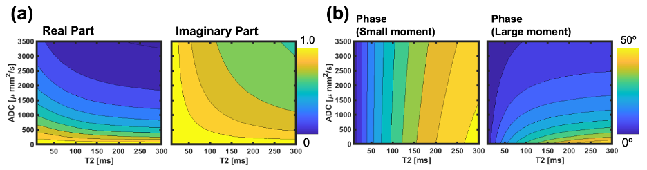
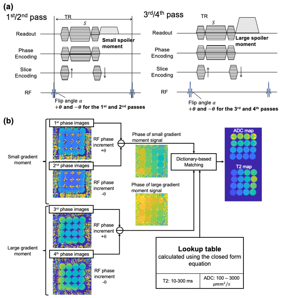
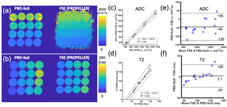
Figure 3: The results of the ADC and T2 measurements of the phantom using SoS-PBD were consistent with those obtained with conventional methods. (a) ADC maps measured using SoS-PBD and PROPELLER-FSE and (b) T2 maps measured using PBD-SoS and a multi-echo spin-echo method were comparable. The overall agreement of (c) ADC and (d) T2 between PBD-SoS and FSE was excellent (R2=0.99). The Bland-Altman plots indicated a minor systematic bias in the (d) ADC (-130-+36ms, mean difference=-45ms) and (f) T2 (-25-+29ms, mean difference=2.1ms) measurements using PBD-SoS, although within the LOA.