2441
Mitigation of Peripheral-Nerve Stimulation with Arbitrary Gradient Waveform Design for Diffusion-Weighted MRI1Department of Radiology, Stanford University, Stanford, CA, United States, 2Division of Radiology, Veterans Administration Health Care System, Palo Alto, CA, United States, 3Cardiovascular Institute, Stanford University, Stanford, CA, United States, 4Department of Bioengineering, Stanford University, Stanford, CA, United States, 5Department of Electrical Engineering, Stanford University, Stanford, CA, United States
Synopsis
Keywords: Diffusion Acquisition, Gradients, Peripheral Nerve Stimulation
Motivation: Peripheral nerve stimulation (PNS) can be problematic on ultra-high-performance gradients systems, especially during diffusion encoding. We developed a gradient waveform optimization approach to mitigate PNS.
Goal(s): Our goal was to investigate the minimum achievable TE (TEmin) using arbitrary gradient waveform design while mitigating PNS for brain, liver and heart DWI.
Approach: We used gradient optimization (GrOpt) to design gradient waveforms for TEmin of different protocols, then imaged a phantom and a volunteer with a brain DWI protocol implemented with Pulseq.
Results: GrOpt consistently reduces TEs compared to conventional approaches and avoids PNS. Image quality was the same in phantom and in vivo studies.
Impact: Ultra high-performance gradient systems increase diffusion sensitivity and resolution, but their application can be constrained due to peripheral nerve stimulation (PNS). We used open-source gradient optimization (GrOpt) to design arbitrary gradient waveforms for minimum time that mitigate PNS.
Introduction
Diffusion-weighted MRI (DWI) non-invasively probes soft tissue microstructure1. Measuring apparent diffusion coefficient (ADC) differences calculated from DWI is clinically valuable, aiding in diagnosing neurodegenerative diseases2, identifying liver lesions3, and assessing myocardial fibrosis4. However, DWI techniques often have low signal-to-noise (SNR) due to long TEs from large diffusion encoding gradients, particularly in body and cardiac applications5-8 where gradient-moment nulling (M1,M2) is required.Ultra-high performance gradient systems (200T/m, 200T/m/s) enable higher b-value and higher resolution acquisitions and are broadly appealing because they can substantially shorten TEs and regain SNR. Using these large gradient amplitudes and slewrates, however, can easily result in peripheral nerve stimulation (PNS). Consequently, ad hoc slewrate derating is typically used to prevent PNS, thereby also prolonging TEs.
Balancing the need for diffusion sensitivity, gradient-moment nulling, PNS mitigation, and TE minimization cannot be optimally resolved by conventional gradient waveform design. Numerical gradient optimization algorithms9,10, such as the Gradient Optimization Toolbox (GrOpt)11 enable time-optimal arbitrarily-shaped gradient waveform design subject to constraints.
The purpose of this work was to evaluate the minimum TE achievable (TEmin) for DWI when using time-optimal gradient waveform design with a PNS threshold constraint.
Methods
We investigated the TEmin with GrOpt in simulation, phantom, and in vivo experiments on a 45/200-System and a 200/200-System (GMax/SRMax). The PNS model was based on the International Engineering Standard (IEC 60601-2-33)12,13 with the following coefficients: α=0.333m, r=23.4T/s, Smin=60T/ms, c=334µs.Simulation Experiments
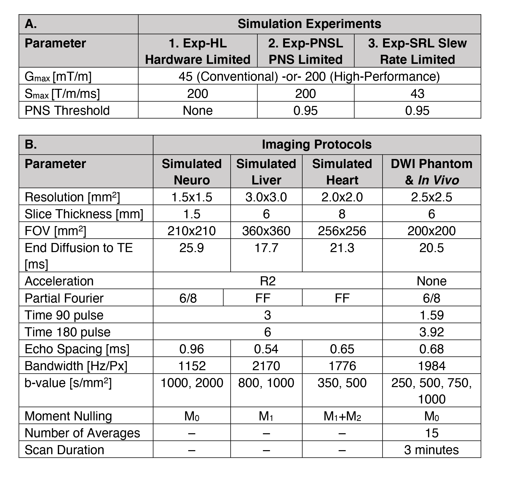
We compared TEmin using GrOpt vs. conventional Stejskal-Tanner trapezoidal (TRAP) waveforms14 with the following three experiments: (1) Hardware Limited (Exp-HWL, using full hardware specifications); (2) PNS Limited (Exp-PNSL, subject to a PNS model constraint); and (3) Slew-Rate Limited (Exp-SRL, subject to derating SRMax to 43 T/m/s). See Table 1A. Protocols were designed to match typical in vivo protocols for neuro, liver, and cardiac DWI (Table-1B).
Phantom Experiments
A diffusion phantom (CaliberMRI, Boulder, CO, USA) was scanned at 3T (Vida Fit, Siemens, 45/200 System) to demonstrate the feasibility of PNS-constrained GrOpt (PNSthresh = 0.95) waveforms compared to TRAP waveforms for a M0-nulled DWI protocol. We programmed the waveforms using open-source Pulseq15 (Pulseq-TRAP & Pulseq-GrOpt - https://github.com/ahannum/gropt-diffusion-pypulseq ). Image parameters are in Table-1B. A vendor DWI protocol (Vendor-TRAP) with closely matched parameters was also acquired.
In Vivo Experiments
In vivo proof-of-concept data was obtained from a 3T brain MRI scan in a volunteer (F, 28 years old, IRB consented/approved) using the same Pulseq-TRAP, Pulseq-GrOpt, and Vendor-TRAP protocols from the phantom experiment.
Analysis

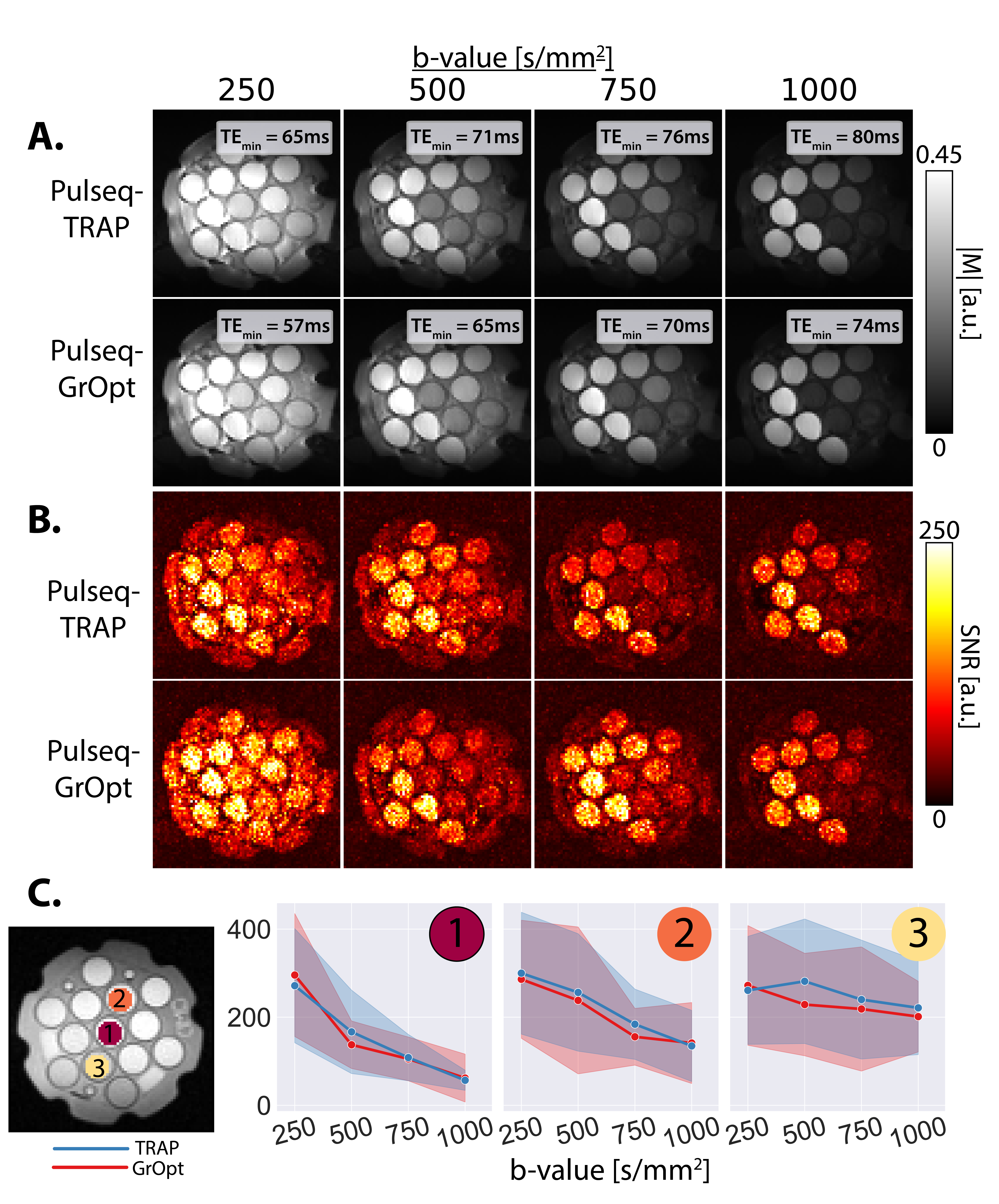
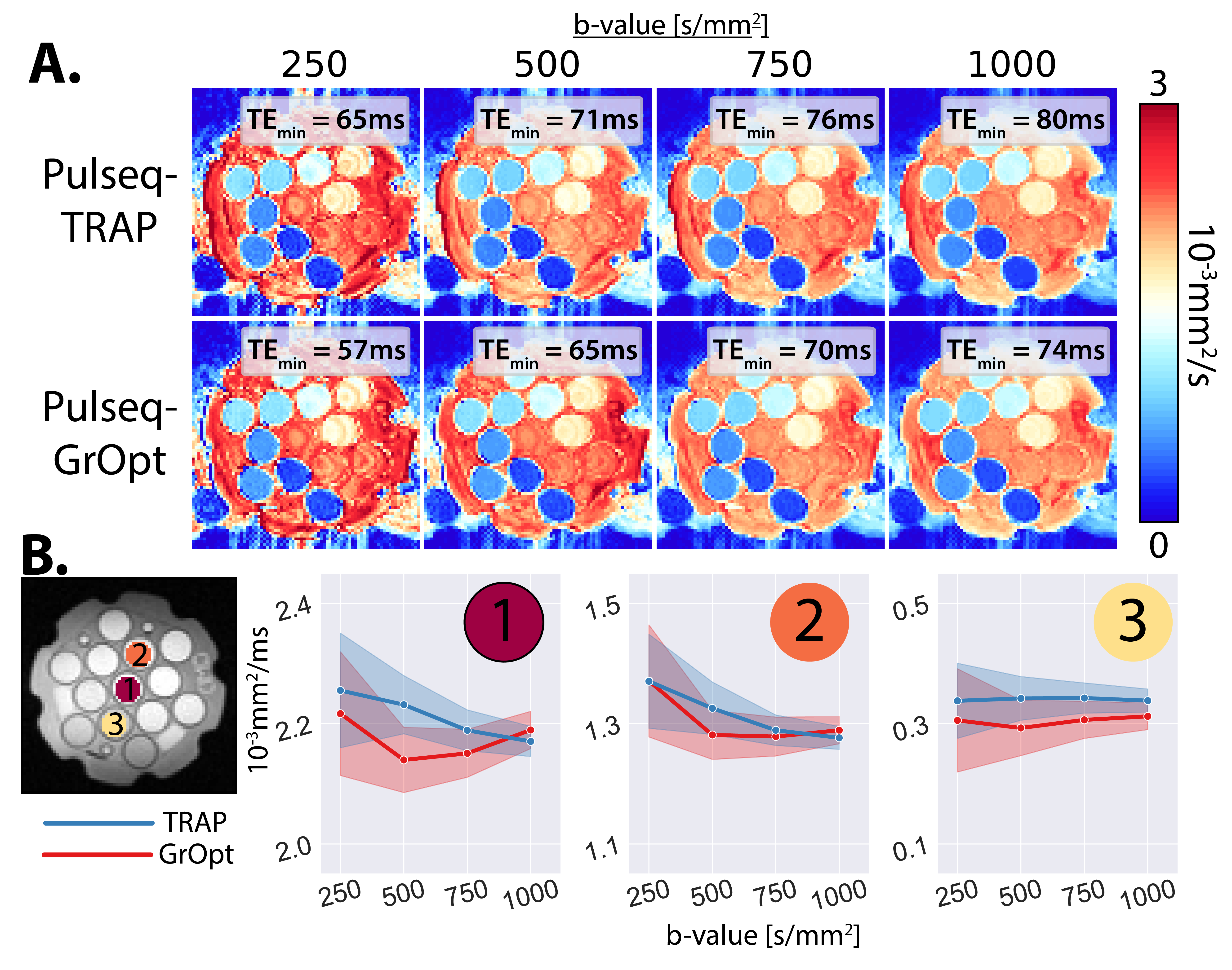
TEmin and TEmin differences (∆TE) were reported between the Pulseq-TRAP and Pulseq-GrOpt Waveforms. Fig-1A shows example gradients, slewrates, and PNS waveforms. For the phantom experiments, temporal SNR (tSNR) was computed across averages for a given direction and averaged for three ROIs. ADC maps were then computed for phantom and in vivo experiments.
Results
Simulations– GrOpt consistently reduces TEmin compared to TRAP waveforms in all three experiments (Fig-1B). Larger reductions in TEmin using GrOpt are observed using a 200/200 System vs. the 45/200 System and in protocols with M1 or M1+M2 gradient moment nulling (liver and heart), with observed TEmin reductions >20 ms. GrOpt utilizes the maximum possible slew-rate, while maintaining PNS below 0.95 (Fig-1A).Phantoms– Proper diffusion-encoding (Fig-2A) was observed with Pulseq-TRAP and Pulseq-GrOpt. Small gains in SNR are observed with Pulseq-GrOpt vs. Pulseq-TRAP, due to TEmin reductions (Fig-2BC). ADC maps (Fig-3) indicate strong agreement between Pulseq-TRAP and Pulseq-GrOpt design for ROIs with unique ADC values.
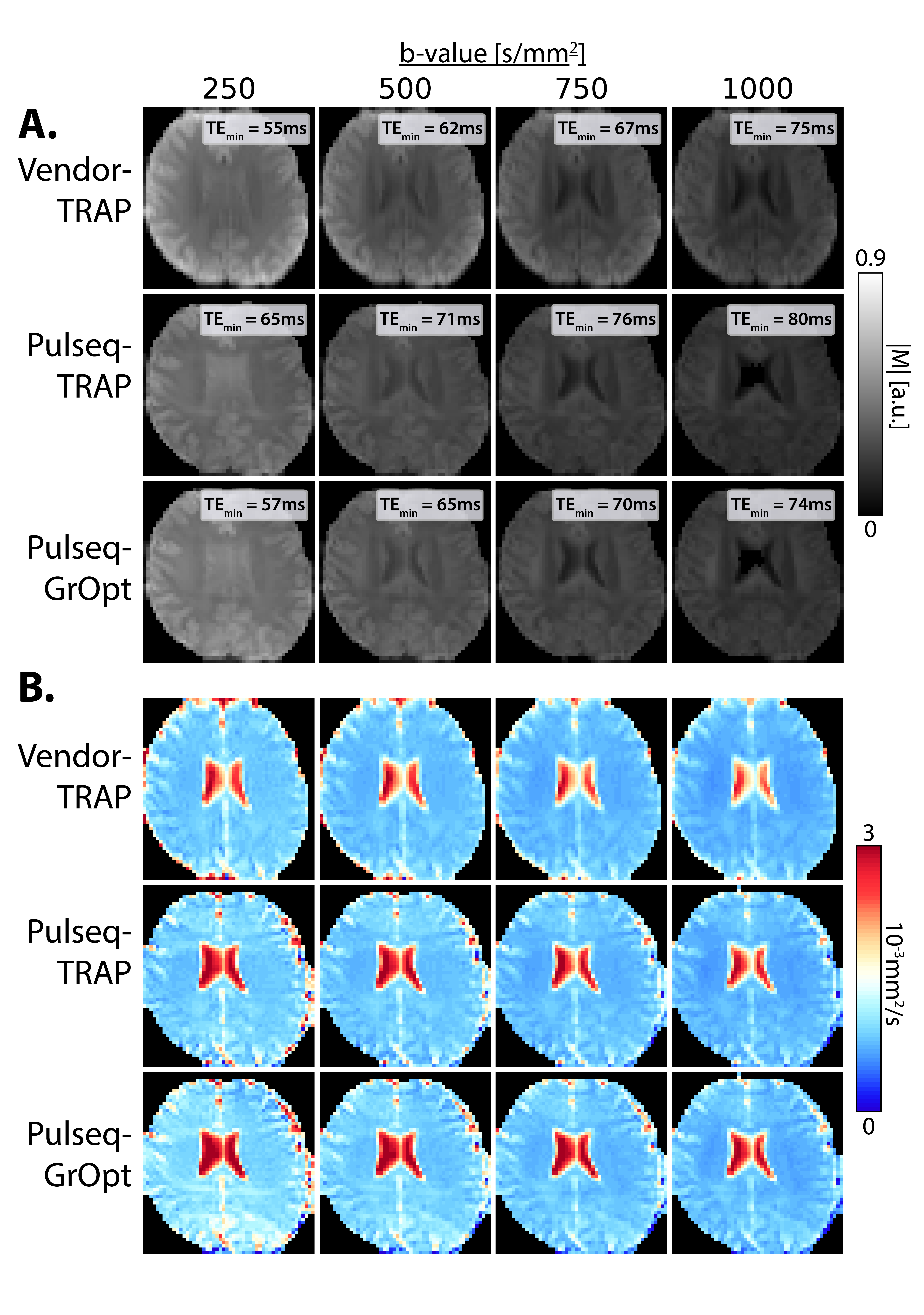
In Vivo– DWIs and the ADC maps are consistent between Pulseq-GrOpt, Pulseq-TRAP, and Vendor-TRAP (Fig-4). The high degree of consistency between the generated images demonstrates that the Pulseq-GrOpt waveform design enables appropriate arbitrary diffusion-encoding while minimizing TEmin and staying below the specified PNS threshold.
Discussion
PNS-constrained GrOpt waveforms provide time-efficient diffusion-encoding. Larger TEmin differences between GrOpt and TRAP waveforms were observed for protocols with longer readouts, moment-nulling, and short T2 tissue. Phantom and in vivo experiments confirmed Pulseq-GrOpt ADC values were consistent with Pulseq-TRAPs and Vendor-TRAPs. Additionally, the use of Pulseq and GrOpt allows an open-source, reproducible, vendor-neutral approach to arbitrary gradient waveform design.Future work will improve the efficiency of the Pulseq implementation of the DWI sequence EPI trajectory. We will also evaluate the effect of GrOpt on Maxwell fields16,17 and eddy currents17,18, which can be additional constraints. We have preliminarily observed eddy current reductions with GrOpt compared to TRAP without applying an eddy current constraint.
Conclusion
Arbitrary gradient waveforms (designed with GrOpt; implemented with Pulseq) enable SNR-efficient, time-optimal, PNS-limited diffusion-encoding. Our PNS-constrained gradient waveform design approach can reduce TEmin by >20ms, especially for moment compensated diffusion encoding gradient waveforms on ultra high-performance gradient systems.Acknowledgements
We would like to acknowledge our funding sources:AHA 23PRE1018442 to AJH
NSF/NIH 2205103 to DBE
NIH R01 HL152256 to DBE
References
[1] Le Bihan D, Breton E. Imagerie de diffusion in-vivo par resonance magnetique nucleaire. CR Acad Sci..1985;301(15):1109–1112.
[2] Le Bihan D. Diffusion MRI: What Water Tells Us About the Brain. EMBO Mol Med. 2014;6(5):569
[3] Saito K, Tajima Y, Harada TL. Diffusion-weighted imaging of the liver: Current applications. World J Radiol. 2016;8(9):785–798.
[4] Nielles-Vallespin Sonia, Scott Andrew, Ferreira Pedro, Khalique Zohya, Pennell Dudley, Firmin David. Cardiac diffusion: technique and practical applications. Journal of Magnetic Resonance Imaging. 2020;52(2):348–368.
[5] Haacke E. M. and Lenz G. W. Improving MR image quality in the presence of motion by using rephasing gradients. American Journal of Roentgenology. 1987; 148(6):1251-1258.
[6] Ozaki M, Inoue Y, Miyati T, Hata H, Mizukami S, Komi S, Matsunaga K, Woodhams R. Motion artifact reduction of diffusion-weighted MRI of the liver: Use of velocity-compensated diffusion gradients combined with tetrahedral gradients. J Magn Reson Imaging. 2013;37(1):172–178.
[7] Stoeck CT, Von Deuster C, GeneT M, Atkinson D, Kozerke S. Second-order motion-compensated spin echo diffusion tensor imaging of the human heart. Magn Reson Med. 2016;75(4):1669-167
[8] Moulin K, Croisille P, Feiweier T, Delattre BMA, Wei H, Robert B, Beuf O, Viallon M. In vivo free-breathing DTI and IVIM of the whole human heart using a real-time slice-followed SE-EPI navigator-based sequence: A reproducibility study in healthy volunteers. Magn Reson Med. 2016;76(1):70-82.
[9] Aliotta E, Wu HH, Ennis DB. Convex optimized diffusion encoding (CODE) gradient waveforms for minimum echo time and bulk motion-compensated diffusion-weighted MRI. Magn Reson Med. 2017;77(2):717-729.
[10] Szczepankiewicz F, Westin CF, Nilsson M. Gradient waveform design for tensor-valued encoding in diffusion MRI. J Neurosci Methods. 2021;348:109007.
[11] Loecher M, Middione MJ, Ennis DB. A gradient optimization toolbox for general purpose time-optimal MRI gradient waveform design. Magn Reson Med. 2020 Dec;84(6):3234-3245.
[12] International Electrotechnical Commission. IEC 60601-2-33. Medical electrical equipment - part 2–33: particular requirements for the basic safety and essential performance of magnetic resonance equipment for medical diagnosis.
[13] Hebrank F, Gebhardt G. SAFE-Model - A New Method for Predicting Peripheral Nerve Stimulations in MRI by F.X. Herbank and M. Gebhardt. Abstract No 2007. Proc. Intl. Soc. Mag. Res. Med. 8, 2000, Denver, Colorado, USA
[14] Stejskal EO, Tanner JE. Spin Diffusion Measurements: Spin Echoes in the Presence of a Time‐Dependent Field Gradient. J Chem Phys. 1965;42:288-292. doi:10.1063/1.1695690.
[15] Layton KJ, Kroboth S, Jia F, et al. Pulseq: A rapid and hardware-independent pulse sequence prototyping framework. Magn Reson Med. 2017;77(4):1544-1552.
[16] Bernstein MA, Zhou XJ, Polzin JA, et al. Concomitant gradient terms in phase contrast MR: analysis and correction. Magn Reson Med. 1998;39(2):300-308.
[17] Middione MJ, Loecher M, Moulin K, Ennis DB. Optimization methods for magnetic resonance imaging gradient waveform design. NMR Biomed. 2020;33(12):e4308.
[18] Aliotta E, Moulin K, Ennis DB. Eddy current-nulled convex optimized diffusion encoding (EN-CODE) for distortion-free diffusion tensor imaging with short echo times. Magn Reson Med. 2018;79(2):663-672. doi:10.1002/mrm.26709
Figures