2440
Spiral interleaving for diffusion encoding and relaxometry (SPIDER)1Key Laboratory for Biomedical Engineering of Ministry of Education, Department of Biomedical Engineering, College of Biomedical Engineering & Instrument Science, Zhejiang University, Hangzhou, China, 2Athinoula A. Martinos Center for Biomedical Imaging, Massachusetts General Hospital, Charlestown, MA, United States, 3Department of Radiology, Harvard Medical School, Boston, MA, United States, 4Department of Radiology, Juntendo University, Tokyo, Japan, 5Department of Radiology, The University of Tokyo, Tokyo, Japan, 6Brigham and Women's Hospital, Harvard Medical School, Boston, MA, United States, 7Harvard/MIT Health Sciences and Technology, Cambridge, MA, United States
Synopsis
Keywords: Diffusion Acquisition, Diffusion Tensor Imaging
Motivation: Current diffusion readout methods have a relatively long echo time and readout duration, which prevent multi-echo imaging.
Goal(s): To implement a diffusion sequence with multiple echoes readout for performing diffusion relaxometry.
Approach: A 3-echo diffusion sequence, SPIDER, with variable density spiral readout was designed.
Results: The proposed SPIDER showed comparable images with reference EPI method at shorter echo time.
Impact: The proposed method showed the ability to acquire 3 echoes for 1mm2 resolution for low b-value, which could help multi-echo diffusion modeling.
Introduction
Diffusion MRI (dMRI) can probe tissue microstructure using motion-sensitizing gradients. It relies on signal decay for diffusion signal modeling. However, this signal decay may not be solely due to diffusion since relaxation effects also contribute to this decay. Thus, it is beneficial to simultaneously probe both relaxation and diffusion [1]. If images are acquired at a single echo time (TE), the effects from relaxation and diffusion cannot be distinguished from each other. Hence, multi-echo acquisitions are desired. For EPI readout, the minimal TE for b-value of 2000s/mm2 at 1mm2 resolution on typical research-dedicated scanners is ~ 70ms, thus later echoes have relatively longer TE (e.g. 140ms) if CPMG condition is satisfied, leading to low SNR and impairing diffusion-relaxometry modeling. Here, we propose SPIDER comprising variable density spirals which have high flexibility for undersampling in multi-echo diffusion imaging.Methods
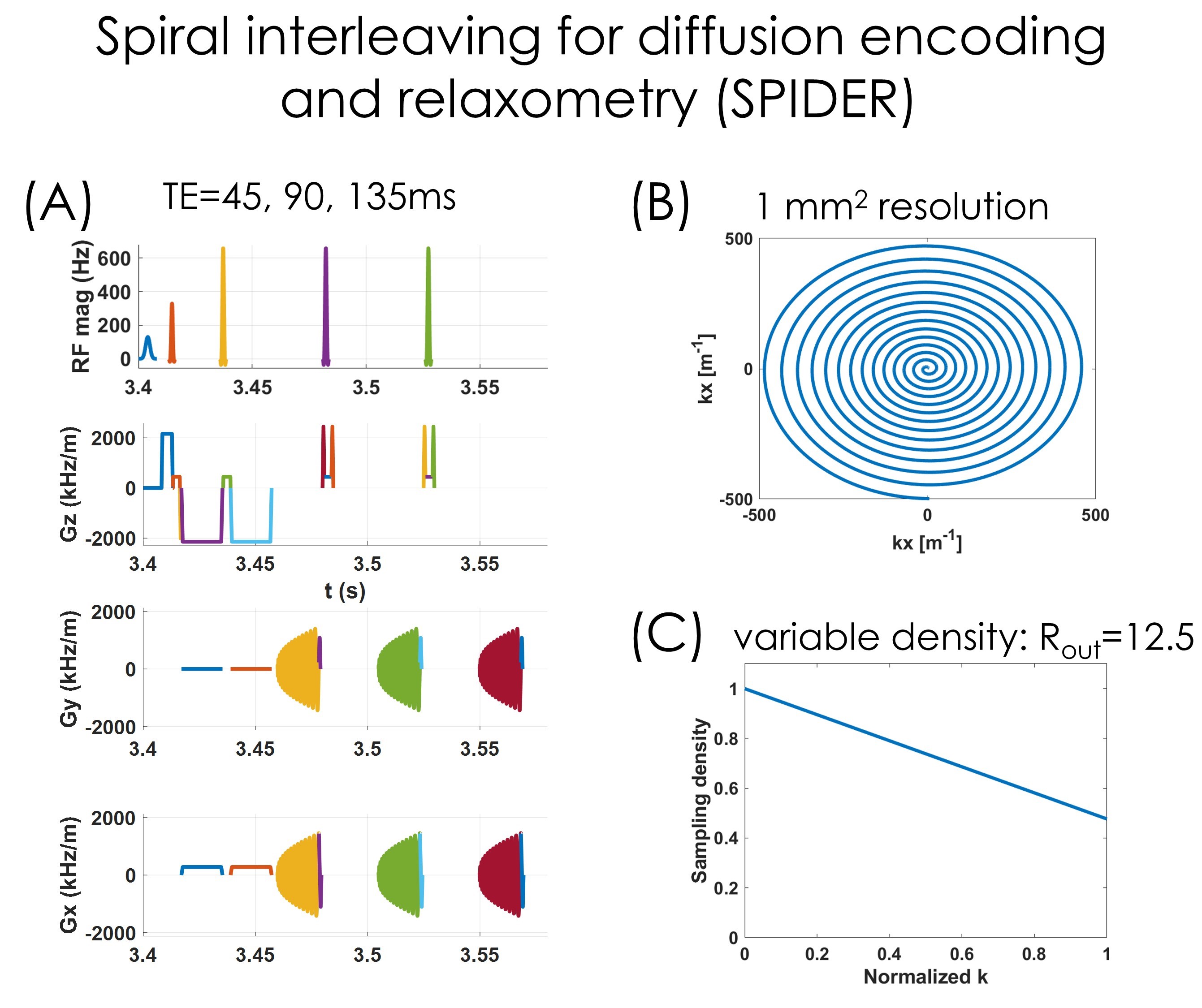
A 3-echo DWI sequence with variable density spiral readout [2] was designed using Pulseq [3]. Equidistant echo spacing was used to avoid secondary echoes. As such, the readout duration of each spiral interleave should be smaller than TE/2. This is achieved by linearly decreasing sampling density from k-space center to periphery, thus reducing readout duration, as shown in Fig1.C. The sampling density at k-space edge was determined by iteratively searching to minimize deadtime while adhering to the TE/2 readout duration constraint.One volunteer was imaged twice on a 3T Siemens Prisma scanner. The acquisition parameters for the proposed SPIDER sequence were TR=3400ms, TE=[45,90,135]ms, resolution=1x1mm2, slice thickness=3mm, #slices=20, R=6, #shots=2 , spiral readout duration=18.5ms, b-value=1000,2000s/mm², #diffusion directions=30.
Diffusion weighted images based on EPI readout were acquired for reference, with TR=4000ms, acceleration factor=3, partial Fourier=6/8, #diffusion directions=64. The diffusion sequence was run twice separately to be able to sample different TE s, once with TE=71ms, a second time with TE=131ms. GRE images were acquired to estimate sensitivity maps. SPIDER images were reconstructed using locally low rank [4] (LLR) regularization. EPI images were reconstructed online using vendor provided software. Fractional anisotropy (FA) maps of both spiral and EPI images were calculated using FSL [5].
Results
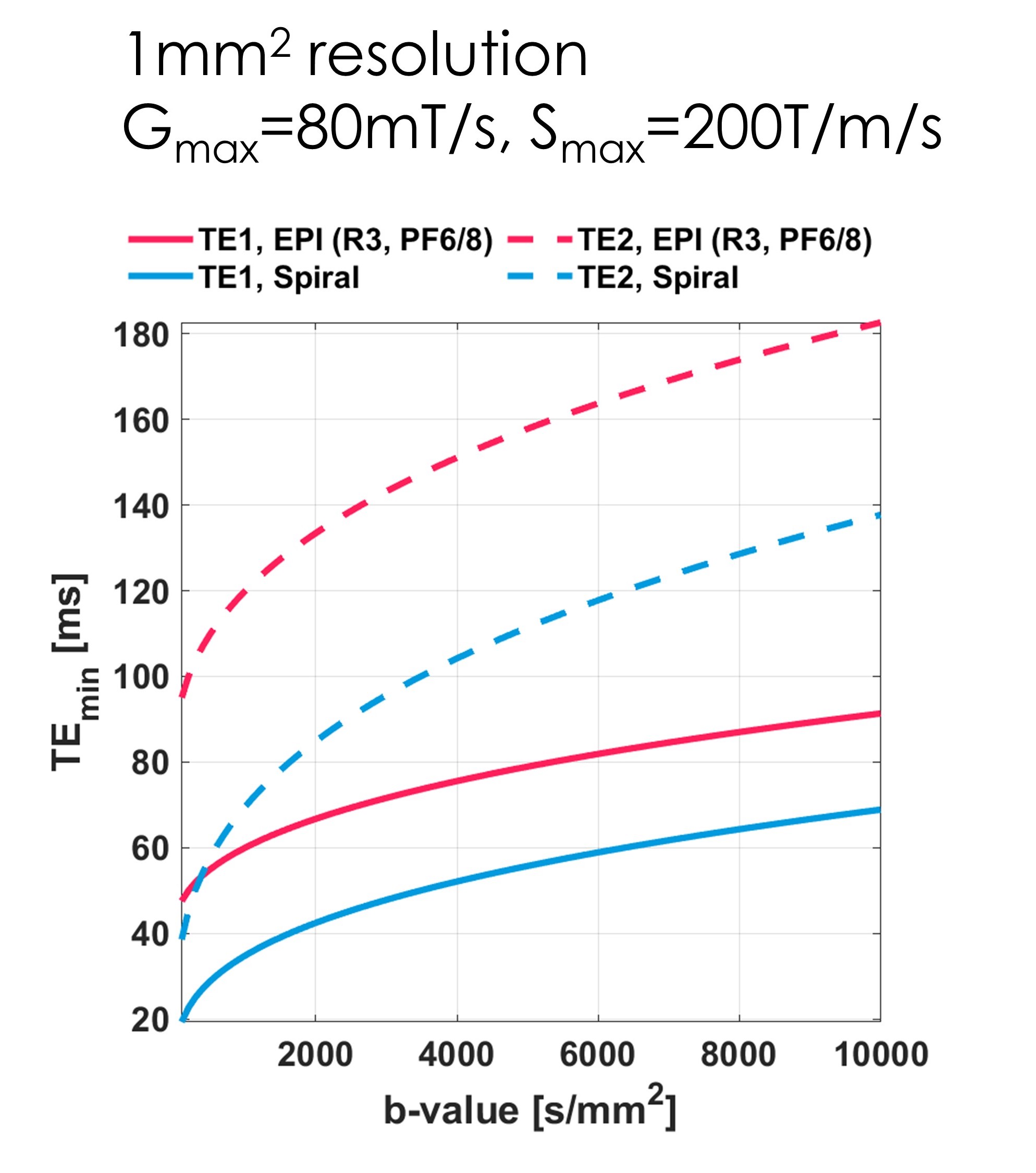
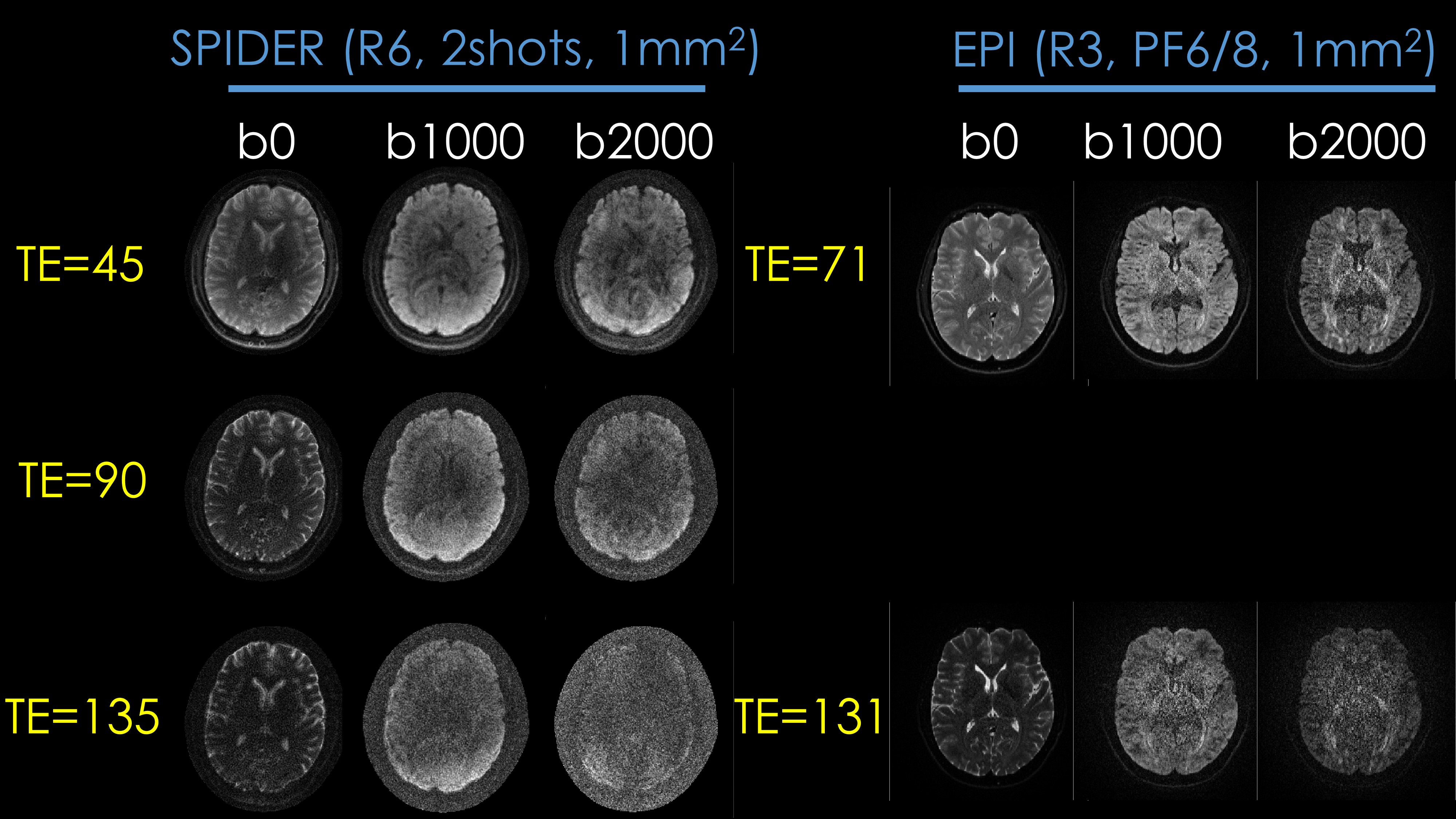
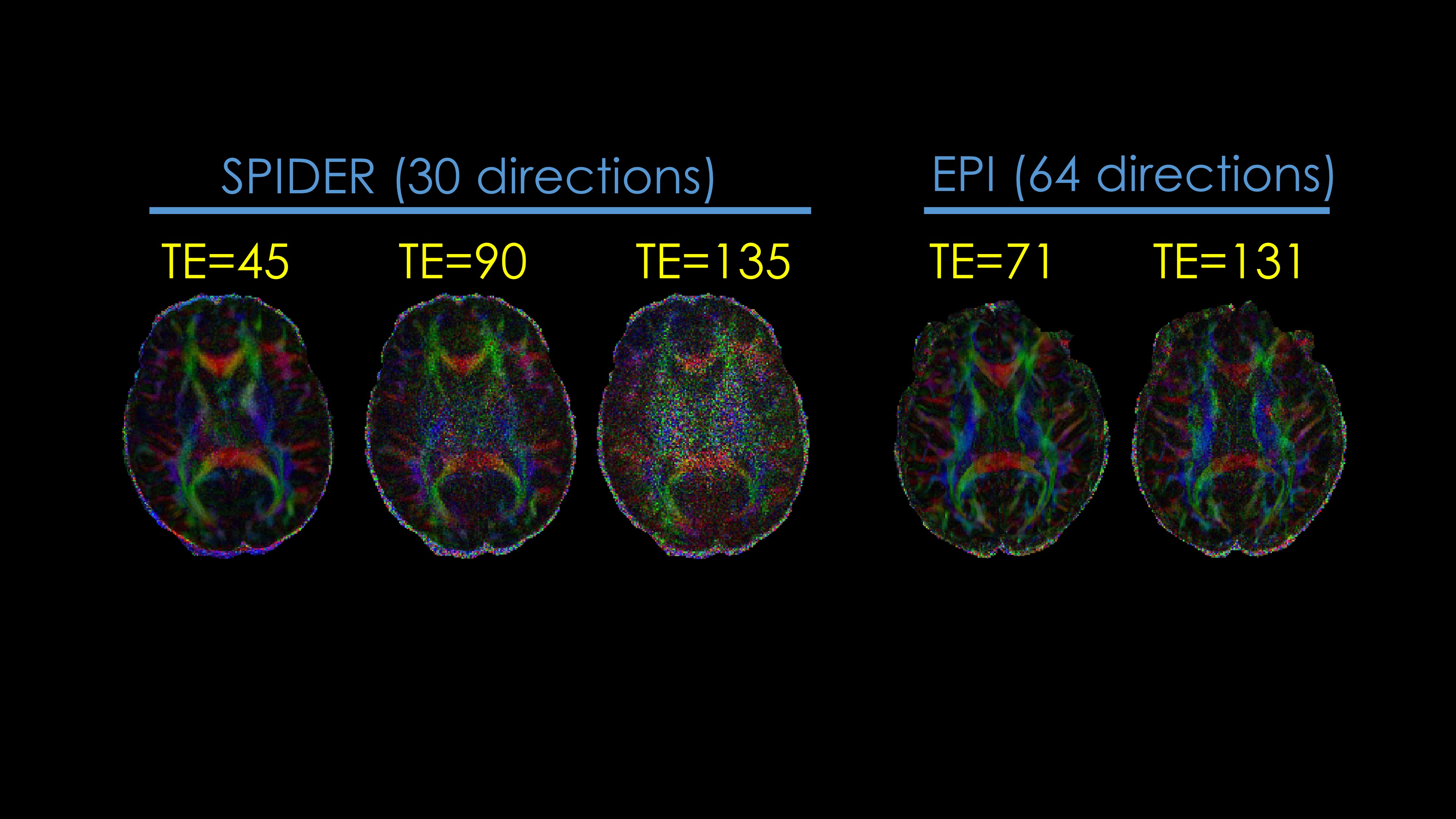
Fig.2 shows the minimal achievable TE as a function of b-value using maximal gradient amplitude of 80mT/m and maximal slew rate of 200T/m/s at 1mm2 resolution. Because spirals can directly start from k-space center, while EPI has tens of lines to acquire before k-space center, EPI requires a 20~30ms longer TE. Consequently, when acquiring multiple echoes, the TE difference of later echoes becomes larger, preventing EPI from acquiring 3 or more echoes. Fig.3 shows images of the proposed SPIDER and reference EPI. At the 1st TE, both methods show clean images. When TE is relatively large, the proposed SPIDER becomes noisier due to highly undersampled k-space periphery and shortened readout. Fig.4 illustrates the FA map at different TEs of both methods from two scan sessions. The proposed method yields a comparable FA to reference method at short TE. At TE=90ms, the proposed method is noisier than EPI@TE=131ms, partly due to smaller number diffusion directions (30 vs. 64).Discussion and conclusion
We proposed a turbo spin echo diffusion sequence with variable density spiral readout for diffusion relaxometry. The multi echo diffusion weighted images showed comparable results to the reference EPI-based diffusion images. To boost SNR, shorter TE is preferred. While adhering to the CPMG condition, spiral readout duration should be less than half of TE. Thus, we iteratively decreased the spiral sampling density to have shorter readout duration. SPIDER provides an efficient strategy for diffusion relaxometry, where multiple echoes are sampled within each TR without necessitating separate acquisitions as in EPI.Acknowledgements
This work was supported by research grants NIH R01 EB028797, U01 EB025162, P41 EB030006, U01 EB026996, R03 EB031175, R01 EB032378, UG3 EB034875, and NVidia Corporation for computing support. National Natural Science Foundation of China: 81971605. Key R&D Program of Zhejiang Province: 2022C04031.References
[1] Slator PJ, Palombo M, Miller KL, Westin CF, Laun F, Kim D, Haldar JP, Benjamini D, Lemberskiy G, de Almeida Martins JP, et al. Combined diffusion-relaxometry microstructure imaging: Current status and future prospects. Magn Reson Med 2021;86(6):2987-3011.
[2] Lee JH, Hargreaves BA, Hu BS, Nishimura DG. Fast 3D imaging using variable-density spiral trajectories with applications to limb perfusion. Magn Reson Med 2003;50(6):1276-1285.
[3] Layton KJ, Kroboth S, Jia F, Littin S, Yu H, Leupold J, Nielsen JF, Stocker T, Zaitsev M. Pulseq: A rapid and hardware-independent pulse sequence prototyping framework. Magn Reson Med 2017;77(4):1544-1552.
[4] Hu Y, Levine EG, Tian Q, Moran CJ, Wang X, Taviani V, Vasanawala SS, McNab JA, Daniel BA, Hargreaves BL. Motion-robust reconstruction of multishot diffusion-weighted images without phase estimation through locally low-rank regularization. Magn Reson Med 2019;81(2):1181-1190.
[5] Jenkinson M, Beckmann CF, Behrens TE, Woolrich MW, Smith SM. Fsl. Neuroimage 2012;62(2):782-790.
Figures