2439
3D Diffusion Image Acquisition with Motion Offsetting and
Navigation-Dependent Segmentation (DIAMONDS)
1Radiology, Clinical Scienes, Sahlgrenska Academy, University of Gothenburg, Gothenburg, Sweden, 2Medical Physics and Biomedical Engineering, Sahlgrenska University Hospital, Gothenburg, Sweden, 3Medical Radiation Sciences, Clinical Scienes, Sahlgrenska Academy, University of Gothenburg, Gothenburg, Sweden, 4Radiology, Sahlgrenska University Hospital, Gothenburg, Sweden, 5Radiology, Brigham and Women's Hospital, Boston, MA, United States
Synopsis
Keywords: Diffusion Acquisition, Diffusion/other diffusion imaging techniques, 3D-DWI; Motion-compensated diffusion imaging; Diffusion acquisition
Motivation: Investigate whether 1D navigator echo phase information can be used to correct residual phase errors in single thick-slab segmented 3D-DWI with first and second order motion-compensated diffusion encoding gradients.
Goal(s): Perform high-resolution single thick-slab segmented 3D-DWI with full brain coverage.
Approach: Extend a first and second order motion-compensated segmented 3D-DWI sequence with three orthogonal 1D navigators, which are utilized to correct for residual phase errors.
Results: Correction of zeroth order phase shifts, which result from translation, demonstrated reduction of ghosting artifacts. First order phase terms, which arise from rotation, were also determined, but require a more complex correction strategy.
Impact: Motion-compensated single thick-slab segmented 3D-DWI with orthogonal 1D navigators is a completely novel and promising approach for high resolution diffusion imaging with full brain coverage.
Introduction
Diffusion-weighted imaging (DWI) predominantly relies on a single-shot 2D data acquisition scheme. However, this approach comes with the cost of low SNR and low spatial resolution, especially in the slice-dimension. For high-resolution diffusion imaging a 3D acquisition compared to a 2D acquisition delivers superior SNR1,2. A diffusion-weighted 3D approach, however, requires a multi-shot acquisition where motion-induced shot-to-shot phase variations result in signal drop-outs and ghosting artifacts. Multi-slab 3D-DWI relies on phase correction with a 2D in-plane phase navigator echo and the assumption that through-slab phase variation is minimal 2–4. While this approach has shown promising results, it is unfortunately prone to slab boundary artifacts 5,6. Increasing the slab thickness would violate the assumption and instead require a more advanced 3D navigator 7, which introduces new problems, such as the inherently low bandwidth in the third sampling direction.The use of first and second order motion-compensated diffusion gradients has been shown to be a viable option to perform single thick-slab 3D-DWI with whole brain coverage 8. However, residual phase variations remain. The present work assumes that these phase shifts predominantly stem from rigid head (jerk) motion, which produces constant and linear phase variations. Under these premises very basic 1D navigator phase information should suffice to correct for the bulk of shot-to-shot phase variations.
Method
Single-slab 3D-DWI brain data were collected on a 3 Tesla scanner (Premier [Gmax = 80mT/m, slew rate = 200 mT/m/ms] GE Healthcare, Milwaukee WI, USA), in 3 volunteers with an in-house developed 3D-DWI sequence with added 1D navigator echoes (Fig. 1). First and second order, i.e., M0=M1=M2=0, motion-compensated diffusion gradients were utilized in all scans. Scan parameters were 1.5 mm3 isotropic resolution, 120 mm slab thickness, full echo, three diffusion encoding directions, in-plane acceleration of 2 using a 48-channel head-coil, and b-values 0 and 1000 s/mm2 (see Table 1). For comparison, 2D-DWI data sets with matching scan time (approximately 3 min) were acquired with b = 0 s/mm2 (NSA=1) and b= 1000 s/mm2 (NSA=5), (see Table 1).The constant phase correction terms, Φ0, x, Φ0, y and Φ0, z, were obtained from the center point in k-space of each navigator. All terms should be equal, as each navigator should hold the same translation-induced phase accumulation. Subsequently, the constant phase terms from the first navigator, i.e., Φ0, x, was used for all constant phase correction and applied by subtracting Φ0, x from each kx-ky plane point for each kz-location along the 3D data stack. All data were processed and reconstructed in MatLab (2021b, Mathworks, Natick, MA, USA). The final reconstruction-step was performed with the BART 9 function pics, which employs a non-uniform Fast Fourier Transform and iterative parallel imaging reconstruction (using the conjugate gradient method) 10 with l2-regularization as, $$argmin_o \| PSFo - m \|^2_2 + \lambda R(o)$$ with sampling operator P, Fourier transform F, coil sensitivities S, unknown images o, acquired k-space data m, regularization parameter λ, and regularization term R (i.e., $$$ R(o)=\| o \|^2_2$$$ for l2-regularization).
Results
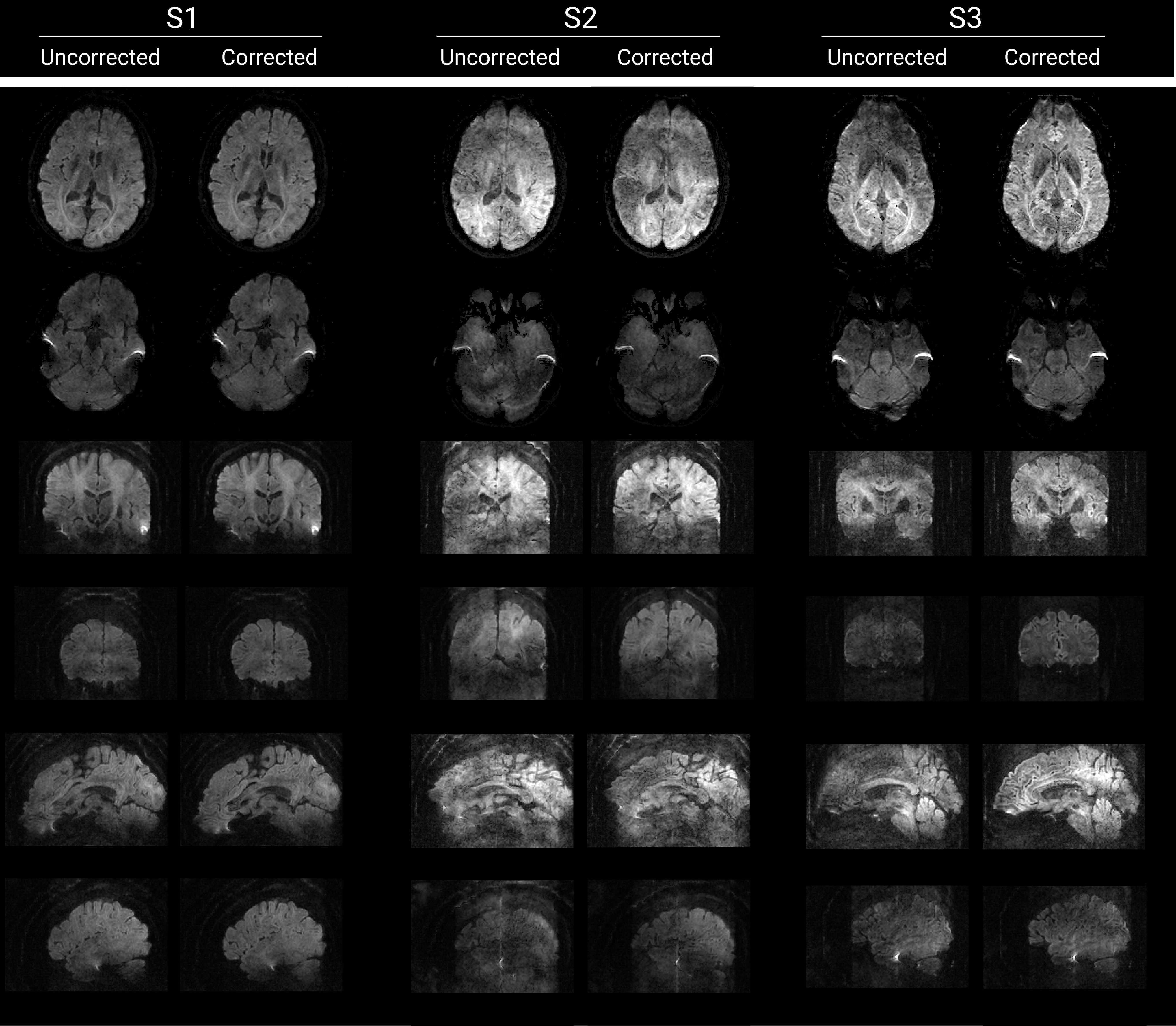
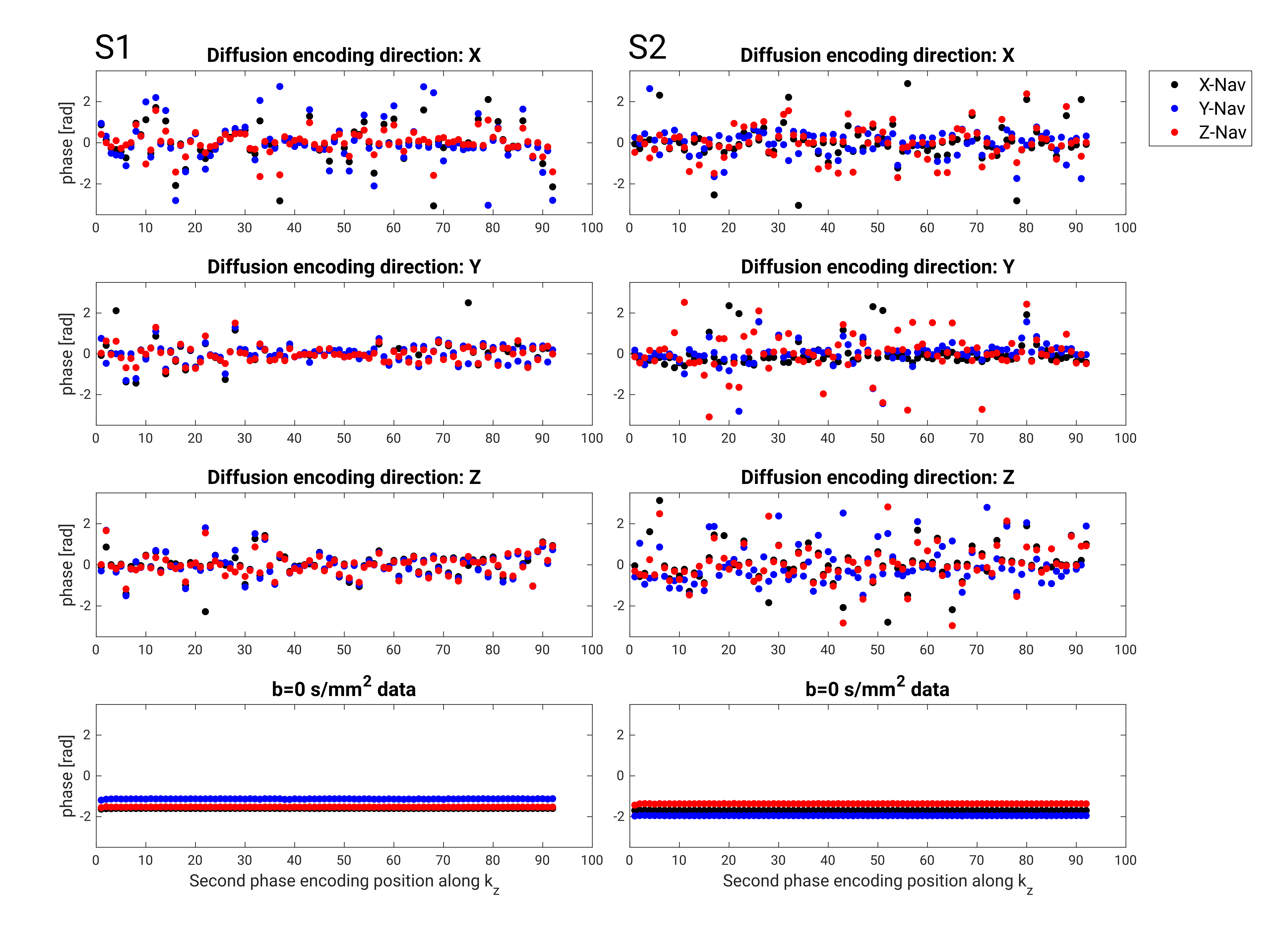
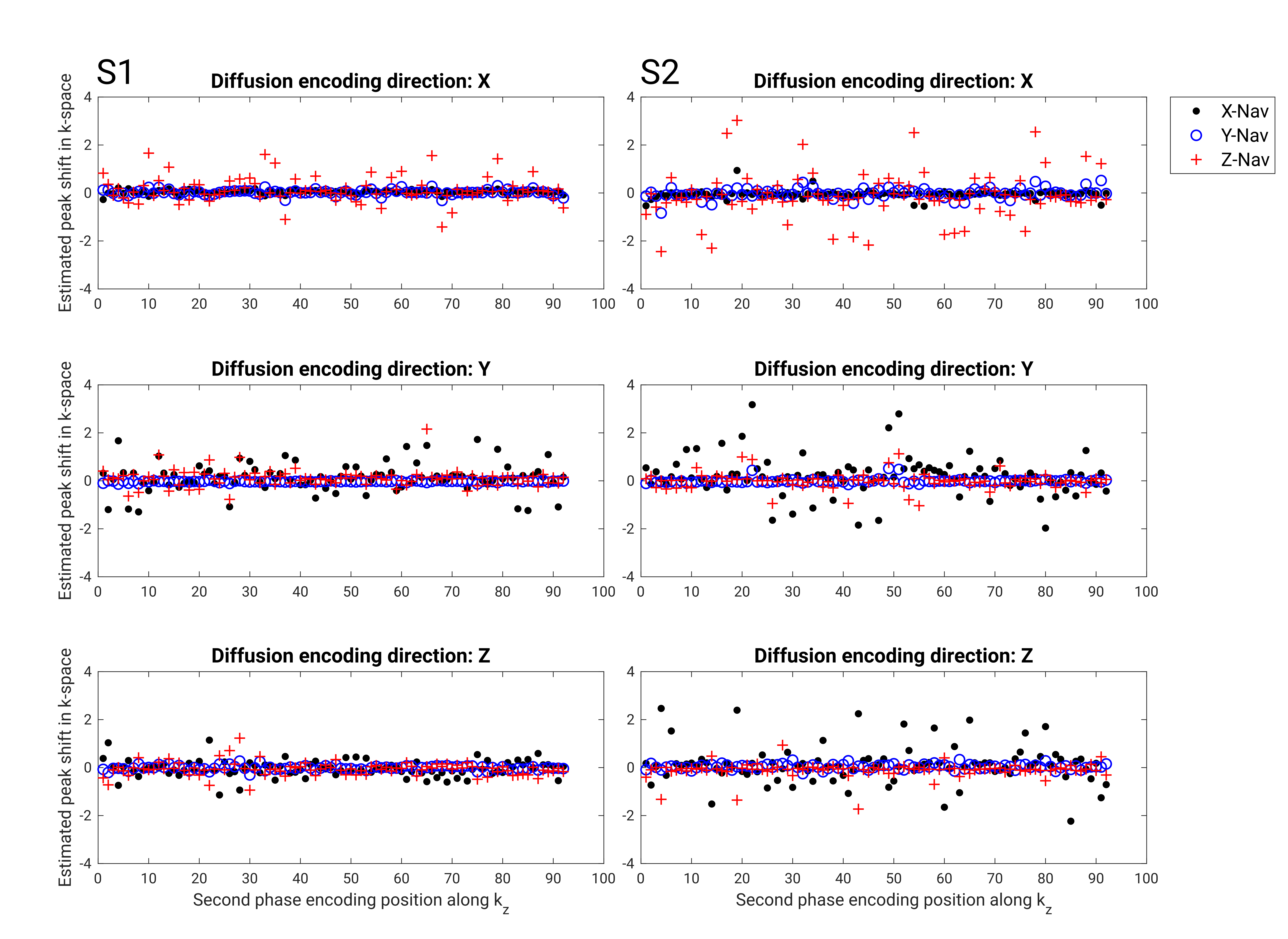
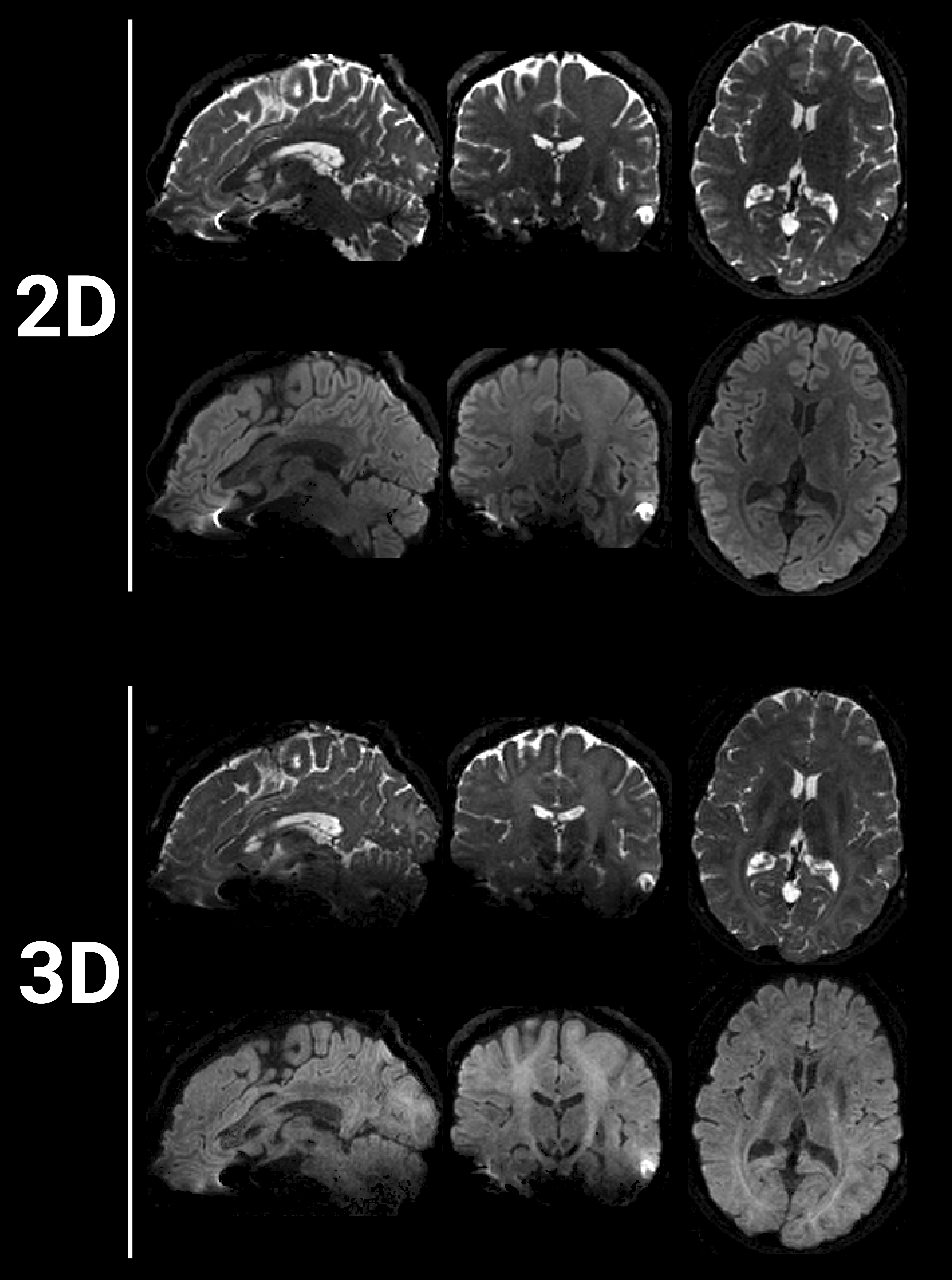
Axial, sagittal, and coronal trace DWI images, from the 3D-DWI scans, reconstructed with and without constant phase correction are shown for all subjects in Fig. 2. The image artifacts for each subject varies but is reduced in all cases with additional constant phase correction. The corresponding constant and linear phase information from each navigator and diffusion encoding direction for subject 1 (S1) and subject 2 (S2) are shown in Fig.3 and 4, respectively. Comparison between the clinical 2D-DWI data and 3D-DWI data shows a higher T1-weightning for the 3D-sequence (Fig. 5).Discussion and Conclusion
We demonstrated that 1D navigators may suffice to monitor and correct for residual phase variations in thick-slab brain 3D-DWI with first and second order motion-compensated diffusion gradients. Though the addition of constant phase correction reduces the ghosting artifacts, adding first order correction is likely to reduce artifacts further. The superior SNR for 3D-DWI, compared to 2D-DWI, can be visually appreciated in Fig. 5 for the b = 0 s/mm2 images. The b = 1000 s/mm2 images would require a more elaborate SNR analysis to decide the SNR advantages.Single thick-slab 3D-DWI with high isotropic resolution and high SNR reduces partial volume effects and could potentially aid in the investigation of small lesions. Unlike multi-slab approaches, this approach collects only one slab and therefore eliminates the problem with slab boundary artifacts. Provided 3D-DWI can produce images with consistent high quality, e.g., by integrating both zeroth and first order phase correction, within a clinically acceptable scan time, it would be of great clinical value.
Acknowledgements
The Swedish state under an agreement between the Swedish government and the country councils (ALFGBG-932648)
The Swedish Research Council (Vetenskapsrådet)
The Swedish Cancer Society (Cancerfonden)
Barncancerfonden
References
1. Engström, M., Mårtensson, M., Avventi, E. & Skare, S. On the signal-to-noise ratio efficiency and slab-banding artifacts in three-dimensional multislab diffusion-weighted echo-planar imaging. Magn Reson Med 73, 718–725 (2015).
2. Engström, M. & Skare, S. Diffusion-weighted 3D multislab echo planar imaging for high signal-to-noise ratio efficiency and isotropic image resolution. Magn Reson Med 70, 1507–1514 (2013).
3. Chang, H. C. et al. Human brain diffusion tensor imaging at submillimeter isotropic resolution on a 3Tesla clinical MRI scanner. Neuroimage 118, 667–675 (2015).
4. Frost, R., Miller, K. L., Tijssen, R. H. N., Porter, D. A. & Jezzard, P. 3D multi-slab diffusion-weighted readout-segmented EPI with real-time cardiac-reordered k-space acquisition. Magn Reson Med 72, 1565–1579 (2014).
5. Wu, W., Koopmans, P. J., Frost, R. & Miller, K. L. Reducing slab boundary artifacts in three-dimensional multislab diffusion MRI using nonlinear inversion for slab profile encoding (NPEN). Magn Reson Med 76, 1183–1195 (2016).
6. Dai, E. et al. A 3D k-space Fourier encoding and reconstruction framework for simultaneous multi-slab acquisition. Magn Reson Med 82, 1012–1024 (2019).
7. Chang, H. C., Hui, E. S., Chiu, P. W., Liu, X. & Chen, N. K. Phase correction for three-dimensional (3D) diffusion-weighted interleaved EPI using 3D multiplexed sensitivity encoding and reconstruction (3D-MUSER). Magn Reson Med 79, 2702–2712 (2018).
8. Johansson, J., Lagerstrand, K. M., Hebelka, H. & Maier, S. E. Segmented thick-slab 3D DWI with first and second order motion-compensated diffusion gradients. in # 3622. Proceedings of ISMRM & ISMRT Annual Meeting & Exhibition (2023).
9. Uecker, M., Tamir, J. I., Ong, F. & Lustig, M. The BART toolbox for computational magnetic resonance imaging. in Proc Intl Soc Magn Reson Med vol. 24 1 (2016).
10. Pruessmann, K. P., Weiger, M., Börnert, P. & Boesiger, P. Advances in sensitivity encoding with arbitrary k-space trajectories. Magn Reson Med 46, 638–651 (2001).
Figures
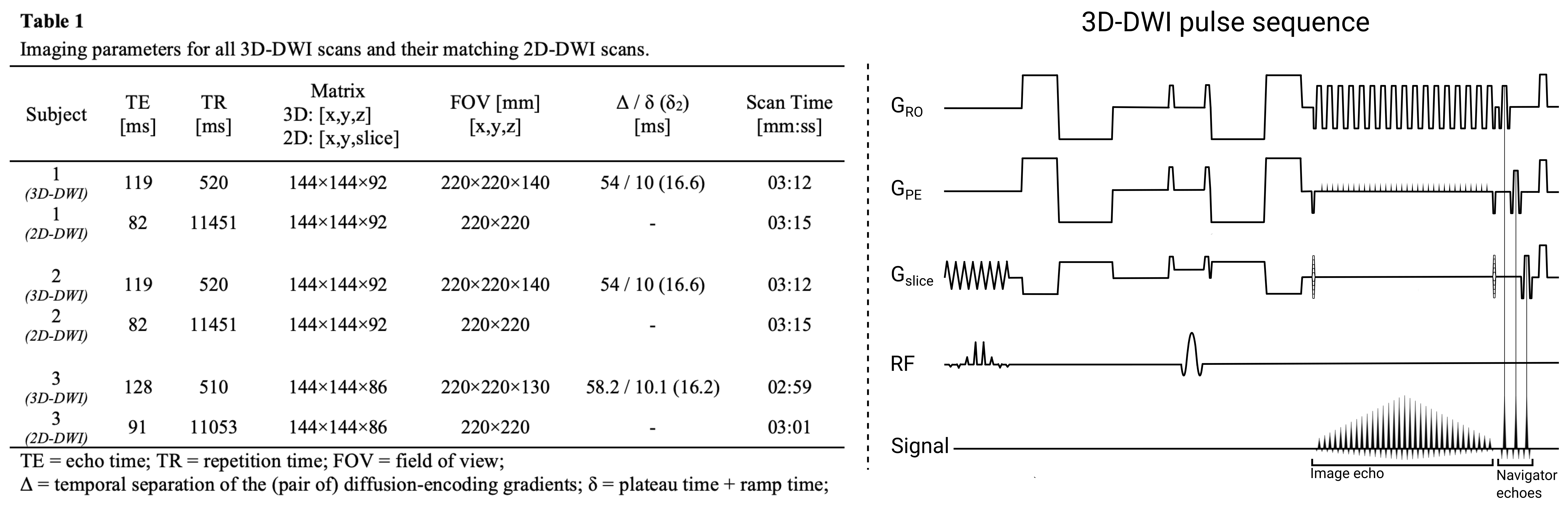
Figure 1: (Left) Table 1. (Right) The 3D-DWI EPI pulse sequence, with first and second order motion compensated diffusion encoding gradients and three orthogonal 1D navigator echoes, obtained after the main EPI train (image echo). The navigator echoes are played out sequentially on the read-out GRO, phase encoding GPH, and slice Gslice, axes. The remaining two axes depict the RF-pulses, and signal echoes.