2438
Diffusion-weighted imaging of the sellar region: A comparison study of TGSE-BLADE and RESOLVE sequences1Department of Radiology, The First Affiliated Hospital of Nanjing Medical University, Nanjing, China, 2Siemens Shenzhen Magnetic Resonance Ltd., Shenzhen, China, 3MR Research Collaboration Team, Siemens Healthineers Ltd., Shanghai, China
Synopsis
Keywords: DWI/DTI/DKI, New Trajectories & Spatial Encoding Methods
Motivation: We aim to investigate the potential of TGSE-BLADE diffusion-weighted imaging(DWI) in mitigating image artifacts and distortions within the sellar region.
Goal(s): Our study involves a comparative analysis of image quality between TGSE-BLADE diffusion-weighted imaging (DWI) and RESOLVE-DWI within the sellar region.
Approach: We compared qualitative (overall imaging quality, artifacts and distortions) and quantitative (signal-to-noise ratio [SNR] and apparent diffusion coefficient [ADC]) parameters between RESOLVE-DWI and TGSE-BLADE-DWI images.
Results: TGSE-BLADE-DWI exhibited higher imaging quality with less artifacts and distortions and a higher SNR than did RESOLVE-DWI (P<0.05). The tissue ADCs did not differ significantly between the groups (P>0.05).
Impact: TGSE-BLADE-DWI offers a solution to diffusion imaging distortion. Comparative analysis highlights its superiority in mitigating sellar region distortion, promising enhanced quality in challenging clinical applications.
Introduction
Several techniques have recently been developed that enable diffusion-weighted imaging (DWI) to overcome the distortion caused by magnetic susceptibility. These techniques include readout-segmented echo-planar imaging (RESOLVE-DWI)[1-3], zoomed echo-planar imaging[4], and periodically rotated overlapping parallel lines with enhanced reconstruction (PROPELLER-DWI)[5-7]. Each technique has advantages and drawbacks that should be considered before application in clinical examinations8. RESOLVE-DWI reduces geometric distortion, T2* blurring, and susceptibility artifacts through segmented acquisition of k-space along the readout direction, reducing echo spacing and echo time[3, 8]. PROPELLER-DWI has a long acquisition time and high specific absorption rate preventing its clinical application[9, 10]. Thus, turbo gradient-echo and spin-echo (TGSE)-BLADE-DWI has been introduced to resolve these issues. TGSE-BLADE-DWI features multi-blade k-space filling and has a shorter acquisition time than that of conventional BLADE DWI[11]. Moreover, the use of gradient echoes and fewer refocusing radiofrequency pulses decrease the specific absorption rate. Additionally, placing gradient echoes and spin echoes into separate blades and removing off-resonance phases reduces off-resonance artifacts[9-11]. To our knowledge, no previous TGSE-BLADE-DWI study has compared distortion and artifacts among sellar regions in healthy volunteers. This study was conducted to compare the quality of TGSE-BLADE with RESOLVE images in healthy volunteers.Methods
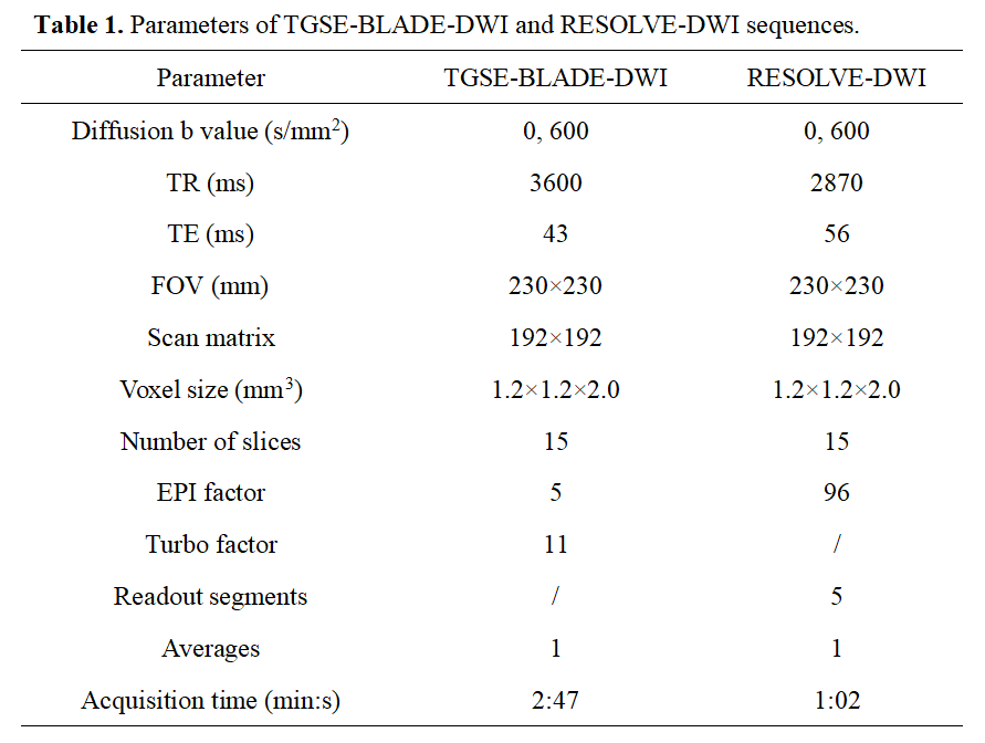
This study included 23 healthy volunteers who underwent magnetic resonance imaging, including morphological T1-weighted imaging, RESOLVE-DWI, and TGSE-BLADE-DWI on a 3T system (MAGNETOM Prisma, Siemens Healthcare, Erlangen, Germany). The detailed sequence scanning parameters of TGSE-BLADE and RESOLVE are summarized in Table 1.Image quality was qualitatively assessed by visualization and classified from 1 (non-diagnostic image quality with severe artifacts and deformations) to 5 (excellent image quality with high-level anatomic details). Image quality was quantitatively assessed by calculating the signal-to-noise ratio (SNR) of the images acquired via TGSE-BLADE-DWI and RESOLVE-DWI. The apparent diffusion coefficients (ADCs) of the pituitary and white matter were also compared.
The Wilcoxon signed rank test was used to determine significant differences in qualitative parameters between TGSE-BLADE and RESOLVE images. Paired t-tests were used to determine significant differences in quantitative parameters between TGSE-BLADE and RESOLVE that conformed to a normal distribution. Interobserver reproducibility for assessing the qualitative parameters was assessed using weighted kappa analysis. P<0.05 was considered statistically significant.
Results
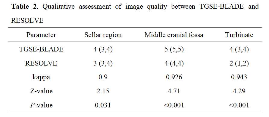
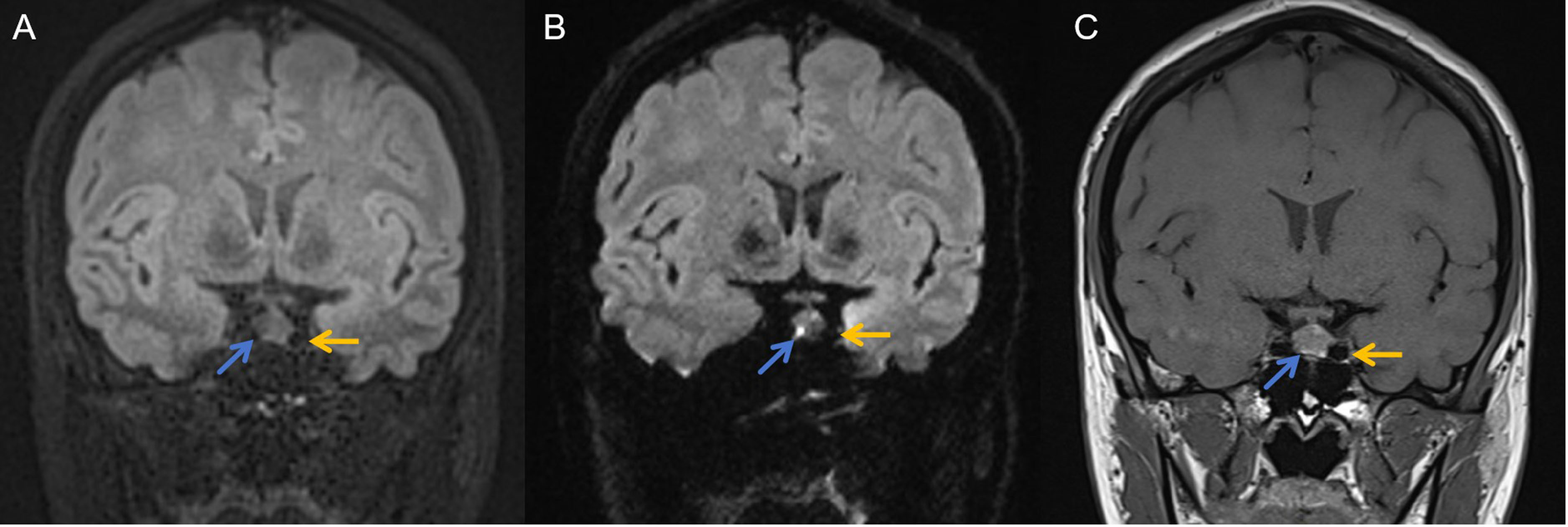
Qualitative assessment of image qualityTwo independent observers found that TGSE-BLADE-DWI exhibited higher overall image quality with fewer artifacts and deformations than did RESOLVE-DWI (Table 2 and Fig. 1). Quality scores of the TGSE-BLADE-DWI and RESOLVE-DWI for the 23 healthy volunteers were sellar region = 4 (3,4) vs 3 (3,4; P<0.05); middle cranial fossa = 5 (5,5) vs 4 (4,4; P<0.001); and turbinate = 4 (3,4) vs 2 (1,2; P<0.001), respectively. Kappa values ranged from 0.9–0.943 with good interobserver reproducibility (Table 2).
Quantitative assessment of image quality
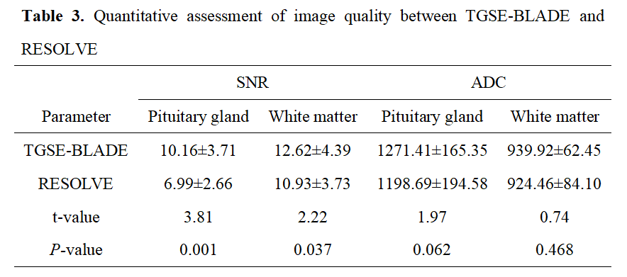
Table 3 shows the detailed quantitative assessment results of the image quality between the two sequence sets. TGSE-BLADE-DWI showed a significantly higher SNR than did RESOLVE-DWI: pituitary gland = 10.16±3.71 vs 6.99±2.66, respectively (P<0.001), and white matter = 12.62±4.39 vs 10.93±3.73, respectively (P<0.05). ADC values for the pituitary gland and white matter did not differ significantly between the two sets (pituitary gland = 1271.41±165.35 vs 1198.69±194.58, respectively; white matter = 939.92±62.45 vs 924.46±84.10, respectively; both P>0.05; Table 3).
Discussion
We compared the quality of TGSE-BLADE-DWI and RESOLVE-DWI in the sellar region, which contains many air-bone interfaces and has large changes in magnetic susceptibility. We adjusted the fields of view, slice thicknesses, gaps, and matrices of each DWI technique to be the same to allow qualitative and quantitative comparisons. The quality of the DWI images in the sellar region acquired via TGSE-BLADE was significantly higher than that acquired via RESOLVE. TGSE-BLADE substantially reduced image artifacts and distortion in the sellar region, with a clinically acceptable scan time.Conclusion
Our preliminary study indicated the feasibility of TGSE-BLADE-DWI for assessing the sellar region. Compared with RESOLVE-DWI, TGSE-BLADE-DWI produced better image quality by reducing susceptibility artifacts and geometric distortion.Acknowledgements
No acknowledgement found.References
[1] Stocker D, Manoliu A, Becker AS, et al. Image Quality and Geometric Distortion of Modern Diffusion-Weighted Imaging Sequences in Magnetic Resonance Imaging of the Prostate. Invest Radiol. 2018. 53(4): 200-206.
[2] Holdsworth SJ, Skare S, Newbould RD, Bammer R. Robust GRAPPA-accelerated diffusion-weighted readout-segmented (RS)-EPI. Magn Reson Med. 2009. 62(6): 1629-40.
[3] Porter DA, Heidemann RM. High resolution diffusion-weighted imaging using readout-segmented echo-planar imaging, parallel imaging and a two-dimensional navigator-based reacquisition. Magn Reson Med. 2009. 62(2): 468-75.
[4] Thierfelder KM, Scherr MK, Notohamiprodjo M, et al. Diffusion-weighted MRI of the prostate: advantages of Zoomed EPI with parallel-transmit-accelerated 2D-selective excitation imaging. Eur Radiol. 2014. 24(12): 3233-41.
[5] Wang FN, Huang TY, Lin FH, et al. PROPELLER EPI: an MRI technique suitable for diffusion tensor imaging at high field strength with reduced geometric distortions. Magn Reson Med. 2005. 54(5): 1232-40.
[6] Fries P, Runge VM, Kirchin MA, et al. Diffusion-weighted imaging in patients with acute brain ischemia at 3 T: current possibilities and future perspectives comparing conventional echoplanar diffusion-weighted imaging and fast spin echo diffusion-weighted imaging sequences using BLADE (PROPELLER). Invest Radiol. 2009. 44(6): 351-9.
[7] Forbes KP, Pipe JG, Karis JP, Heiserman JE. Improved image quality and detection of acute cerebral infarction with PROPELLER diffusion-weighted MR imaging. Radiology. 2002. 225(2): 551-5.
[8] Bergen RV, Ryner L, Essig M. Field-map correction in read-out segmented echo planar imaging for reduced spatial distortion in prostate DWI for MRI-guided radiotherapy applications. Magn Reson Imaging. 2020. 67: 43-49.
[9] Li Z, Pipe JG, Lee CY, Debbins JP, Karis JP, Huo D. X-PROP: a fast and robust diffusion-weighted propeller technique. Magn Reson Med. 2011. 66(2): 341-7.
[10] Fu Q, Kong XC, Liu DX, et al. Clinical comparison of single-shot EPI, readout-segmented EPI and TGSE-BLADE for diffusion-weighted imaging of cerebellopontine angle tumors on 3 tesla. Magn Reson Imaging. 2021. 84: 76-83.
[11] Hu HH, McAllister AS, Jin N, et al. Comparison of 2D BLADE Turbo Gradient- and Spin-Echo and 2D Spin-Echo Echo-Planar Diffusion-Weighted Brain MRI at 3 T: Preliminary Experience in Children. Acad Radiol. 2019. 26(12): 1597-1604.
Figures