2437
Submillimeter Isotropic Whole Brain DTI at 3T with 2D Multi-band Multi-shot EPI Acquisition and Deep Learning Reconstruction1GE Healthcare, Waukee, WI, United States, 2GE Healthcare, Houston, TX, United States, 3Duke University Medical Center, Durham, NC, United States, 4GE Healthcare, Boston, MA, United States, 5GE Healthcare, Calgary, AB, Canada, 6GE Healthcare, Menlo Park, CA, United States
Synopsis
Keywords: Tractography, Brain Connectivity
Motivation: High resolution diffusion tensor imaging (DTI) is associated with low intrinsic sensitivity and therefore it is difficult to achieve simultaneously high resolution and high SNR.
Goal(s): To acquire submillimeter DTI to map human brain structural connectivity networks with improved accuracy and detail than standard resolution DTI.
Approach: We combine a deep learning reconstruction method with MB-MUSE to further enhance the image quality and demonstrate improved quantification of submillimeter isotropic (0.8×0.8×0.8 mm3) whole brain DTI at 3T with approximately 1 minute per diffusion direction scan time.
Results: The exceptional delineation and clarity of fiber tracking was achieved from submillimeter isotropic whole brain DTI.
Impact: Submillimeter isotropic whole brain DTI with approximately 1 minute per diffusion direction scan time at 3T with high SNR, low distortion, provides us with a powerful tool to map whole brain structural connectivity networks with exceptional clarity and quantification.
Introduction
Comparing to conventional DTI (e.g., 2×2×2 mm3 resolution), submillimeter DTI can map human brain structural connectivity networks with greater details1,2. However, it remains challenging to simultaneously achieve high SNR, high resolution, and minimal artifact (distortion, Gibbs ringing, high background noise, etc.) within reasonable acquisition times at 3T. In principle, multi-shot EPI can reduce image distortion at the expense of scan time, simultaneous multi-slice acquisition can accelerate the acquisition and noise reduction reconstruction methods can improve the SNR for mitigating these challenges. In this study we aim to further integrate deep learning-based reconstruction methods3,4,5 with 2D multi-band multiplexed sensitivity-encoding (MB-MUSE)6,7 acquisition to improve the image quality and quantification of submillimeter isotropic (0.8×0.8×0.8 mm3) whole brain DTI at 3T by: 1) reducing noise amplification associated with MB-MUSE reconstruction, 2) improving SNR and in-plane resolution, and 3) reducing Gibbs ringing artifacts and background noise4.Methods
Reconstruction: The proposed reconstruction included the conventional MB-MUSE integrated with a deep learning-based phase image reconstruction (DL Phase) for the phase images per shot and a deep learning-based MR image reconstruction (DL Recon) pipeline3 which removes both noise and ringing artifacts for the final diffusion weighed images (DWI).Acquisition: DTI images were acquired on a GE 3T SIGNA MRI research scanner with a Nova 32-channel head coil and an ultra-high-performance (UHP) gradient (max. strength 100mT/m, max. slew rate=200T/m/s). The images were acquired at 0.8×0.8×0.8 mm3 resolution, b=1000 s/mm2, directions=16, 150 slices per direction, partial Fourier = 0.75, bands = 2, shots = 4. To optimize other scan parameters to reduce noise amplification associated with MB-MUSE, 4 datasets were acquired with different FOV, TR, TE scan matrix combinations. Here CAIPI shift of FOV/2 was used in MB excitation to reduce g-factor penalty8 and scan matrix was adjusted to keep uniform pixel size.
FOV(cm): 21 27.6 32 42
TR(s): 10 11 12 13.5
TE(ms): 64.3 64.3 65 69.6
Scan matrix: 256×256 336×336 384×384 512×512
Scan time (minute): 12.67 13.93 15.2 17.1
Processing: MRtrix3 (https://www.mrtrix.org/) was used to generate FA map and fiber tracking.
Results and Discussion
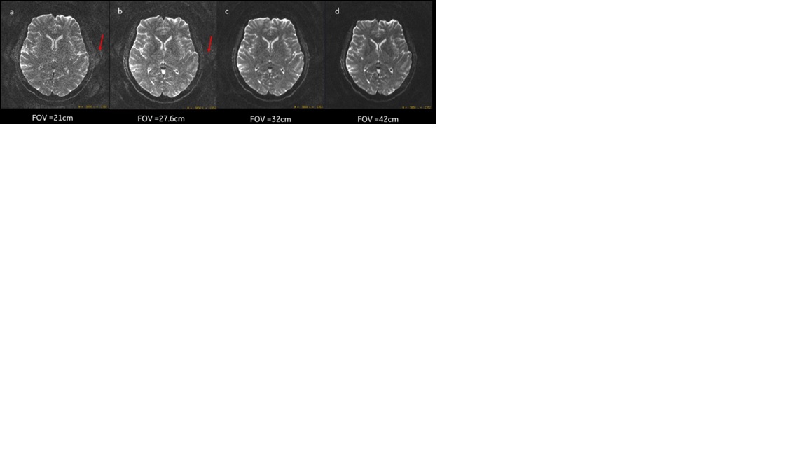
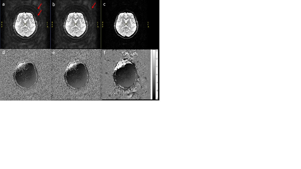
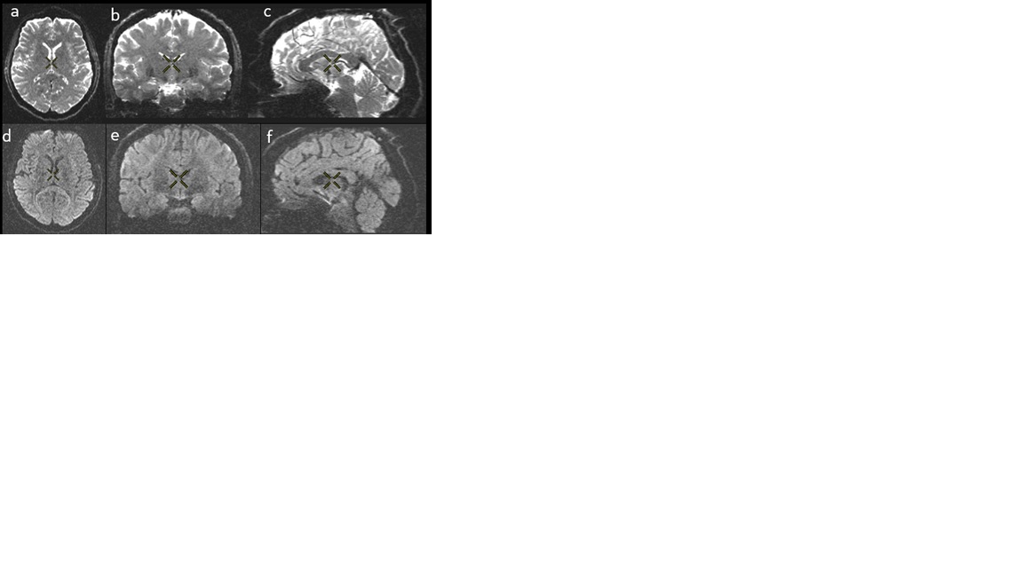
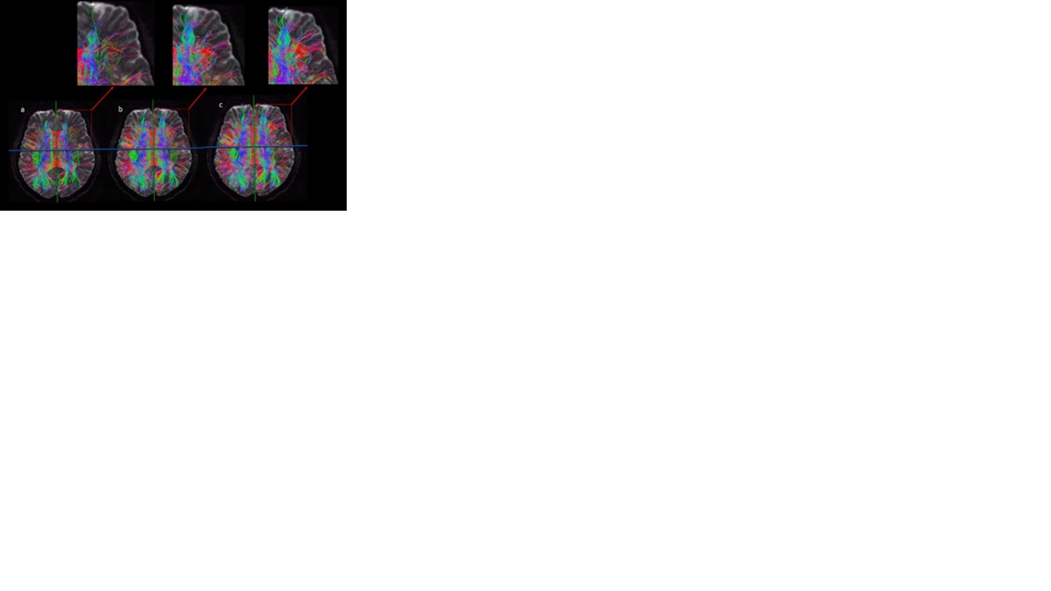
Figure 1 shows the b0 images acquired at 0.8mm isotropic resolution with different FOVs and reconstructed using the MB-MUSE without DL on phase image per shot and without DL on final image. For the extended FOV scans (32cm and 42cm, Fig. 1c and 1d), SNR was significantly improved, and artifacts caused by noise amplification associated with MB-MUSE were largely removed since extended FOV provided better MB slice separation8. However, artifacts still exist in figure1a and Figure1b (as indicated by the red arrow in Figure1a and Figure1b) for datasets with FOVs at 21cm and 27.6cm. For the 32cm FOV, there were minimal TE penalty and small TR increase, which were offset by greatly improved SNR and reduced artifacts. This dataset was chosen to further evaluate the effects of DL algorithms. Here three methods were evaluated: fermi filter to smooth the map, DL recon3 and DL phase5. Figure 2 shows the FOV32 B0 images (2a-2c) using these 3 methods; a higher window level is applied here to show background noise and artifact outside of brain structure. There was only minor improvement for DL recon method as compared to the traditional Fermi filter smoothing. But for images using DL phase, the background noise was greatly reduced and ringing artifact removed. Figure 2d-2e are the corresponding phase maps from the first shot for each method. It is worth noting that DL recon was trained for denoising and reducing Gibbs ringing artifacts within image area, while DL phase was trained for generating high quality phase map. Figure 3 is the 3-plane view of the final images generated by MB-MUSE with DL phase recon on phase image per shot and DL Recon on final output, acquisition was in axial plane and reformed for sagittal and coronal slices. Figure 4 shows the fiber tracking generated from MRtrix3 for the fiber >100mm with MB-MUSE only (Figure4a), MB-MUSE with DL phase (Figure4b), and MB-MUSE with DL phase and DL recon (Figure4c). The improved delineation and clarity of fiber tracking is illustrated in Figure4c for the MB-MUSE Deep Learning Reconstruction.Conclusion
With the MB-MUSE DL reconstruction, we can acquire submillimeter isotropic whole brain diffusion tensor imaging with approximately 1 minute per diffusion direction scan time at 3T with high SNR, low distortion, which could provide us with a powerful tool to map whole brain structural connectivity networks with improved clarity and quantification.Acknowledgements
We would like to thank Iain P. Bruce for providing the original MB-MUSE code.References
1. Miller KL, Stagg CJ, et al. Diffusion imaging of whole, post-mortem human brains on a clinical MRI scanner. Neuroimage, 2011;57:167-181.
2. Chang HC, Sundman M, et al. Human brain diffusion tensor imaging at submillimeter isotropic resolution on a 3 Tesla clinical MRI scanner. Neuroimage, 2015;118:667-675.
3. Lebel, R.M. Performance characterization of a novel deep learning-based MR image reconstruction pipeline. August 2020, http://arxiv.org/abs/2008.06559
4. Wang X, Yang B, et al. High-resolution Diffusion Tensor Imaging at 7T with Multi-band Multi-shot EPI acquisition and Deep Learning Reconstruction. ISMRM Annual Meeting, London UK. 2022; Abstract #3966.
5. Xinzeng Wang, Dan Litwiller, Arnaud Guidon et al. Robust Complex Signal Averaging for Diffusion Weighted Imaging. ISMRM Annual Meeting, Toronto, ON, Canada 2023; Abstract #7566.
6. Chen NK, Guidon A, et al. A robust multi-shot scan strategy for high-resolution diffusion weighted MRI enabled by multiplexed sensitivity-encoding (MUSE). Neuroimage. 2013; 72: 41–47.
7. Bruce IP, Chang HC, et al. 3D-MB-MUSE: A robust 3D multi-slab, multi-band and multi-shot reconstruction approach for ultrahigh resolution diffusion MRI. Neuroimage, 2017; 159:46-56.
8. Setsompop K, Gagoski BA, et al. Blipped-controlled aliasing in parallel imaging for simultaneous multislice echo planar imaging with reduced g-factor penalty. Magnetic Resonance in Medicine 2012;67 : 1210–1224.
Figures