2436
Ultra-high resolution b-tensor encoding using gSlider on a clinical scanner1Brigham and Women's Hospital, Harvard Medical School, Boston, MA, United States, 2School of Biomedical Engineering, Southern Medical University, Guangzhou, China, 3Department of Radiology, Stanford University, Stanford, CA, United States, 4Department of Electrical Engineering, Stanford University, Stanford, CA, United States, 5Fetal-Neonatal Neuroimaging and Developmental Science Center, Boston Children’s Hospital, Harvard Medical School, Boston, MA, United States, 6Department of Biomedical Engineering, Case School of Engineering, Case Western Reserve University, Cleveland, OH, United States, 7Division of Medical Physics, Department of Radiology, University Medical Center Freiburg, Freiburg, Germany, 8fMRI Laboratory and Biomedical Engineering, University of Michigan, Ann Arbor, MI, United States, 9Athinoula A. Martinos Center for Biomedical Imaging, Massachusetts General Hospital, Charlestown, MA, United States, 10Department of Radiology, Harvard Medical School, Boston, MA, United States
Synopsis
Keywords: Diffusion Acquisition, Diffusion/other diffusion imaging techniques
Motivation: Spherical tensor encoding (STE) reveals microstructural information about tissues, which is hidden in conventional diffusion MRI techniques. However, existing STE acquisition techniques have low spatial resolution which masks intricate anatomical details.
Goal(s): To increase the spatial resolution of STE using the SNR-efficient gSlider sequence.
Approach: Isotropic b-tensor encoding diffusion-gradient waveforms were synergistically combined with the gSlider sequence using Pulseq vendor-neutral sequence development platform to obtain high resolution data.
Results: We demonstrate in-vivo results with the highest spatial resolution to-date of 1 mm isotropic voxels using the proposed STE-gSlider sequence.
Impact: High spatial resolution for b-tensor encoding will enable investigation of microstructural information in intricate detail in the brain in health and disease. Further, the proposed open-source sequence can be used on any vendor platform.
introduction
Spherical tensor encoding (STE) also referred to as q-space trajectory imaging (QTI) can capture the complex microstructural properties of tissues in diffusion MRI (dMRI) by using orientation-varying diffusion gradient waveforms which is not achievable by conventional dMRI techniques.1 Nevertheless, this advantage comes with a trade-off in spatial resolution due to signal-to-noise ratio (SNR) losses from strong diffusion gradients and prolonged echo times. To address this challenge, one potential solution involves obtaining low-resolution images from different perspectives, achieved through either shifting2,3 or rotating4, and subsequently applying a super-resolution reconstruction (SRR) algorithm. Generalized SLIce Dithered Enhanced Resolution (gSlider) sequence is an advanced dMRI sequence that instead uses multiple RF encodings to obtain high-resolution acquisition.5–7 Using RF-encoding with different subslice profiles, gSlider provides high SNR-efficiency. In this study, we demonstrate the feasibility of integrating STE gradients with the gSlider sequence to acquire ultra-high-resolution, isotropic b-tensor encoded images on a clinical scanner. The proposed QTI-gSlider sequence was implemented in the Pulseq open source vendor neutral sequence development platform.8Methods
The complex gradient waveforms for spherical tensor encoding were obtained using Numerical Optimization of gradient Waveforms (NOW) toolbox.9–12 During the optimization, the maximum gradient was set to 78 mT/m, the maximum slew rate was set to 100 T/m/s, with 1st-order motion compensation, and the total duration of the STE waveform was limited to 100ms (to ensure sufficient SNR considering the echo time), with a maximum b-value=1400 s/mm2. The generated gradient waveforms were then integrated with the gSlider sequence implemented in the Pulseq platform.8This study follows approval from the local IRBs. A healthy male volunteer was scanned after signing consent forms. The experiment was performed on a Siemens whole-body clinical scanner, Prisma, with a maximum gradient of 80 mT/m, and a maximum gradient slew rate of 200 T/m/s per axis. The following scanning protocol was used for the QTI-gSlider sequence: TE/TR/echo spacing=138/5500/0.93 ms, voxel size = 1 mm × 1 mm, GRAPPA = 3, Partial Fourier factor = 6/8. For a particular b-tensor, the diffusion gradient waveforms were scaled to acquire a group of b-values, including 0, 50, 100, 150, 700, 1000, and 1400, respectively. With six repetitions for each b-value, the total scan time was 21 minutes.
The raw k-space gSlider data was reconstructed offline in MATLAB using GRAPPA, after which the gSlider reconstruction and T1 and B1+ corrections were applied to get the 1 mm isotropic super-resolution images. The data from six repetitions first went through FSL TOPUP and eddy for distortion and motion correction and then averaged. The ADC map was calculated by mono-exponential fitting from data of all b-values and b0.
Results
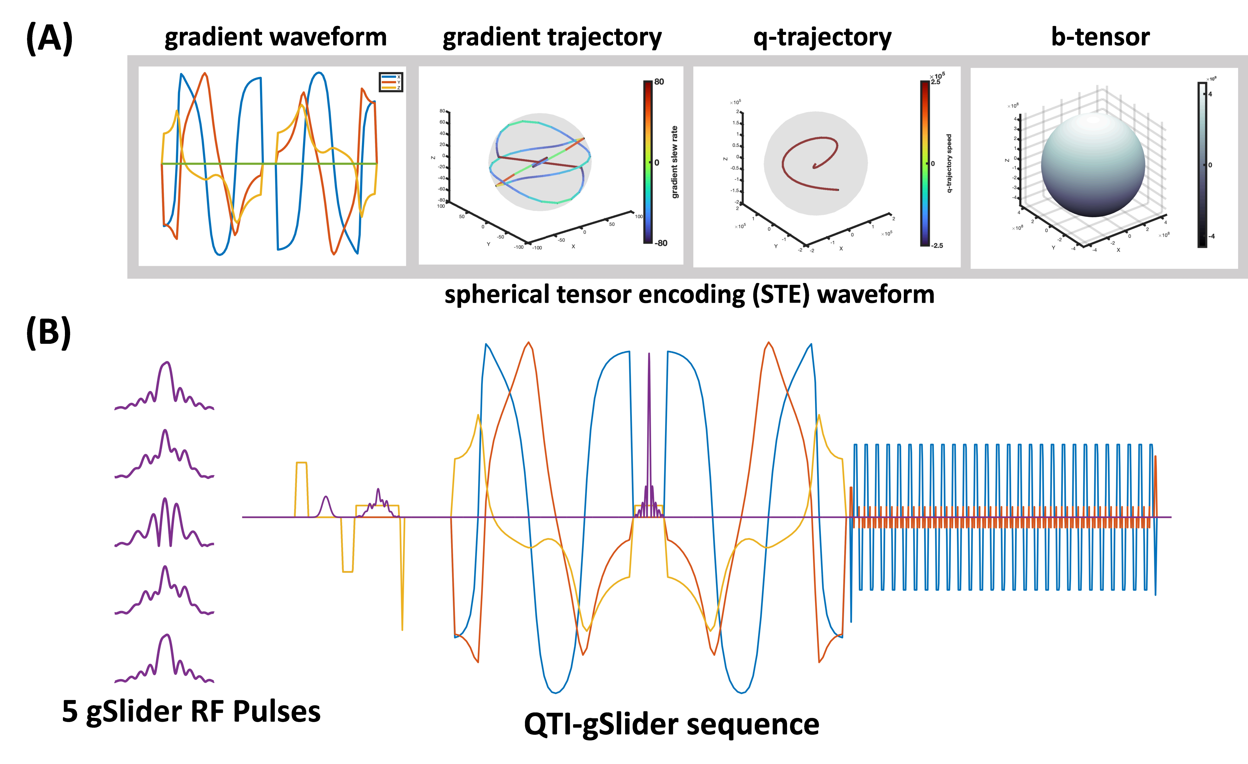
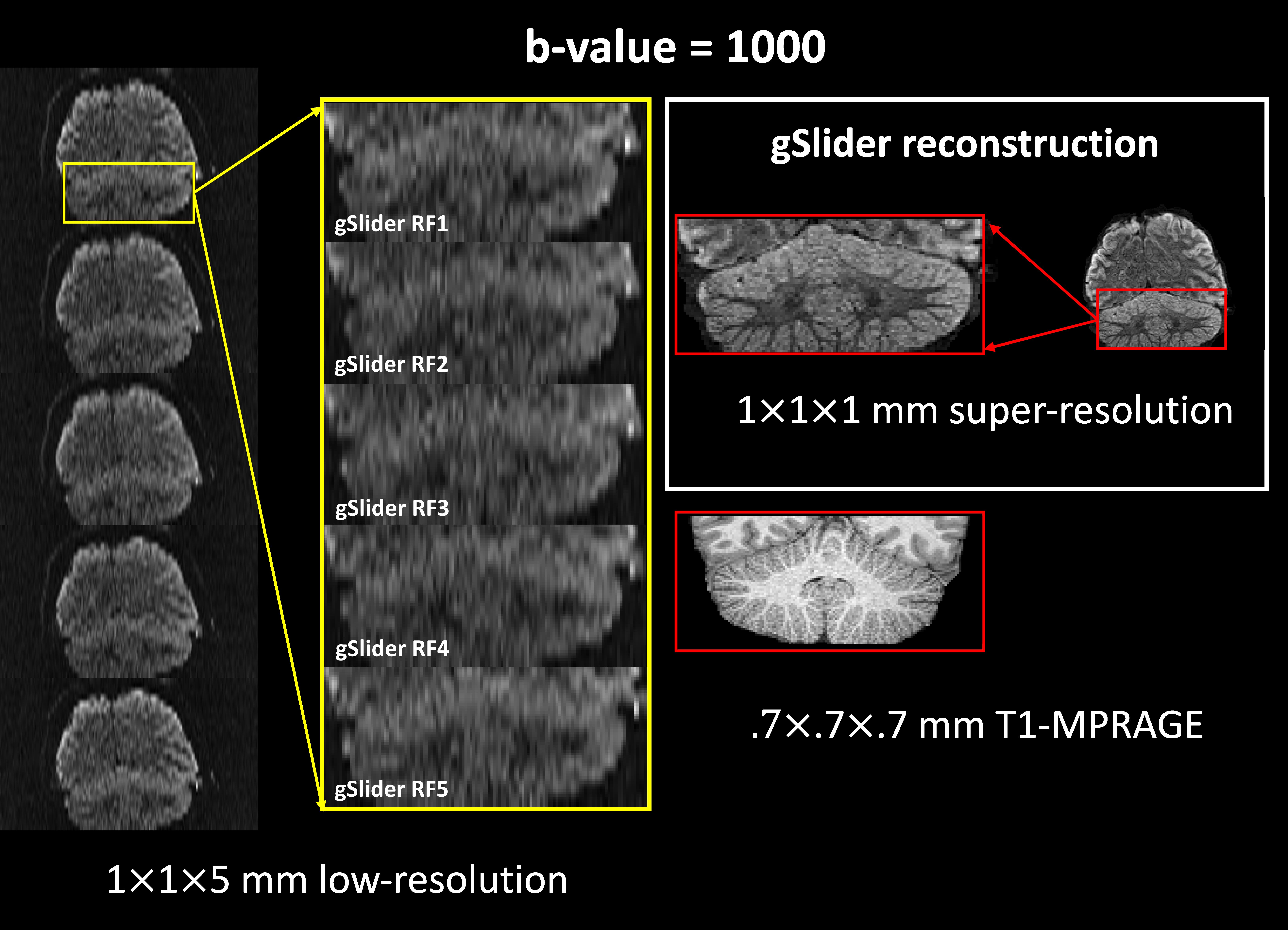
Figure 1 displays the sequence diagram of the implemented QTI-gSlider sequence. Figure 1(A) shows the gradient waveforms and their trajectories, the q-space trajectory, and the b-tensor; Figure 1 (B) shows the Pulseq-gSlider sequence with five different gSlider-RFs.In Figure 2, the QTI-gSlider low-resolution images for the 5 RF encodings are shown along with SRR (coronal view). Notably, when using T1-MPRAGE as an anatomical reference, detailed cerebellar features become discernible in 1 mm isotropic resolution images.
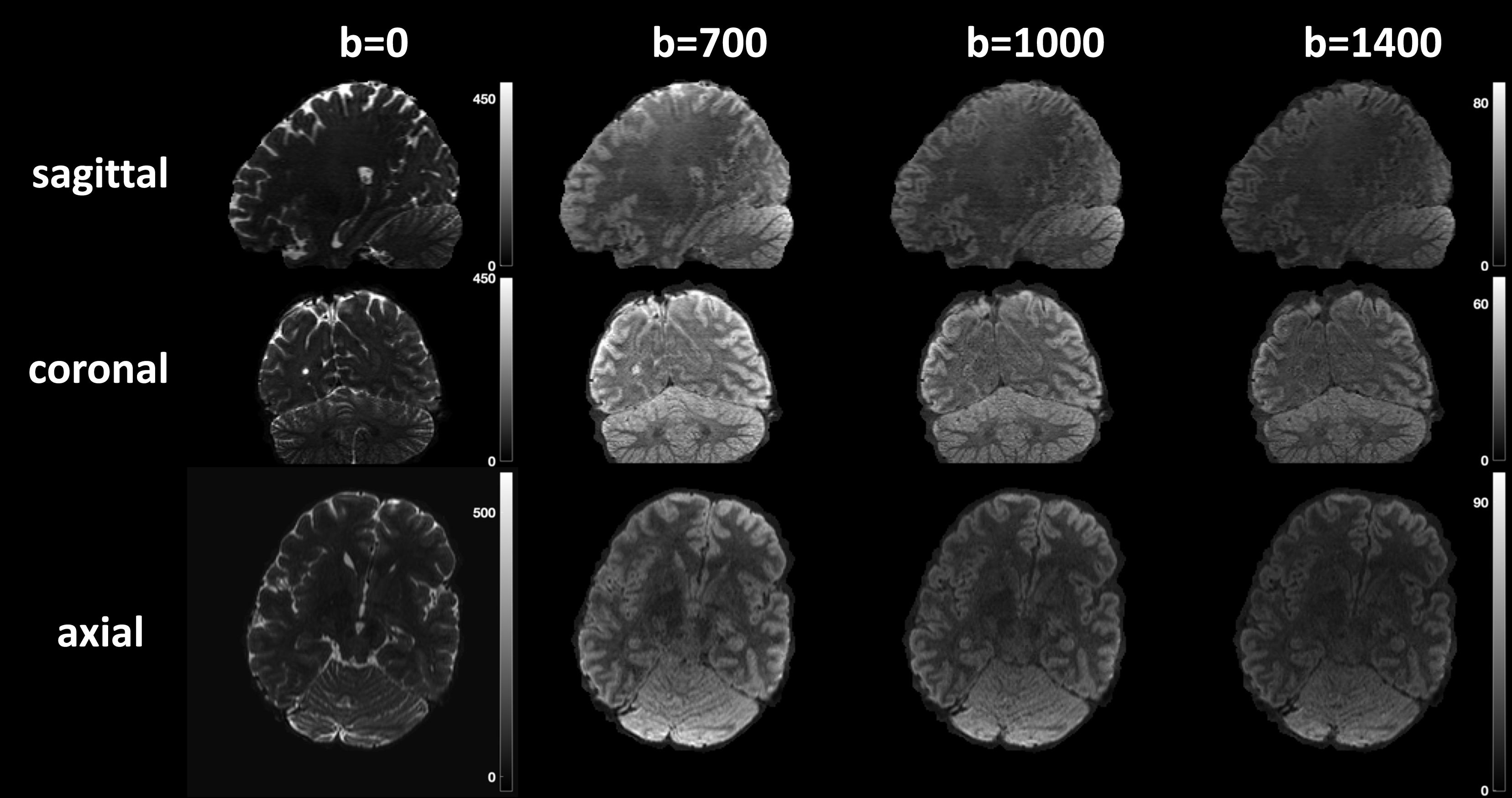
Figure 3 shows b0 images and isotropic b-tensor encoded images at b-values of [700, 1000, & 1400]. These images reveal high contrast in cortical regions in the diffusion-encoded images.
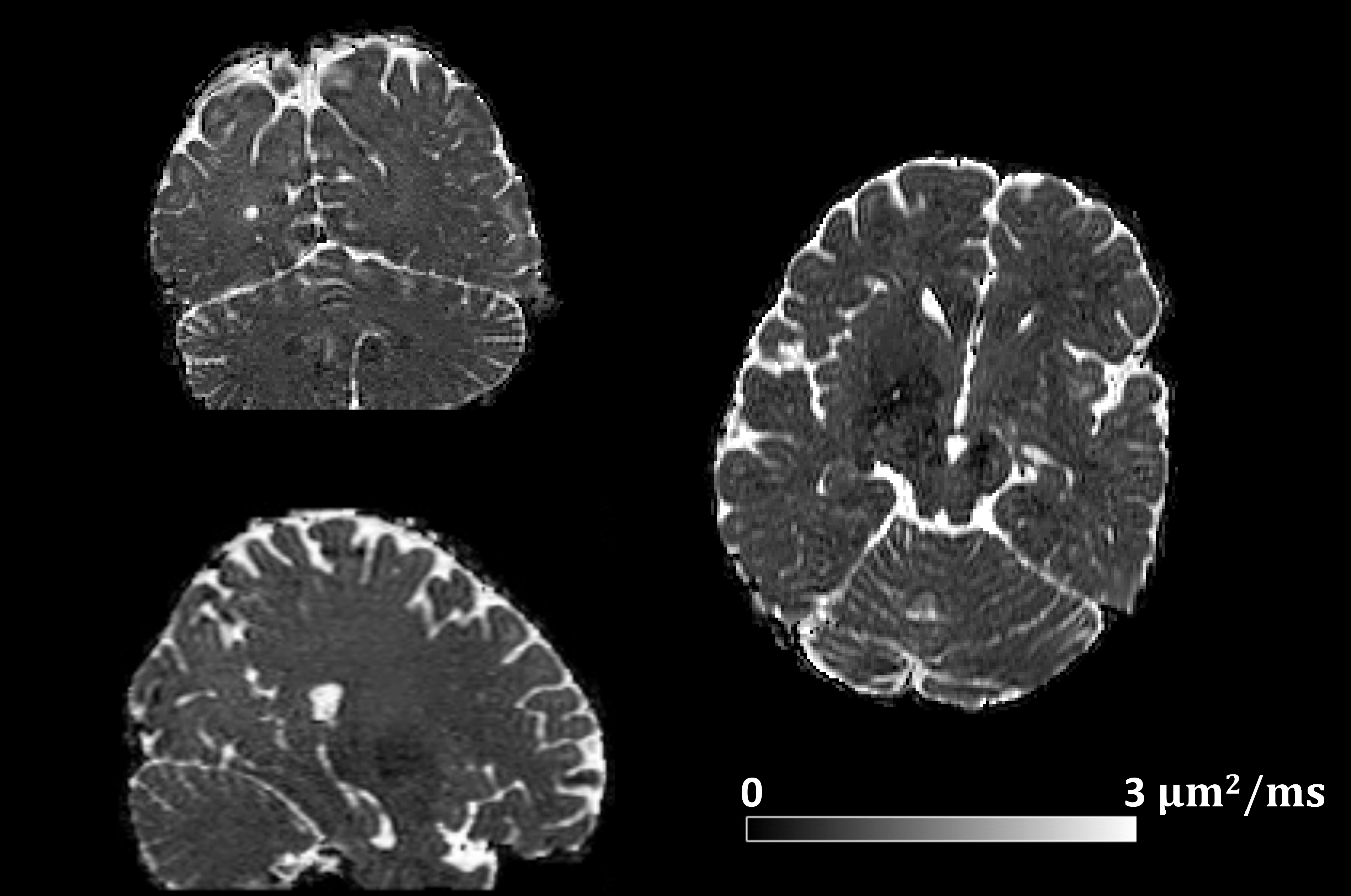
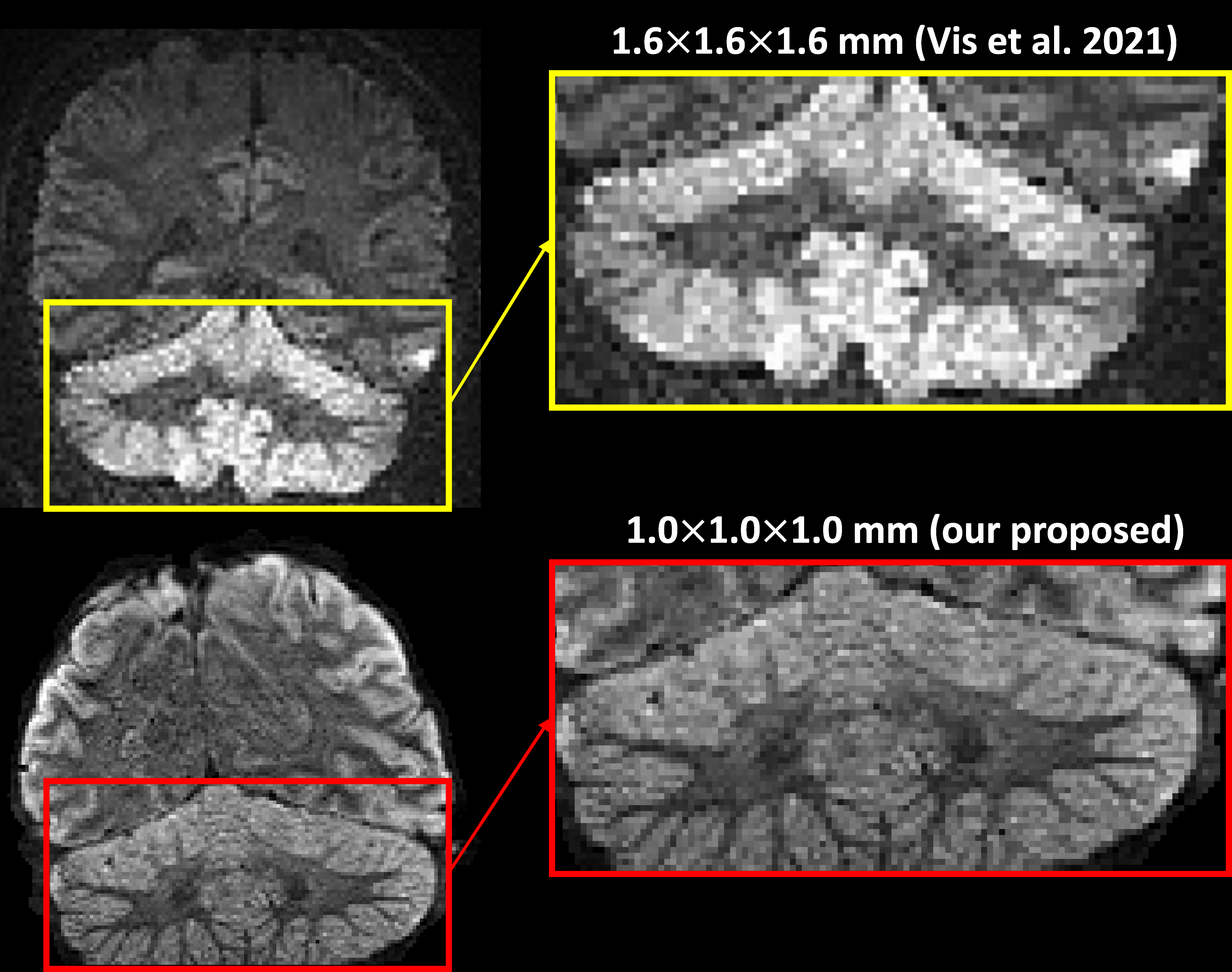
Figure 4 showcases the ADC map, and in Figure 5, we compare it to existing literature4 with the highest resolution of 1.6 mm with a rotation of eight views. This comparison highlights the superior image quality achieved with higher resolution, offering sharper boundaries and enhanced visualization of finer anatomical structures.
Discussion and Conclusion
In this study, we successfully increased the resolution of QTI to 1 mm by employing the gSlider sequence on a clinical scanner. This enhancement was due to ~3.4x higher SNR gain achieved through gSlider RF-encoding. To mitigate artifacts arising from concomitant gradients, we employed symmetric gradients. Furthermore, for TE reduction, we can consider introducing asymmetric gradient waveforms with Maxwell-compensated properties.11 Furthermore, our experiment was conducted on a whole-body clinical scanner, which is constrained by the peripheral nerve stimulation (PNS) threshold, leading to limitations in the achievable b-value and longer TE. However, since our sequence was implemented in Pulseq, it can be immediately tested on high-performance head-only GE MAGNUS or Siemens Connectome 2.0 scanners to attain higher spatial resolution and b-values.13Acknowledgements
This study is supported by NIH grants R01MH116173, R01MH125860, R01EB032378, and R01NS125781.References
1. Westin, C. F. et al. Q-space trajectory imaging for multidimensional diffusion MRI of the human brain. Neuroimage 135, 345–362 (2016).
2. Greenspan, H., Oz, G., Kiryati, N. & Peled, S. MRI inter-slice reconstruction using super-resolution. (2002).
3. Vu, A. T., Beckett, A., Setsompop, K. & Feinberg, D. A. Evaluation of SLIce Dithered Enhanced Resolution Simultaneous MultiSlice (SLIDER-SMS) for human fMRI. (2017).
4. Vis, G., Nilsson, M., Westin, C. F. & Szczepankiewicz, F. Accuracy and precision in super-resolution MRI: Enabling spherical tensor diffusion encoding at ultra-high b-values and high resolution. Neuroimage 245, (2021).
5. Liao, C. et al. Distortion-free, high-isotropic-resolution diffusion MRI with gSlider BUDA-EPI and multicoil dynamic B0 shimming. Magn Reson Med 86, 791–803 (2021).
6. Liao, C. et al. High-fidelity, high-isotropic-resolution diffusion imaging through gSlider acquisition with B1+ and T1 corrections and integrated ΔB0/Rx shim array. Magn Reson Med 83, 56–67 (2020).
7. Setsompop, K. et al. High-resolution in vivo diffusion imaging of the human brain with generalized slice dithered enhanced resolution: Simultaneous multislice (gSlider-SMS). Magn Reson Med 79, 141–151 (2018).
8. Layton, K. J. et al. Pulseq: A rapid and hardware-independent pulse sequence prototyping framework. Magn Reson Med 77, 1544–1552 (2017).
9. Szczepankiewicz, F. et al. Motion-compensated gradient waveforms for tensor-valued diffusion encoding by constrained numerical optimization. Magn Reson Med 85, 2117–2126 (2021).
10. Szczepankiewicz, F. & Sjölund, J. Cross-term-compensated gradient waveform design for tensor-valued diffusion MRI. Journal of Magnetic Resonance 328, (2021).
11. Szczepankiewicz, F., Westin, C. F. & Nilsson, M. Maxwell-compensated design of asymmetric gradient waveforms for tensor-valued diffusion encoding. Magn Reson Med 82, 1424–1437 (2019).
12. Sjölund, J. et al. Constrained optimization of gradient waveforms for generalized diffusion encoding. Journal of Magnetic Resonance 261, 157–168 (2015).
13. Chakwizira, A. et al. Diffusion MRI with free gradient waveforms on a high-performance gradient system: Probing restriction and exchange in the human brain. Neuroimage 283, 120409 (2023).
Figures