2431
Pushing the boundaries of in-vivo human diffusion MRI using Romer-EPTI: initial results on the Connectome 2.0 scanner1Athinoula A. Martinos Center for Biomedical Imaging, Massachusetts General Hospital, Charlestown, MA, United States, 2Department of Radiology, Harvard Medical School, Boston, MA, United States, 3Institute of Medical Physics and Radiation Protection, TH Mittelhessen University of Applied Science, Giessen, Germany, 4Harvard-MIT Division of Health Sciences and Technology, Massachusetts Institute of Technology, Cambridge, MA, United States
Synopsis
Keywords: Diffusion Acquisition, Data Acquisition, Diffusion Acquisition
Motivation: The progress in in-vivo high-resolution and high-b-value dMRI has advanced the investigation of structural connectivity and tissue microstructure.
Goal(s): We aim to further push the spatial resolution and b-value of in-vivo diffusion MRI.
Approach: A high-SNR(√25x), distortion-free, motion-robust Romer-EPTI technique that addresses the challenges in dMRI acquisition with further enhanced capabilities by taking advantage of the cutting-edge Connectome 2.0 scanner.
Results: We demonstrate motion-robust distortion-free in-vivo mesoscale dMRI (~485um-istropic) on clinical 3T&7T scanners, and further improvements in SNR on Connectome2.0 scanner enabling high-SNR, high b-value dMRI and a spatial resolution up to 400um-iso.
Impact: Romer-EPTI achieves high-SNR motion-robust distortion-free in-vivo mesoscale dMRI (~485 um) with exceptional quality on clinical 3T&7T scanners. Its capabilities were further enhanced on the Connectome 2.0 scanner, enabling high-SNR high b-value dMRI and a spatial resolution up to 400um-iso.
Introduction
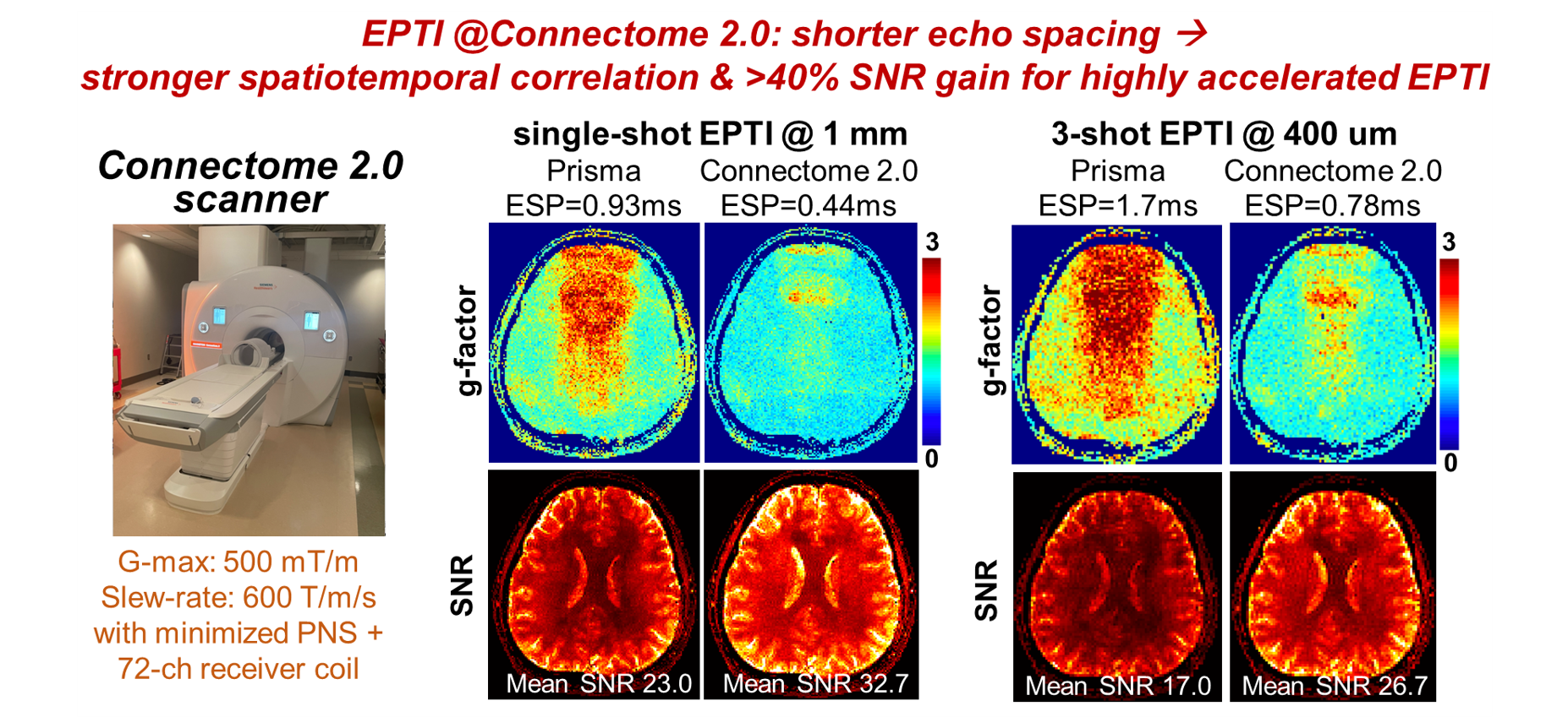
The advancement in in-vivo dMRI has provided exciting insights into the structural connectivity and tissue microstructure of the human brain. Notably, high-spatial-resolution dMRI techniques1-7 have enabled the investigation of brain’s fine-scale structures by imaging crucial but small fibers1 and the cytoarchitecture of the thin cortical layers7. Furthermore, the use of stronger diffusion gradients8-13 has become available due to hardware advances14-16, combined with advanced diffusion models17-22, providing more precise information about tissue microstructure. However, further advancing dMRI in these directions faces several major technical challenges, which become increasingly pronounced as we seek higher resolution or diffusion gradients: i) reduced SNR as voxel-size is diminished and/or b-values are raised; ii) severe image degradation (distortion and T2*-blurring); iii) motion sensitivity.Here, we address these challenges of in-vivo dMRI by maximizing the capabilities of our recently-developed Romer-EPTI23-25 technique in conjunction with the newly-built Connectome 2.0 scanner (C2.0-scanner)26. Romer-EPTI is a novel dMRI technique that provides significantly higher SNR efficiency (e.g.,~√25x) with high motion robustness. Moreover, it provides distortion-free images with minimized blurring and slab-boundary artifacts. Romer-EPTI was able to achieve, for the first time, in-vivo dMRI at mesoscale ~500um resolution at both 3T and 7T23,25, and high-SNR ultra-high-b-value and time-dependent dMRI24. In this work, we first further characterize Romer-EPTI’s performance on clinically-available scanners, and demonstrate its high motion robustness and image-quality at mesoscale resolution up to 485-um-iso (~0.1 mm3). Then, to further enhance Romer-EPTI’s capabilities, we pursue its development on the ultra-high-gradient-strength C2.0-scanner, equipped with Gmax=500mT/m and maximal slew-rate=600T/m/s—about 5x gradient performance over modern MRI systems26. We demonstrated the improved SNR of Romer-EPTI on C2.0 derived from the shorter echo-spacing and therefore stronger spatiotemporal correlation. We then showed that Romer provides significant SNR gain compared to EPI on C2.0-scanner and can enable spatial resolution up to 400um-iso (0.064mm3, 2x-smaller voxel than 500um-iso).
Methods
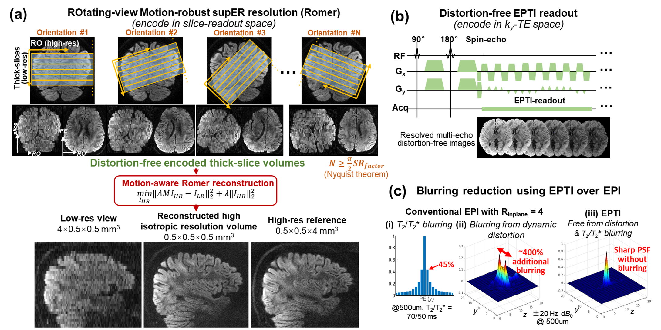
Romer-EPTI integrates the efficient ky-t EPTI27-30 encoding with a rotating-view thick-slice super-resolution acquisition25,31-34 that encodes the kx-slice dimensions (Fig.1). It resolves high-SNR distortion-free high-isotropic-resolution volumes from multiple encoded volumes with different slice orientations using a motion-aware reconstruction25. The significant SNR gain of Romer-EPTI (e.g.,√25x) comes from: i) EPTI encoding that enables continuous readout with minimal dead-time, minimal TE, and optimal readout length (~1.3xSNR), and ii) short-TR thick-slice acquisition combined with SMS (e.g., additional √14.6xSNR). Romer-EPTI’s motion robustness stems from: i) its motion-aware reconstruction which models the motion between different Romer-encoded volumes; ii) the distortion-free EPTI readout which eliminates geometric inconsistency between volumes caused by motion-induced field changes. This avoids blurring/artifacts on the reconstructed high-resolution images, a problem that plagued conventional EPI (Fig.1c,~400% blurring in EPI @500μm even with 4x in-plane-acceleration). In addition, EPTI images are free from T2*-decay blurring, eliminating additional blurring seen in EPI (e.g., 45% @500um). Slab-boundary artifacts are minimized in Romer with modeled slice profiles.We validate the approach in high-quality mesoscale in-vivo dMRI data acquired on a Siemens 3T-Prisma and 7T-Terra as well as high-b-value and ultra-high-resolution data on the C2.0-scanner (MAGNETOM Connectom.X, Siemens Healthineers) with a custom-built 72-channel head-coil. The C2.0-scanner’s gradient performance provided 2x shorter echo-spacings, strengthening the spatiotemporal correlation within EPTI readout. This, in turn, improves reconstruction conditioning and further improves SNR (in addition to that from minimized-TE and optimal readout-length).
Results
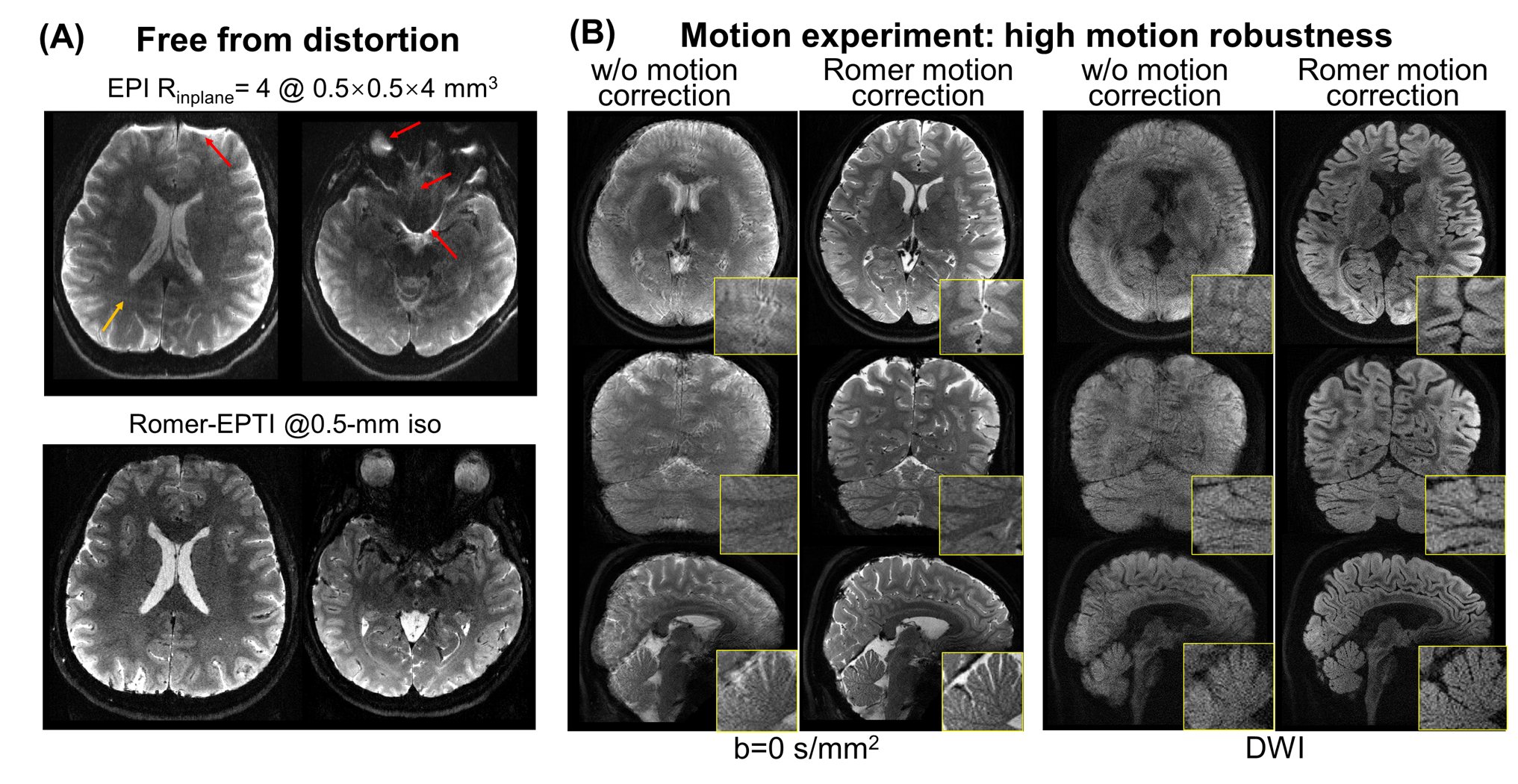
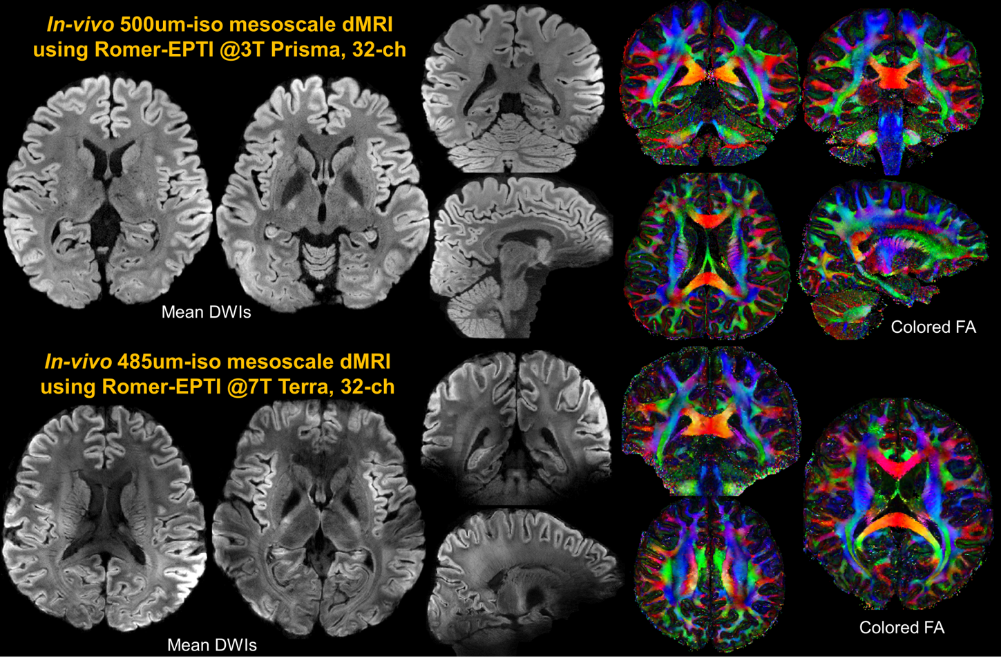
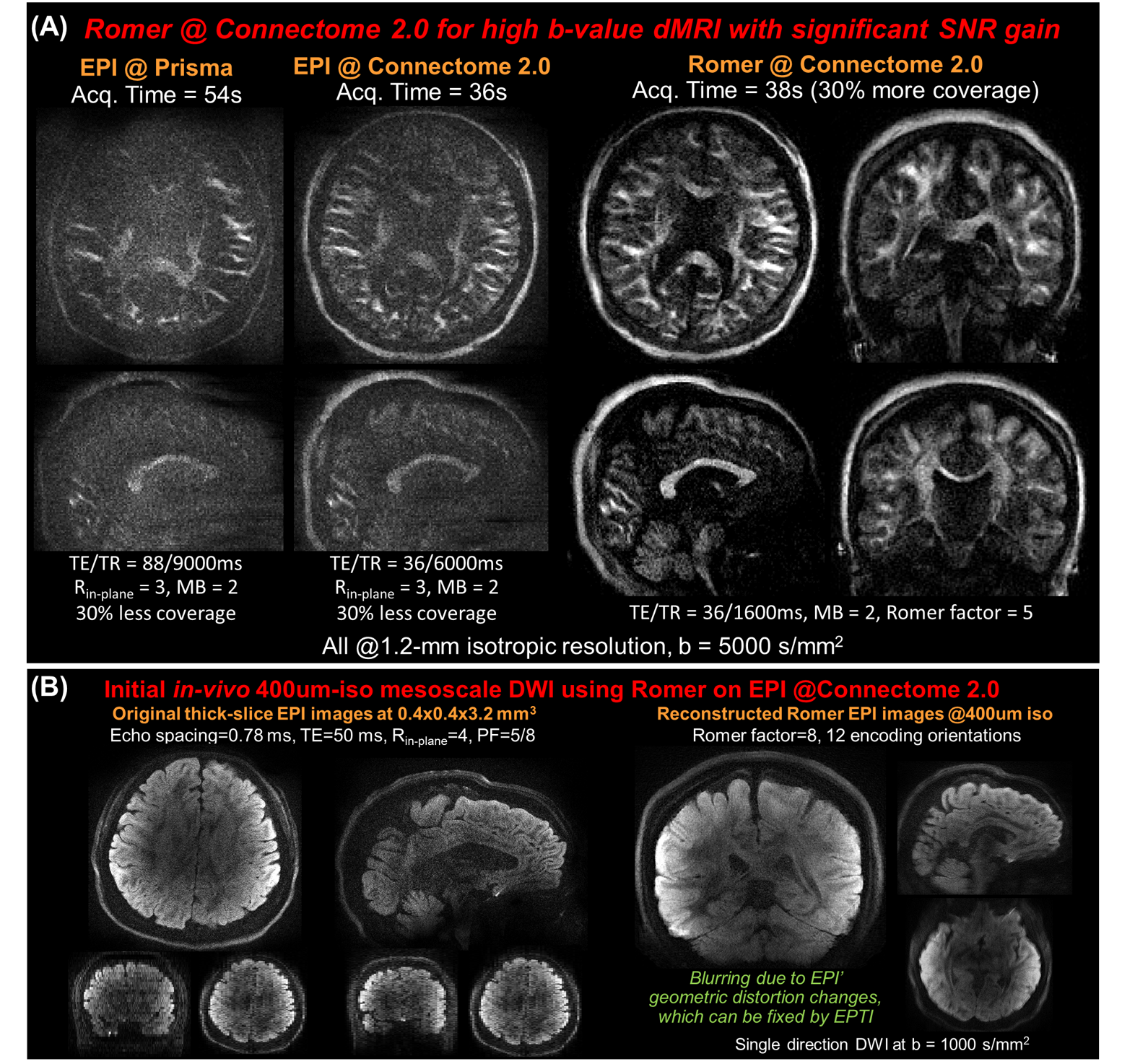
Fig.2A demonstrates Romer-EPTI provides distortion-free images at 500um compared to the severe distortions of EPI (even when using a high in-plane-acceleration of 4). Fig.2B highlights the high motion robustness of Romer-EPTI, as it successfully recovers detailed structures through motion-aware reconstruction. Romer-EPTI images from both 3T&7T clinical scanners reveal fine-scale anatomical structures in exceptional quality at 500um-iso and 485um-iso resolutions (Fig.3), characterized by their distortion-free, high SNR, and minimal motion and slab-boundary artifacts.Moving to C2.0-scanner, >2x shorter echo-spacings can be achieved, resulting in >40% g-factor reduction and SNR boost for EPTI when using high accelerations compared to 3T-Prisma. In Fig.5A, SNR was improved for EPI at b=5000s/mm2 and 1.2-mm resolution on C2.0 thanks to shortened TEs (36ms vs. 88ms). Using Romer, additional significant SNR gain was achieved from the SNR-efficient acquisition. Moreover, an initial experiment combining Romer-acquisition and conventional EPI achieves 400um-iso resolution. While Romer offers high SNR, image blurring results from the EPI readout, which will be fixed by EPTI in ongoing work.
Conclusion
We demonstrated Romer-EPTI’s ability to provide high-SNR, distortion-free, motion-robust in-vivo dMRI at mesoscale resolution on clinical scanners, and further enhanced its performance by taking advantage of the C2.0-scanner to achieve high-resolution high-b-value, and mesoscale dMRI at 400um-iso.Acknowledgements
This work was supported by the NIH BRAIN Initiative (U24NS129893), NIH (K99AG083056, U01EB026996, R01-EB019437, P41EB030006), and the instrumentation Grants (S10OD023637).References
[1] Song, A. W., Chang, H.-C., Petty, C., Guidon, A. & Chen, N.-K. Improved delineation of short cortical association fibers and gray/white matter boundary using whole-brain three-dimensional diffusion tensor imaging at submillimeter spatial resolution. Brain Connect. 4, 636–640 (2014).
[2] Chang HC, Sundman M, Petit L, Guhaniyogi S, Chu ML, Petty C, Song AW, Chen NK. Human brain diffusion tensor imaging at submillimeter isotropic resolution on a 3 Tesla clinical MRI scanner. Neuroimage. 2015 Sep 1;118:667-75.
[3] Setsompop K, Fan Q, Stockmann J, Bilgic B, Huang S, Cauley SF, Nummenmaa A, Wang F, Rathi Y, Witzel T, Wald LL. High‐resolution in vivo diffusion imaging of the human brain with generalized slice dithered enhanced resolution: Simultaneous multislice (g S lider‐SMS). Magnetic resonance in medicine. 2018 Jan;79(1):141-51.
[4] Wang F, Bilgic B, Dong Z, Manhard MK, Ohringer N, Zhao B, Haskell M, Cauley SF, Fan Q, Witzel T, Adalsteinsson E. Motion‐robust sub‐millimeter isotropic diffusion imaging through motion corrected generalized slice dithered enhanced resolution (MC‐gSlider) acquisition. Magnetic resonance in medicine. 2018 Nov;80(5):1891-906.
[5] Liao C, Stockmann J, Tian Q, Bilgic B, Arango NS, Manhard MK, Huang SY, Grissom WA, Wald LL, Setsompop K. High‐fidelity, high‐isotropic‐resolution diffusion imaging through gSlider acquisition with and T1 corrections and integrated ΔB0/Rx shim array. Magnetic resonance in medicine. 2020 Jan;83(1):56-67.
[6] Wu W, Poser BA, Douaud G, Frost R, In MH, Speck O, Koopmans PJ, Miller KL. High-resolution diffusion MRI at 7T using a three-dimensional multi-slab acquisition. NeuroImage. 2016 Dec 1;143:1-4.
[7] McNab JA, Polimeni JR, Wang R, Augustinack JC, Fujimoto K, Stevens A, Janssens T, Farivar R, Folkerth RD, Vanduffel W, Wald LL. Surface based analysis of diffusion orientation for identifying architectonic domains in the in vivo human cortex. Neuroimage. 2013 Apr 1;69:87-100.
[8] Clark CA, Le Bihan D. Water diffusion compartmentation and anisotropy at high b values in the human brain. Magnetic Resonance in Medicine: An Official Journal of the International Society for Magnetic Resonance in Medicine. 2000 Dec;44(6):852-9.
[9] Jensen JH, Helpern JA, Ramani A, Lu H, Kaczynski K. Diffusional kurtosis imaging: the quantification of non‐gaussian water diffusion by means of magnetic resonance imaging. Magnetic Resonance in Medicine: An Official Journal of the International Society for Magnetic Resonance in Medicine. 2005 Jun;53(6):1432-40.
[10] Jespersen SN, Kroenke CD, Østergaard L, Ackerman JJ, Yablonskiy DA. Modeling dendrite density from magnetic resonance diffusion measurements. Neuroimage. 2007 Feb 15;34(4):1473-86.
[11] Alexander DC, Hubbard PL, Hall MG, Moore EA, Ptito M, Parker GJ, Dyrby TB. Orientationally invariant indices of axon diameter and density from diffusion MRI. Neuroimage. 2010 Oct 1;52(4):1374-89.
[12] Fan Q, Nummenmaa A, Witzel T, Zanzonico R, Keil B, Cauley S, Polimeni JR, Tisdall D, Van Dijk KR, Buckner RL, Wedeen VJ. Investigating the capability to resolve complex white matter structures with high b-value diffusion magnetic resonance imaging on the MGH-USC Connectom scanner. Brain connectivity. 2014 Nov 1;4(9):718-26.
[13] Fan Q, Witzel T, Nummenmaa A, Van Dijk KR, Van Horn JD, Drews MK, Somerville LH, Sheridan MA, Santillana RM, Snyder J, Hedden T. MGH–USC Human Connectome Project datasets with ultra-high b-value diffusion MRI. Neuroimage. 2016 Jan 1;124:1108-14.
[14] McNab JA, Edlow BL, Witzel T, Huang SY, Bhat H, Heberlein K, Feiweier T, Liu K, Keil B, Cohen-Adad J, Tisdall MD. The Human Connectome Project and beyond: initial applications of 300 mT/m gradients. Neuroimage. 2013 Oct 15;80:234-45.
[15] Setsompop K, Kimmlingen R, Eberlein E, Witzel T, Cohen-Adad J, McNab JA, Keil B, Tisdall MD, Hoecht P, Dietz P, Cauley SF. Pushing the limits of in vivo diffusion MRI for the Human Connectome Project. Neuroimage. 2013 Oct 15;80:220-33.
[16] Huang SY, Nummenmaa A, Witzel T, Duval T, Cohen-Adad J, Wald LL, McNab JA. The impact of gradient strength on in vivo diffusion MRI estimates of axon diameter. Neuroimage. 2015 Feb 1;106:464-72.
[17] Zhang H, Schneider T, Wheeler-Kingshott CA, Alexander DC. NODDI: practical in vivo neurite orientation dispersion and density imaging of the human brain. Neuroimage. 2012 Jul 16;61(4):1000-16.
[18] Palombo M, Ianus A, Guerreri M, Nunes D, Alexander DC, Shemesh N, Zhang H. SANDI: a compartment-based model for non-invasive apparent soma and neurite imaging by diffusion MRI. Neuroimage. 2020 Jul 15;215:116835.
[19] Novikov DS, Jensen JH, Helpern JA, Fieremans E. Revealing mesoscopic structural universality with diffusion. Proceedings of the National Academy of Sciences. 2014 Apr 8;111(14):5088-93.
[20] Burcaw LM, Fieremans E, Novikov DS. Mesoscopic structure of neuronal tracts from time-dependent diffusion. NeuroImage. 2015 Jul 1;114:18-37.
[21] Fieremans E, Burcaw LM, Lee HH, Lemberskiy G, Veraart J, Novikov DS. In vivo observation and biophysical interpretation of time-dependent diffusion in human white matter. Neuroimage. 2016 Apr 1;129:414-27.
[22] Lee HH, Papaioannou A, Kim SL, Novikov DS, Fieremans E. A time-dependent diffusion MRI signature of axon caliber variations and beading. Communications biology. 2020 Jul 7;3(1):354.
[23] Dong Z, Polimeni JR, Wald LL, Wang F. Mesoscale distortion-free in-vivo dMRI at 7T using ROtating-view Motion-robust supEr Resolution EPTI (Romer-EPTI). In Proceedings of the 30th Annual Meeting of ISMRM, 2023. p. 0541.
[24] Wang F, Dong Z, Lee H, Huang SY, Polimeni JR, Wald LL. High-SNR whole-brain microstructure diffusion MRI using Romer-EPTI. In Proceedings of the 30th Annual Meeting of ISMRM, 2023. p. 0690.
[25] Dong Z, Polimeni J, Wald LL, Wang F. SuperRes-EPTI: in-vivo mesoscale distortion-free dMRI at 500μm-isotropic resolution using short-TE EPTI with rotating-view super resolution. In Proceedings of the 30th Annual Meeting of ISMRM, 2022, p. 3488.
[26] Huang SY, Witzel T, Keil B, Scholz A, Davids M, Dietz P, Rummert E, Ramb R, Kirsch JE, Yendiki A, Fan Q. Connectome 2.0: Developing the next-generation ultra-high gradient strength human MRI scanner for bridging studies of the micro-, meso-and macro-connectome. Neuroimage. 2021 Nov 1;243:118530.
[27] Wang F, Dong Z, Reese TG, Bilgic B, Katherine Manhard M, Chen J, Polimeni JR, Wald LL, Setsompop K. Echo planar time‐resolved imaging (EPTI). Magnetic resonance in medicine. 2019 Jun;81(6):3599-615.
[28] Dong Z, Wang F, Reese TG, Bilgic B, Setsompop K. Echo planar time‐resolved imaging with subspace reconstruction and optimized spatiotemporal encoding. Magnetic resonance in medicine. 2020 Nov;84(5):2442-55.
[29] Dong, Z. et al. Tilted‐CAIPI for highly accelerated distortion‐free EPI with point spread function (PSF) encoding. Magn. Reson. Med. 81, 377–392 (2019).
[30] SNR-efficient distortion-free diffusion relaxometry imaging using accelerated echo-train shifted echo-planar time-resolving imaging (ACE-EPTI). Dong Z, Wang F, Wald L, Setsompop K. Magn Reson Med. 2022 Jul;88(1):164-179.
[31] Shilling RZ, Robbie TQ, Bailloeul T, Mewes K, Mersereau RM, Brummer ME. A super-resolution framework for 3-D high-resolution and high-contrast imaging using 2-D multislice MRI. IEEE transactions on medical imaging. 2008 Oct 31;28(5):633-44.
[32] Plenge E, Poot DH, Bernsen M, Kotek G, Houston G, Wielopolski P, van der Weerd L, Niessen WJ, Meijering E. Super‐resolution methods in MRI: can they improve the trade‐off between resolution, signal‐to‐noise ratio, and acquisition time?. Magnetic resonance in medicine. 2012 Dec;68(6):1983-93.
[33] Van Steenkiste, G., Jeurissen, B., Veraart, J., den Dekker, A.J., Parizel, P.M., Poot, D.H.J. and Sijbers, J. (2016), Super-resolution reconstruction of diffusion parameters from diffusion-weighted images with different slice orientations. Magn. Reson. Med., 75: 181-195. https://doi.org/10.1002/mrm.25597
[34] Vis G, Nilsson M, Westin CF, Szczepankiewicz F. Accuracy and precision in super-resolution MRI: Enabling spherical tensor diffusion encoding at ultra-high b-values and high resolution. NeuroImage. 2021 Dec 15;245:118673.
Figures