2430
Multi-shot 3D diffusion MRI sequence for a fast and high-resolution imaging at 3T1McGill University, Montreal, QC, Canada, 2McConnell Brain Imaging Center, Montreal Neurological Institute, McGill University, Montreal, QC, Canada, 3Department of Neurology and Neurosurgery, McGill University, Montreal, QC, Canada
Synopsis
Keywords: Diffusion Acquisition, Brain
Motivation: Several multi-shot 2D acquisition techniques have been developed to improve the resolution of diffusion MRI (dMRI) of the brain, but require long scan times.
Goal(s): To develop a 3D dMRI sequence with improved SNR efficiency to achieve high-resolution scans in a reasonable time.
Approach: We implemented a 3D spin echo sequence with an inversion pulse before the excitation to improve SNR at short repetition times and a TURBINE readout trajectory. Projection-based image reconstruction is employed to avoid artifacts caused by shot-to-shot phase errors.
Results: Phantom and human scans at 3T show an improvement in SNR efficiency over 2D dMRI.
Impact: We designed a multi-shot 3D dMRI sequence that is SNR efficient and robust to motion artifacts between shots. This sequence will enable high-resolution diffusion-weighted imaging of the whole brain in short scan times.
Introduction
Several imaging techniques have been developed to improve the signal-to-noise ratio (SNR) and resolution of diffusion-weighted MRI (dMRI) [e.g., 1-3]. Most of these 2D multi-slice, multi-shot techniques require long scan times, limiting their use in patients or in advanced protocols where many different diffusion encodings are required. 3D sequences have several advantages over 2D, including a higher SNR efficiency at high-resolution. However, the use of shorter repetitions times (TR) in 3D imaging saturates the signal. Most 3D dMRI techniques use a navigator and/or cardiac gating to correct phase errors between shots which lengthen the scan time [e.g., 4], and advanced offline image reconstruction, which further limit their use. We designed and implemented a fast, multi-shot 3D dMRI sequence that has a similar scan time to 2D DWI with multi-band acceleration and fast image reconstruction.Methods
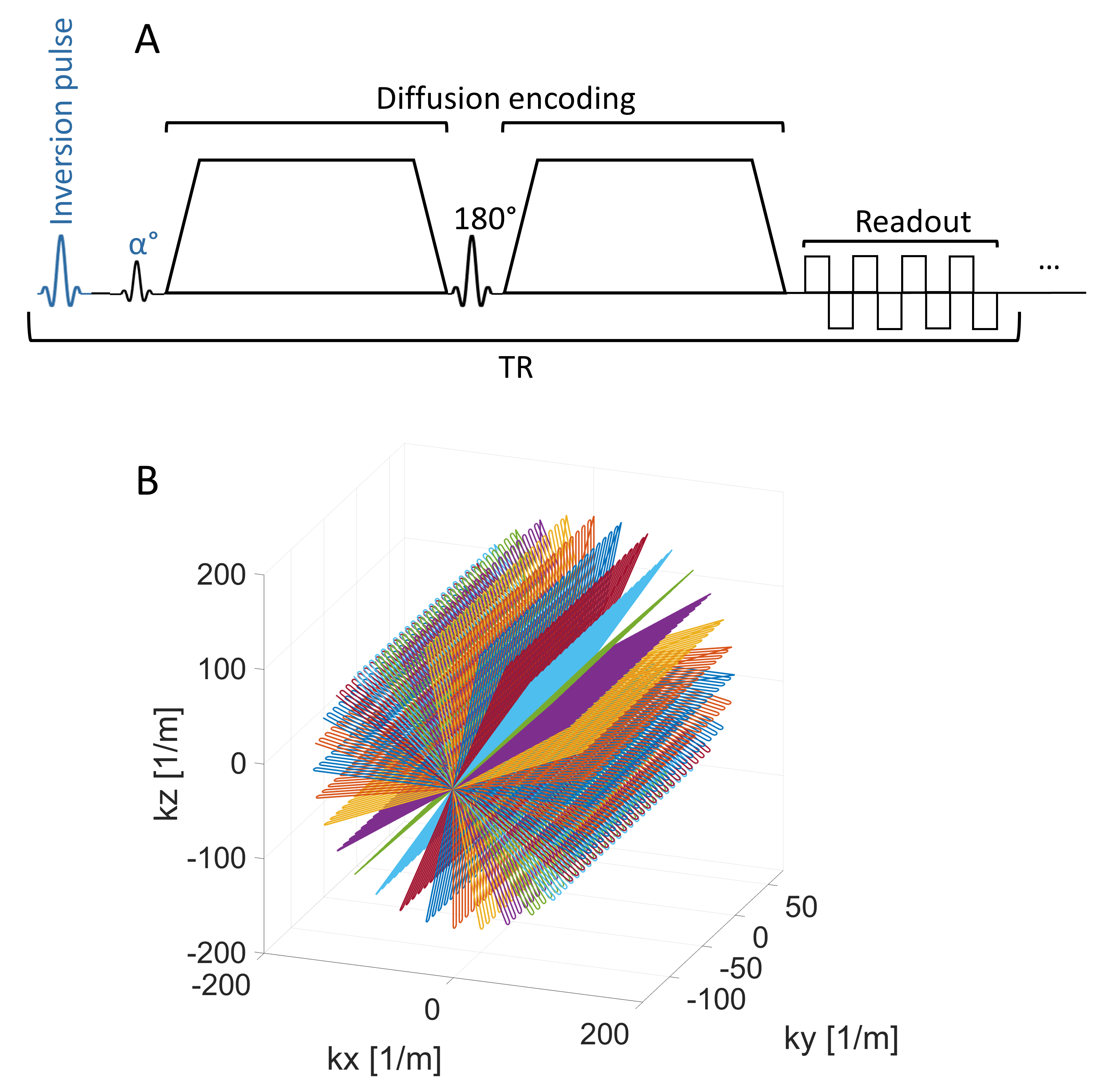
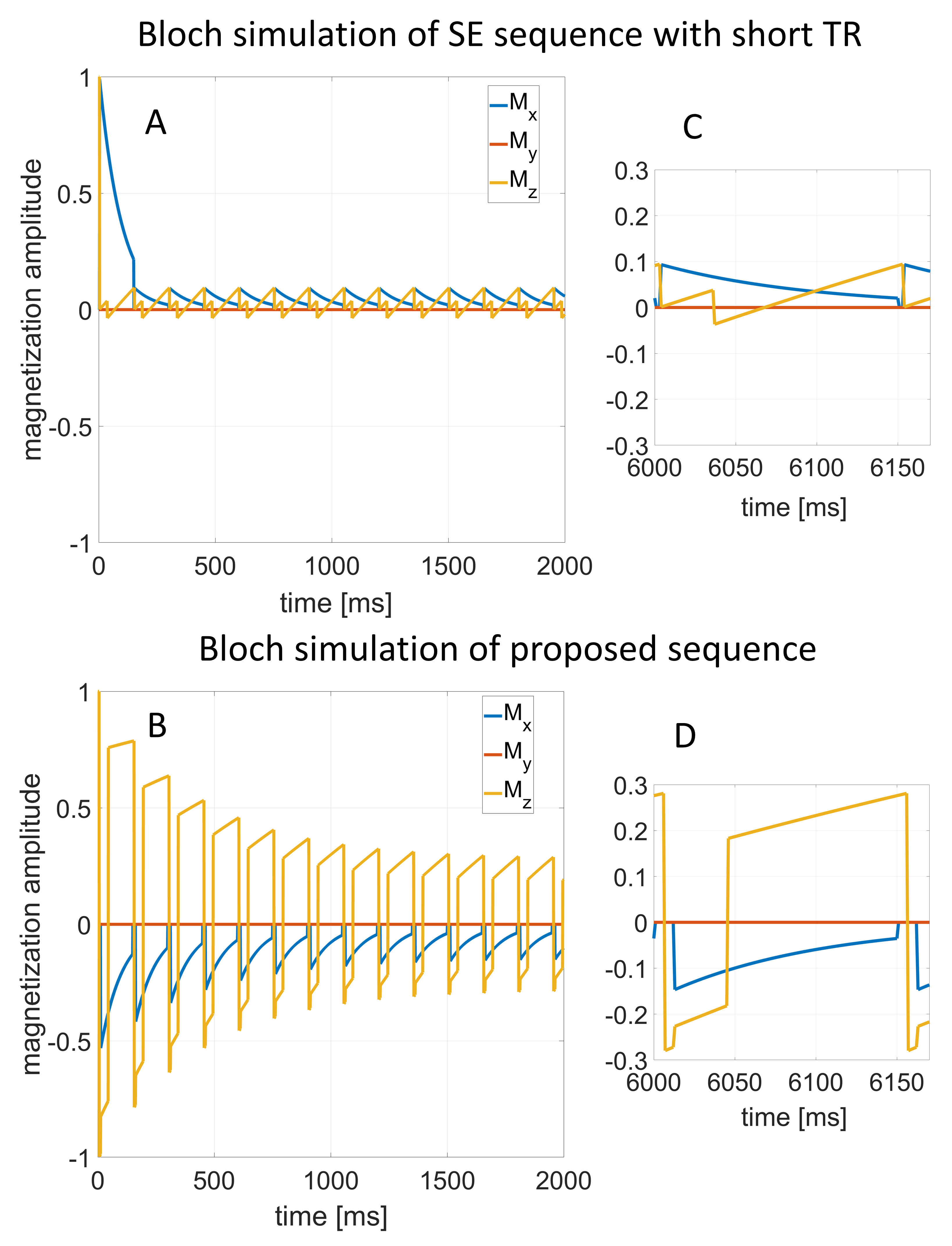
The proposed 3D diffusion sequence diagram is shown in Figure 1A. The longitudinal magnetization is inverted before the excitation to reduce signal saturation, and the excitation flip angle is reduced. Bloch simulations were used to quantify the increase in steady-state signal compared to a typical spin-echo (SE) sequence. 3D k-space is acquired using a Trajectory Using Radially Batched Internal Navigator Echoes (TURBINE) trajectory [4] in multiple shots using a short TR (Figure 1B). A complete 3D volume is acquired within a time similar to the TR of a 2D dMRI sequence. After phase correction of the raw data [5], each projection is reconstructed separately from a k-space plane using the fast Fourier transform. Then, all projections are combined using back-projection (i.e., a radon transform with Hanning filtering to reduce ringing) to reconstruct the 3D image. The use of EPI in TURBINE allows us to use eddy and topup post-processing tools to correct for eddy current and B0 inhomogeneity effects [6]. To demonstrate feasibility, a phantom and a human subject were scanned using the 3D sequence on a 3T Siemens PrismaFit scanner. The phantom and human subject were scanned using the 3D dMRI EPI sequence with the following parameters: FOV= 240 mm3, resolution= 2 mm3, bandwidth-per-pixel=1666 Hz, TE=74 ms, TR=151 ms, number of shots = 190, R=1, partial Fourier factor=6/8, and b-value=0, 1000, and 1500 s/mm2 in a single direction. A 2D dMRI EPI sequence with the same parameters, a TR of 5500 ms, and a multi-band factor of 3 was run for comparison. Scan time per volume was 28 and 16.5 seconds for 3D and 2D dMRI sequences, respectively. To calculate SNR-per-unit-time, 15 repetitions were acquired for both phantom and human scans.Results
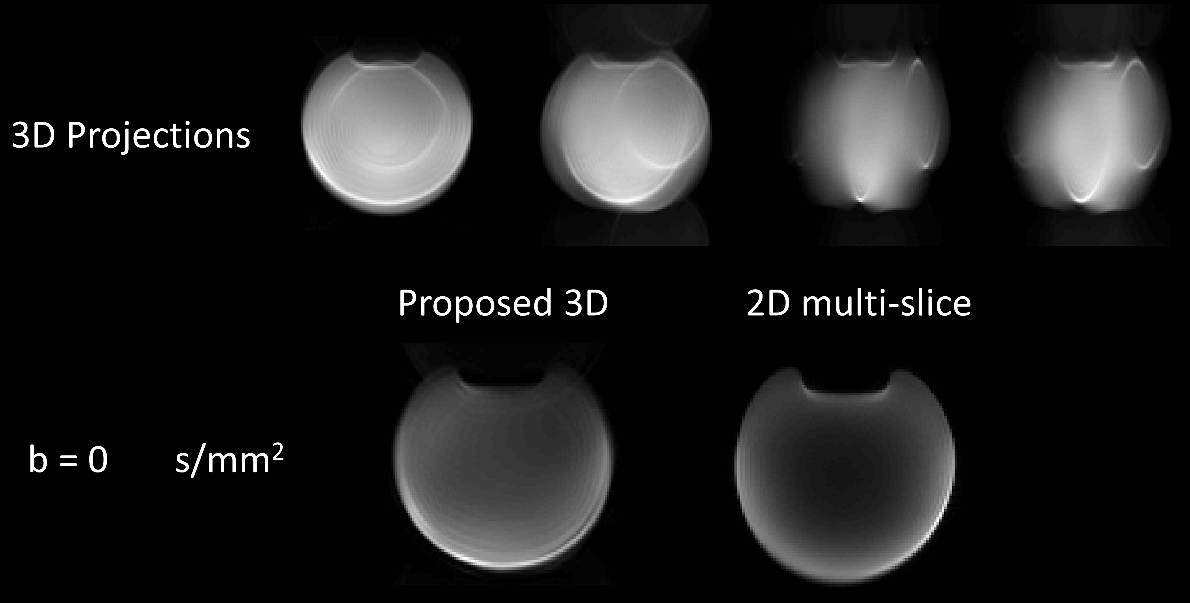
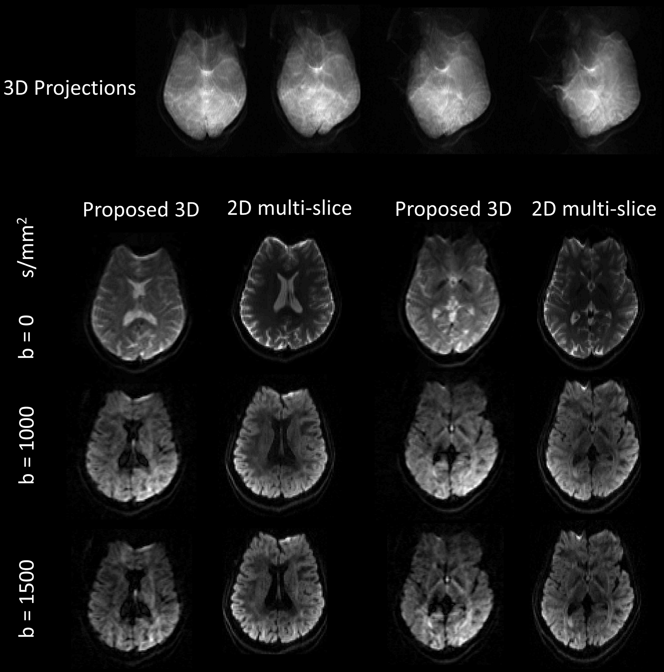
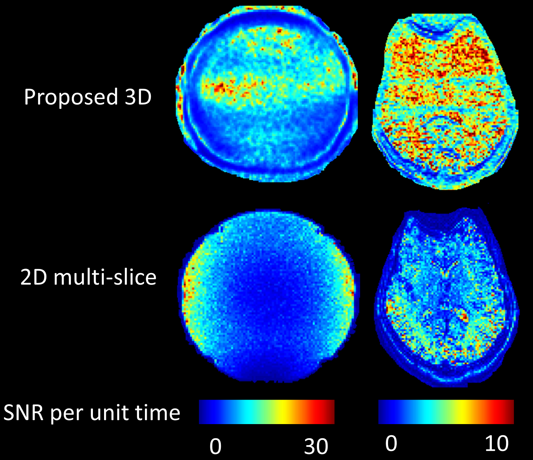
The steady state signal derived using the Bloch equations for the SE sequence with 90° flip angle is ~9.3%, while using the proposed sequence the steady state signal reaches to ~14.7%, corresponding to a ~58% increase (Figure 2). Reconstructed projections and images of the phantom and human brain at different b-values are shown in Figures 3 and 4, respectively. There is a difference in tissue contrast between the 2D and 3D images due to the enhanced T1-weighting resulting from the short TR in the 3D sequence. Artifacts caused by B0 non-uniformity are similar in both sequences and can be corrected. The 3D image quality can be improved by using a more advanced back-projection reconstruction technique [7]. The 3D sequence has a higher SNR efficiency throughout the brain (Figure 5).Discussion
The inclusion of the inversion pulse in the 3D sequence significantly increased the steady state signal for short TRs, which allows fast imaging without reduction in SNR due to signal saturation. A major concern in multi-shot dMRI is the extreme sensitivity to motion, which causes artifacts and signal loss. By using the TURBINE trajectory and back-projection based reconstruction, phase inconsistencies are avoided since the 3D images are reconstructed from the magnitude of the projection images. Here, we used a simple technique for image reconstruction that caused some blurring in the reconstructed images. The use of more advanced techniques will improve image quality [7]. We did not implement k-space undersampling in this proof-of-concept. Future work will include undersampling in-plane and across angles to shorten the echo time and total scan time, respectively, with GRAPPA image reconstruction for high-resolution imaging.Conclusion
We propose a 3D spin echo sequence with an inversion pulse prior to excitation and a TURBINE readout trajectory to enhance the SNR efficiency of dMRI. The Bloch simulations and preliminary phantom and human images suggest that this 3D sequence is an ideal candidate for high-resolution dMRI of the brain at 3T within reasonable scan times.Acknowledgements
Authors would like to thank Marcus Couch (Siemens Collaboration Scientist) and Ilana Leppert (McConnell Brain Imaging center) for their technical support, and David Costa, Ronaldo Lopez, and Soheil Mollamohseni Quchani for helping with human scans. This work was supported by: the Montreal Neurological Institute, the Brain Canada Foundation, Healthy Brains for Healthy Lives, Fonds de Recherche du Québec - Santé, Fonds de Recherche du Québec - Nature et technologie, and the Natural Sciences and Engineering Research Council of Canada.References
[1] Y. Lee et al., “On the signal-to-noise ratio benefit of spiral acquisition in diffusion MRI,” Magn Reson Med, Dec. 2020, doi: 10.1002/mrm.28554.
[2] T.-K. Truong and A. Guidon, “High-Resolution Multi-Shot Spiral Diffusion Tensor Imaging with Inherent Correction of Motion-Induced Phase Errors,” Magn Reson Med, vol. 71, no. 2, pp. 790–796, Feb. 2014, doi: 10.1002/mrm.24709.
[3] Haldar JP et al., Fast submillimeter diffusion MRI using gSlider-SMS and SNR-enhancing joint reconstruction. Magn Reson Med. 2020 Aug;84(2):762-776. doi: 10.1002/mrm.28172. Epub 2020 Jan 10. PMID: 31919908; PMCID: PMC7968733.
[4] McNab, J. A., Gallichan, D., & Miller, K. L. (2010). 3D steady‐state diffusion‐weighted imaging with trajectory using radially batched internal navigator echoes (TURBINE). Magnetic Resonance in Medicine: An Official Journal of the International Society for Magnetic Resonance in Medicine, 63(1), 235-242.
[5] Heid, O. (2000). U.S. Patent No. 6,043,651. Washington, DC: U.S. Patent and Trademark Office.
[6] Andersson, J. L., Skare, S., & Ashburner, J. (2003). How to correct susceptibility distortions in spin-echo echo-planar images: application to diffusion tensor imaging. Neuroimage, 20(2), 870-888.
[7] Zhang, S., & Xia, Y. (2021). CT image reconstruction algorithms: A comprehensive survey. Concurrency and Computation: Practice and Experience, 33(8), e5506.
Figures