2426
Deep learning constrained compressed sensing reconstruction for diffusion-weighted imaging in patients with breast cancers: a plot study1Department of Radiology, West China Hospital, Sichuan University, Chengdu, China, Cheng du, China, 2Clinical Science, Philips Healthcare,Chengdu,China, Chengdu, China
Synopsis
Keywords: Diffusion Reconstruction, Diffusion/other diffusion imaging techniques, Breast cancer, Deep learning
Motivation: The challenges such as image quality and long scan time limitations have degraded the diffusion-weighted imaging (DWI) of breast cancer in clinical practice.
Goal(s): This study aims to investigate the application of deep learning constrained compressed sensing (CS) reconstruction in DWI to overcome existing limitations.
Approach: Quantitative and qualitative image quality of DWI and value apparent diffusion coefficient (ADC) of using CS (DWI-CS) and deep learning constrained CS (DWI-DLCS) were compared.
Results: The results of DWI-DLCS exhibited better contrast, contrast-to-noise ratio (CNR), lesion detectability and diagnostic confidence. There were no differences regarding the signal intensity values of the apparent diffusion coefficient (ADC).
Impact: Our study showcases the potential of deep learning constrained reconstruction in enhancing the quality and efficiency of DWI. This approach offers a promising clinical implementation to obtain high-quality DWI images while reducing scan time.
Introduction
Breast cancer is one of the most common types of cancer among women. It accounts for most new cancer diagnoses and cancer-related deaths among women. Early detection and accurate diagnosis are crucial for improving breast cancer patient outcomes. Diffusion-weighted imaging (DWI) enhances the specificity in multiparametric breast MRI that measures the rate of microscopic water molecule diffusion in tissues, thus reflecting tissue cellularity and cell membrane integrity [1,2]. However, its examination time and related image quality are considered as critical issue in clinics. The compressed sensing (CS) reconstruction is an approach to reduce scan time [3]. Despite promising results, DWI with CS still suffers from some loss of image detail and reduced image quality when it is conducted large acceleration factors due to insufficient noise removal with CS [4]. Recently, deep-learning-based reconstruction involves the use of neural networks with multiple layers to learn complex representations from rawdata, which have shown enormous potential and highly effective to optimize the image quality [5]. Therefore, this study aims to explore the feasibility of a novel deep-learning based compressed sensing reconstruction algorithm on breast DWI to improve image quality.Methods
This study was approved by the institutional ethics committee and written informed consent was obtained. Twenty-four subjects were enrolled. The breast MRI examination was performed on a Ingenia Elition 3.0T X system (Philips Healthcare, Best, The Netherlands) equipped with an 8-channel bilateral breast coil. Bilateral breast imaging protocol consisted of fat-suppressed T2-weighted sequence, DWI, non-fat-suppressed T1-weighted sequence and fat-suppressed 3D T1weighted imaging before and after contrast agent injection. DWI sequence with compressed sensing acceleration factor 2.5 was performed pre-contrast with two b values, 0 s/mm2 and 1000 s/mm2, for creating apparent diffusion coefficient (ADC) maps. Final DWI images with deep learning reconstruction (DWI-DLCS) and without (DWI-CS) were generated immediately on the scanner when the raw data was acquired. Observers reviewed the images and drew the region of interest randomly and blindly. The contrast, signal-to-noise ratio (SNR), contrast-to-noise ratio (CNR), tumor ADC values (ADCtumor) and normal tissue ADC values (ADCnormal) were quantitatively evaluated; the overall image quality, sharpness, artifacts, lesion detectability, and diagnostic confidence were qualitatively assessed using 5-point scale. The statistics analysis was performed using SPSS version 23.0 and GraphPad Prism. The quantitative parameters were compared using paired t-test. The agreement of ADC value was tested using Bland-Altman-Plot. The qualitative parameters were compared using Wilcoxon sighed rank test.Results
As shown in Figure 1, the lesion contrast and CNR was significantly higher in DWI-DLCS than DWI-CS (lesion contrast: 3.11 ± 1.74 vs. 2.80 ± 1.31, p = 0.03; CNR: 2.72 ± 1.27 vs. 2.30 ± 1.05, p = 0.02). The SNR was similar in the DWI-DLCS (5.75 ± 3.86 vs. 4.62 ± 1.43, p = 0.2). The ADC values of the tumor (1.23 ± 0.52 vs. 1.21 ± 0.49, p = 0.10) and normal tissue (1.49 ± 0.39 vs. 1.45 ± 0.44, p = 0.13) showed no difference between the two sequences. From the Bland-Altman-Plot, the measurable ADC value showed no significant bias (ADCtumor: 0.04 and ADCnormal: 0.03, respectively) between DWI-DLCS and DWI-CS (Figure 2). As Figure 3 showed, there were no differences in overall image quality and artifact scores between the two sequences (all p>0.05). The scores of lesion detectability (4.84 ± 0.37 vs. 4.58 ± 0.51, p = 0.03) and diagnostic confidence (4.63 ± 0.50 vs. 4.26 ± 0.56, p = 0.02) in DWI-DLCS were higher than DWI-CS (Figure 4). However, the sharpness of DWI -DLCS was significantly lower than that of DWI-CS (4.00 ± 0.58 vs 4.74 ± 0.45, p < 0.01).Discussion
In the present study, DWI-DLCS was compared with DWI-CS in breast cancer. The results showed that DWI-DLCS had a higher contrast, CNR, scores of lesion detectability and diagnostic confidence, which indicated the deep learning algorithm resulted in improved image quality and reduced noise and artifacts. This DWI-DLCS can improve the accuracy and sensitivity of DWI in detecting and characterizing breast cancer lesions. Although the sharpness of DWI-DLCS was lower, it does not affect the detection of lesions. The value of ADC kept consistent between DWI-DLCS and DWI-CS. Similar to that observed in previous studies [5,6], deep learning reconstruction can improve the detection and diagnostic accuracy of abnormal regions by radiologists.Conclusion
This study demonstrated the feasibility and effectiveness of deep learning constrained compressed sensing reconstruction for improving DWI image quality of breast cancers. Further research and validation on larger patient cohorts are warranted to establish the clinical utility and generalizability of deep learning reconstruction in DWI for breast cancer diagnosis and management.Acknowledgements
We thank Hans Peeters (Philips, Best Netherlands) for the technical help of this study.References
1. Ei Khouli RH, Jacobs MA, Mezban SD, et al. Diffusion-weighted imaging improves the diagnostic accuracy of conventional 3.0-T breast MR imaging. Radiology 2010;256:64-73.
2. N.S. White, C. McDonald, N. Farid, et al., Diffusion-weighted imaging in cancer: physical foundations and applications of restriction spectrum imaging, Cancer Res 2014; 74:4638–4652.
3. Wessling D, Gassenmaier S, Olthof SC, et al. Novel deep-learning-based diffusion weighted imaging sequence in 1.5 T breast MRI. Eur J Radiol. 2023;166:110948.
4. Y. Kuroki, K. Nasu, Advances in breast MRI: diffusion-weighted imaging of the breast, Breast Cancer 2008; 15 (3): 212–217.
5. Pezzotti N, Yousefi S, Elmahdy MS, et al. An Adaptive Intelligence Algorithm for Undersampled Knee MRI Reconstruction. IEEE Access 2020; 8:204825-204838
6. Kapsner LA, Balbach EL, Folle L, et al. Image quality assessment using deep learning in high b-value diffusion-weighted breast MRI. Sci Rep 2023; 29;13(1):10549.
Figures
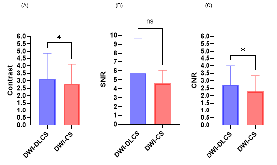
Figure 1. The comparison of contrast (A), SNR (B) and CNR (C) between DWI-DLCS and DWI-CS.

Figure 2. The Bland-Altman-Plot of the agreement of ADCtumor value (A) and ADCnormal value (B) between DWI-DLCS and DWI-CS
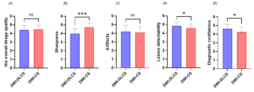
Figure 3. The comparison of the overall image quality (A), sharpness (B), artifacts (C), lesion detectability (D), and diagnostic confidence (E) between DWI-DLCS and DWI-CS.
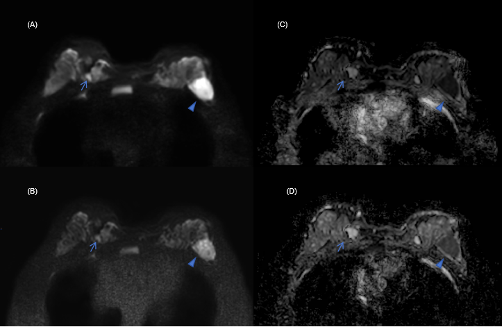
Figure 4. Breast MR images in a 30-year-old woman with two invasive ductal carcinomas with 3 cm (arrowheads) and 0.7 cm (arrows) in the breast. (A) b = 1000 images of DWI-DLCS, (B) b = 1000 images of DWI-CS, (C) ADC map of DWI-DLCS, (D) ADC map of DWI-CS. Lesion contrast on DWI-DLCS was better than that of DWI-CS. Especially for the small lesion (arrows), diagnostic confidence was higher on DWI-DLCS.