2425
Efficient High-Resolution Cardiac DTI with Spiral Keyhole Sampling (SPIK) and Joint Spatial-Angular Sparse Representation1School of Computing, University of Leeds, Leeds, United Kingdom, 2Leeds Institute of Cardiovascular and Metabolic Medicine (LICAMM), School of Medicine, University of Leeds, Leeds, United Kingdom, 3School of Health Sciences, University of Manchester, Manchester, United Kingdom
Synopsis
Keywords: Simulation/Validation, Pulse Sequence Design, Cardiac Diffusion MRI
Motivation: In cardiac diffusion MRI, spatial resolution is often limited by the available SNR and scan time. However, reducing voxel size enables precise microstructure characterisation.
Goal(s): To develop and evaluate the feasibility of a new sampling/reconstruction scheme that takes advantage of similarities between diffusion-weighted images in (k,q) space in order to provide high spatial resolution with significantly reduced scan times.
Approach: A new spiral-based sampling scheme is proposed and its benefits when combined with an efficient reconstruction is demonstrated through simulations.
Results: Evaluations showed that the proposed technique provides high-quality diffusion parameter maps comparable to fully sampled reference data with RMSEs less than 4%.
Impact: We introduce a novel sampling technique for accelerating high-resolution cardiac diffusion tensor imaging (cDTI). Harnessing the improved spatial resolution and scan time reduction has the potential to make significant contributions to practicability of more complex diffusion models in the heart.
Introduction
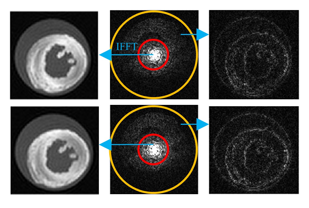
Cardiac diffusion tensor imaging (cDTI) has emerged as a valuable tool for the non-invasive assessment of myocardial microstructure in health and disease. However, the need for high spatial and directional resolution results in long scan times, which is prohibitive for its use in clinical practice. The fact that (i) the DW images are acquired from the same anatomical structure and (ii) the prior knowledge that the contrast (e.g., diffusion) information is mainly encoded in the central k-space can be exploited to make the cDTI data acquisition more efficient. We can strategically allocate our limited scan time to prioritise the acquisition of data in the distinct/unique k-space regions of the DW images, and at the same time minimise the acquisition of redundant data from similar areas, where information related to anatomy is encoded (Figure 1). Specifically, similarities can be leveraged to achieve higher sparsity levels in the data representation. Here, we investigate the feasibility of a new joint k-q sampling scheme, referred to as Spiral with Keyhole (SPIK), to enable high-resolution cDTI. We present the advantage of combining SPIK sampling with efficient reconstruction using a sparse dictionary of the joint k-q space. Simulations are performed to ensure that the diffusion information is sufficiently preserved and that the DTI parameter maps are reconstructed with high accuracy.Methods
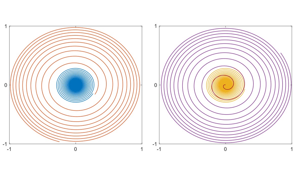
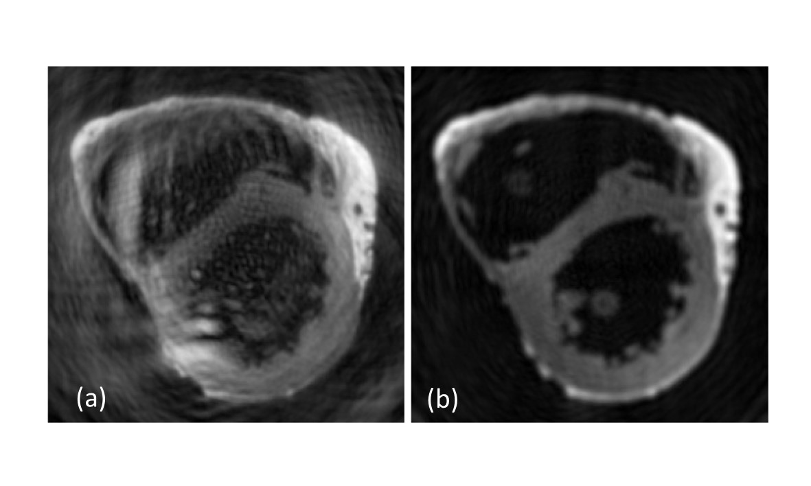
Inspired by the keyhole imaging technique [1], the proposed SPIK sampling utilises a customised (optimised) variable density (VD) spiral to densely capture the central k-space of every DW image. Higher spatial frequencies are sampled in a segmented manner with a reversed VD spiral such that the entire periphery of the k space is fully covered for every fifth DW scan, as illustrated in Figure 2. The overlapped region of the two spirals (depicted in red in Figure 2) can potentially be used to account for possible phase inconsistencies and were discarded for the reconstruction. The configuration of the VD spiral design parameters and the radius of the keyhole region were carefully optimised through pilot experiments (not shown here) to yield the least artifacts and RMSE in the DW images (illustrated in Figure 3). We used a new generic parametrisation for the spiral which takes into account the field-of-view, desired resolution, and gradient hardware specifications, making it easily adaptable to MRI systems and various datasets.The DW signal $$$S_{x \times q}$$$ can be represented with a higher sparsity in the combined spatial ($$$x$$$) and diffusion-directional ($$$q$$$) domain through an appropriate joint spatial-angular dictionary $$$\Phi$$$ [2]. We consider the separability of the dictionary to reduce computational complexity. The reconstruction problem is formulated in matrix form as
$$ C^{*} = \arg \min_{C} \| \mathcal{A}(\Psi^T C \Gamma) - B \|_{F}^2 + \lambda \|C\|_1 $$
where $$$\mathcal{A}$$$ is the encoding matrix, $$$B$$$ is the undersampled measurement matrix, $$$C$$$ denotes the spatial-angular coefficients, and $$$\Psi^T$$$ and $$$\Gamma$$$ are sparse dictionaries of spatial and angular basis functions, respectively. We used a modified FISTA algorithm to solve Eq. 1. We defined the angular dictionary using singular value decomposition (SVD) of the magnitude of the initial reconstruction. For the choice of spatial dictionary, the spatial Haar wavelets were used. We conducted retrospective experiments on three DTI datasets from an isolated human [3] and two rat hearts, comprising 15, 61, and 27 DW images, respectively. The raw k-space data were retrospectively gridded onto the proposed SPIK sampling, and then the images were recovered through the proposed reconstruction. We used typical values of hardware specifications for both clinical (in human data) and preclinical (in rat data) settings, and the readout duration $$$T_{ro}$$$ was set to 15 ms.
Results
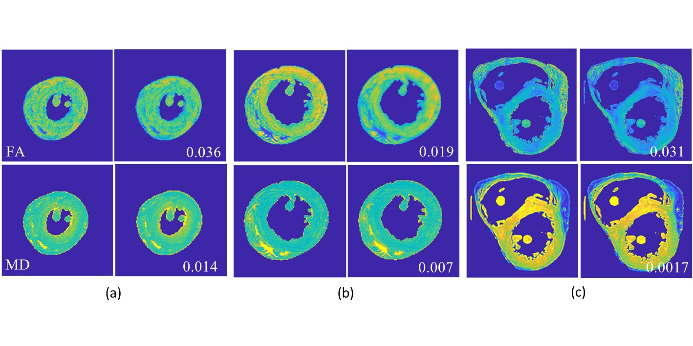
Figure 4 shows the FA and MD maps, obtained from human data (a), and the two rat data (b,c) retrospectively resampled onto the SPIK trajectory and reconstructed by the proposed method. The blurred structures were effectively recovered through joint reconstruction. The mean RMSE in the reconstructed DW images is improved by about 40% compared to the initial reconstruction using regridding, leading to enhanced diffusion parameter maps. We observed that the choice of the angular dictionary can severely affect the reconstruction quality, and more sophisticated dictionaries could lead to higher accuracy. The SPIK sampling allows for efficiently capturing the image information without severe artifacts.Conclusion
The proposed approach represents a novel sampling strategy for cDTI that considerably reduces the scan time while preserving the spatial resolution. The method can benefit more complex diffusion weighting schemes, such as multishell and DSI and may facilitate practical applications. Experimental verification of the proposed approach and extension to more complex diffusion models is subject to future work.Acknowledgements
AFF and JES joint senior authors. This work received funding from (BQ-MINDED H2020-MSCA-ITN-2017-764513). IT acknowledges funding from British Heart Foundation (PG/19/1/34076).References
1. Zaitsev et al. Magn. Reson. Med. 2001;45:109–117.
2. Schwab et al. Med. Image Anal. 2018;48:25-42.
3. Helm, et al. Magn. Reson. Med., 2005;54:850–859.
Figures