2422
Whole-Brain Mesoscale 0.7 millimeter-Isotropic-Resolution Diffusion MRI at Ultra-High Fields: The JETS-NAViEPI Approach.1Artificial Intelligence in Biomedical Engineering, Friedrich-Alexander-University Erlangen-Nürnberg, Erlangen, Germany, 2Siemens Healthcare GmbH, Erlangen, Germany, 3Institute of Radiology, University Hospital Erlangen, Friedrich-Alexander-Universität Erlangen-Nürnberg (FAU), Erlangen, Germany
Synopsis
Keywords: DWI/DTI/DKI, High-Field MRI
Motivation: Diffusion weighted MRI (DWI) may blur structures due to limited resolution. Increasing to submillimeter resolution reduces partial volume effects but sacrifices SNR.
Goal(s): We seek to demonstrate submillimeter isotropic DWI at 7T in 3 minutes, and enhanced high-resolution DTI maps.
Approach: Data acquisition is done via NAViEPI, enforcing consistent echo spacing between the imaging and the navigator echo, rendering minimal distortion mismatch. Image reconstruction is done using JETS, benefiting from complementary k-q-space sampling and regularization.
Results: We show a 3-scan trace acquisition with a scan time of 3:19 minutes, and offer high-resolution DTI measurement leading to Mean Diffusivity and colored Fractional Anisotropy maps.
Impact: This work demonstrates submillimeter isotropic diffusion MRI at ultra-high field 7T with improved image quality and reduced scan duration. The JETS-NAViEPI method has the potential to advance high-resolution diffusion MRI, enhancing both spatial and temporal resolution.
Introduction
Diffusion MRI plays an important role in understanding the microstructure of the brain. These structures are very small and therefore regions with more complex fiber arrangements such as crossing, fanning and branching are in a single voxel. Increasing the resolution to submillimeter-isotropic has shown to reduce partial volume effects 1-4 and it can also help detect small micro infarcts in cerebral small vessel disease5. Moreover, higher resolution has shown to improve brain parcellation6-8.However, increasing spatial resolution comes with decreasing signal-to-noise ratio which limits the ability to reconstruct the fiber tracks9,10. Often times, high resolution has been achieved at the expense of scan time or by using thick slices. Increasing the SNR would be possible by utilizing the dependence of SNR on the strength of B0. Imaging at 7 T has the potential to provide the necessary SNR, but it comes at the cost of artifacts from phase variations caused by B0 inhomogeneity.
In this work we show submillimeter isotropic DWI at ultra-high field 7 T with a scan time of little over 3 min as well as high resolution DTI parameter maps.
Methods
We used an adapted navigator based interleaved EPI (iEPI) method originally developed by Tan et al.11 The primary objective of Joint Reconstruction for Shift-Encoded Navigator-based Interleaved Echo Planar Imaging (JETS-NAViEPI) is to enforce consistent effective echo spacing (ESP) between the imaging and the navigator echos. By achieving this, NAViEPI mitigates distortion discrepancies between these two echos, simplifying the correction of shot-to-shot phase variations and enabling undersampled iEPI acquisition. To maintain image quality and reduce distortions, we introduced ky-shift encoding along the diffusion encoding direction. This approach allows for more comprehensive data sampling in k-q-space. Combined with a joint reconstruction method with overlapping locally low-rank regularization, implemented with integrated SigPY12 and PyTorch features, this reduces noise and artifacts by exploiting signal redundancies along the diffusion encoding directions. The coil sensitivity estimation was done using ESPIRIT13. NAViEPI facilitates the acquisition of a large number of shots with undersampled iEPI, which enables the implementation of high-resolution mesoscale sub-millimeter protocols in a clinically feasible scan time.We acquired diffusion data on two healthy volunteers on a clinical 7T scanner (Siemens Magnetom Terra, Siemens Healthineers, Erlangen, Germany). Using NAViEPI, we conducted a 3-scan trace acquisition with the b- value 1000s/mm2 and a DTI acquisition with 20 diffusion encoding directions and the b-value 1000s/mm2. The field of view covered 200x200mm2 with 176 slices.
Two-fold in-plane acceleration and 5/8 partial Fourier with a multiband factor of 2 and a resolution of 0.7mm isotropic with 2-shot EPI were used, resulting in a scan time of 3:19 min for three diffusion encoding directions and 15 minutes for the DTI scan, respectively.
The reconstructed diffusion-weighted images were processed in DIPY14 to generate color-coded fiber anisotropy (FA) maps.
Results
Our approach demonstrated improved signal-to-noise ratio (SNR) compared to the state-of-the-art MUSE reconstruction method15. Figure 1 illustrates the comparison between images reconstructed using MUSE and our JETSNAVi method, showing consistently higher SNR and overall better image quality with JETSNAVi.In Figure 2, the DTI images, including Mean Diffusivity (MD), mean diffusion-weighted imaging (DWI), and colored Fractional Anisotropy (cFA) maps are presented. Figure 3 shows more detailed cFA maps. Notably, the FA maps reconstructed with JETSNAVi exhibited reduced noise.
Discussion & Conclusion
Obtaining 0.7 mm isotropic diffusion imaging has been a longstanding challenge, particularly due to concerns related to signal-to-noise ratio (SNR) and scan duration. Our results emphasize the potential significance of JETS-NAViEPI in advancing the pursuit of high spatial-angular-temporal resolution.We demonstrated the ability to complete a whole brain coverage 3D diffusion weighted scan in just over 3 minutes, marking a notable improvement in scan efficiency. Importantly, JETS-NAViEPI consistently produced images with superior SNR, reduced distortion, and reduced scan time when compared to the state-of-the-art single-shot EPI and MUSE. This not only enables higher isotropic resolution but also contributes to more clinically practical scan duration.
Furthermore, our method extended to acquiring a full diffusion tensor imaging (DTI) scan with 20 diffusion encoding directions in a reasonable 15-minute time frame. The JETSNAVi method, with its focus on maintaining consistent echo-spacing between imaging and navigator echoes, significantly improved image reconstruction quality, particularly evident in the fractional anisotropy maps.
These findings underscore the potential impact of the JETSNAVi method in enhancing diffusion imaging, representing a significant step in the pursuit of higher spatial-temporal resolution in diffusion MRI.
To support open and reproducible science, our source code is available on GitHub [https://github.com/ZhengguoTan/sigpy], and interactive demonstrations are provided at [https://github.com/ZhengguoTan/demo_jets_diffusion_mri_7t].
Acknowledgements
This work was supported by Deutsche Forschungsgemeinschaft (DFG) -- Research Unit 500888779 / RU5534: Fast Mapping of Quantitative and Metabolic MRI-Fingerprints in Ultra-High Magnetic Fields. In addition, we gratefully acknowledge the scientific support and HPC resources provided by the Erlangen National High Performance Computing Center (NHR@FAU) of Friedrich-Alexander-University Erlangen-Nuremberg (FAU) under the NHR project b143dc. NHR funding is provided by federal and Bavarian state authorities. NHR@FAU hardware is partially funded by the German Research Foundation (DFG) -- 440719683.References
1. Assaf Y. Imaging laminar structures in the gray matter with diffusion MRI. Neuroimage. 2019;197:677-688. doi:10.1016/j.neuroimage.2017.12.096
2. Wang F, Dong Z, Tian Q, et al. In vivo human whole-brain Connectom diffusion MRI dataset at 760 µm isotropic resolution. Sci Data. 2021;8(1):122. Published 2021 Apr 29. doi:10.1038/s41597-021-00904-z
3. Gulban OF, De Martino F, Vu AT, Yacoub E, Uğurbil K, Lenglet C. Cortical fibers orientation mapping using in-vivo whole brain 7 T diffusion MRI. Neuroimage. 2018;178:104-118. doi:10.1016/j.neuroimage.2018.05.010
4. McNab JA, Polimeni JR, Wang R, et al. Surface based analysis of diffusion orientation for identifying architectonic domains in the in vivo human cortex. Neuroimage. 2013;69:87-100. doi:10.1016/j.neuroimage.2012.11.065
5. Misquitta K, Daou M, Conklin J, et al. Detecting Silent Acute Microinfarcts in Cerebral Small Vessel Disease Using Submillimeter Diffusion-Weighted Magnetic Resonance Imaging: Preliminary Results. Stroke. 2022;53(7):e251-e252. doi:10.1161/STROKEAHA.122.039723
6. Johansen-Berg H, Behrens TE, Sillery E, et al. Functional-anatomical validation and individual variation of diffusion tractography-based segmentation of the human thalamus. Cereb Cortex. 2005;15(1):31-39. doi:10.1093/cercor/bhh105
7. Leuze CW, Anwander A, Bazin PL, et al. Layer-specific intracortical connectivity revealed with diffusion MRI. Cereb Cortex. 2014;24(2):328-339. doi:10.1093/cercor/bhs311
8. Bianciardi M, Toschi N, Edlow BL, et al. Toward an In Vivo Neuroimaging Template of Human Brainstem Nuclei of the Ascending Arousal, Autonomic, and Motor Systems. Brain Connect. 2015;5(10):597-607. doi:10.1089/brain.2015.0347
9. Fillard P, Descoteaux M, Goh A, et al. Quantitative evaluation of 10 tractography algorithms on a realistic diffusion MR phantom. Neuroimage. 2011;56(1):220-234. doi:10.1016/j.neuroimage.2011.01.032
10. Schilling K, Gao Y, Janve V, Stepniewska I, Landman BA, Anderson AW. Can increased spatial resolution solve the crossing fiber problem for diffusion MRI?. NMR Biomed. 2017;30(12):10.1002/nbm.3787. doi:10.1002/nbm.3787
11. Tan, Zhengguo et al. “Accelerated Diffusion Weighted Magnetic Resonance Imaging at 7 T: Joint Reconstruction for Shift-Encoded Navigator-based Interleaved Echo Planar Imaging (JETS-NAViEPI)”. Manuscript submitted for publication, 2023, Erlangen, Germany.
12. Ong F, Lustig M. SigPy: A Python package for high performance iterative reconstruction. Proc. ISMRM 2019;27:4819.
13. Uecker M, Lai P, Murphy MJ, et al. ESPIRiT--an eigenvalue approach to autocalibrating parallel MRI: where SENSE meets GRAPPA. Magn Reson Med. 2014;71(3):990-1001. doi:10.1002/mrm.24751
14. Garyfallidis E, Brett M, Amirbekian B, Rokem A, van der Walt S, Descoteaux M, Nimmo-Smith I, Dipy Contributors. DIPY, a library for the analysis of diffusion MRI data. Front Neuroinform 2014;8:1-17. doi: 10.3389/fninf.2014.00008.
15. Chen NK, Guidon A, Chang HC, Song AW. A robust multi-shot scan strategy for high-resolution diffusion weighted MRI enabled by multiplexed sensitivity-encoding (MUSE). Neuroimage. 2013;72:41-47. doi:10.1016/j.neuroimage.2013.01.038
Figures
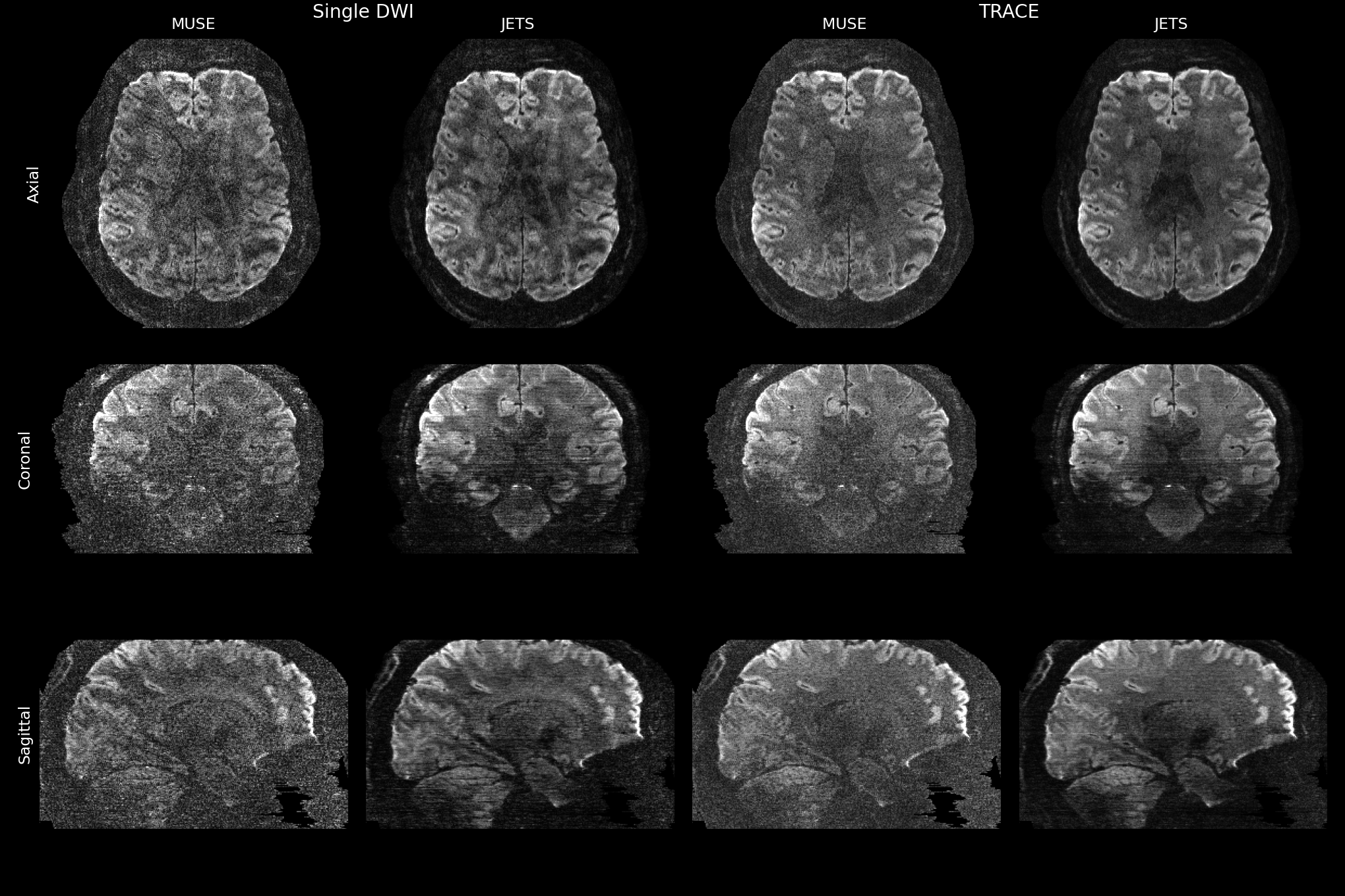
Figure 1: Single diffusion encoding direction and the corresponding TRACE images reconstructed using the MUSE and JETS methods in a 3-scan-trace dataset. The images are presented in axial, coronal, and sagittal views.
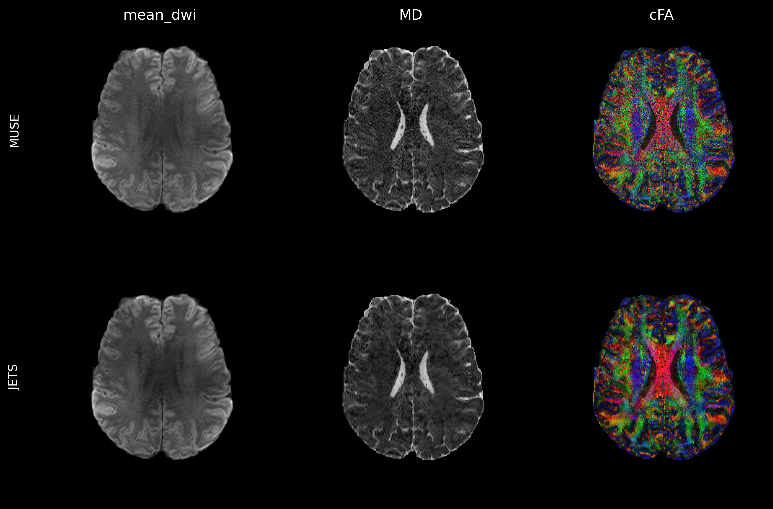
Figure 2: DTI image with a 0.7mm isotropic resolution, showing mean diffusion-weighted MRI (mean_dwi), Mean Diffusivity (MD), and colored Fractional Anisotropy (cFA) maps reconstructed using both the state-of-the-art MUSE reconstruction and the JETS reconstruction. The JETS reconstruction consistently yields images with improved signal-to-noise ratio and overall image quality compared to the state-of-the-art MUSE reconstruction.
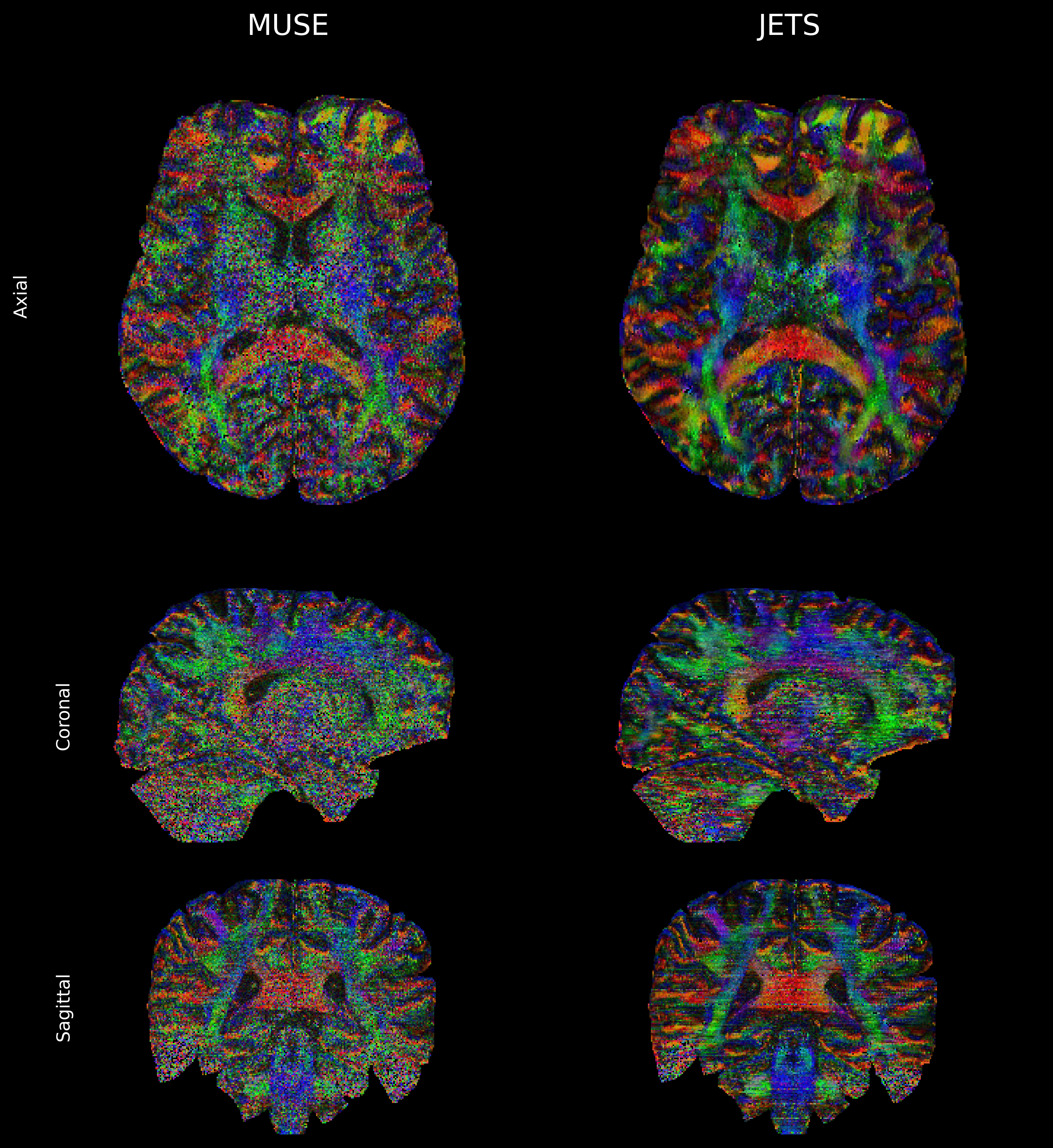
Figure 3: Fractional Anisotropy maps using both, the state-of-the-art MUSE reconstruction, and the JETS reconstruction. The axial, sagittal, and coronal views are presented. The JETS reconstruction demonstrates better Signal-to-Noise Ratio (SNR) and reveals more complex structural details.