2421
Navigator-free, high-resolution prostate diffusion imaging: Multi-Shot EPI with Multi-Scale Low-Rank Reconstruction1C.J. Gorter MRI Center, Department of Radiology, LUMC, Leiden, Netherlands, 2Philips, Best, Netherlands, 3Centre for Medical Imaging, University College London, London, United Kingdom, 4Philips Research Hamburg, Hamburg, Germany
Synopsis
Keywords: Diffusion Reconstruction, Prostate
Motivation: Prostate DWI is an important contrast for the diagnosis of prostate cancer.
Goal(s): The aim is to achieve prostate DW images and ADC maps with less-geometric-distortion, high-resolution and high signal-to-noise ratio (SNR).
Approach: A novel multi-scale low-rank reconstruction approach is introduced to improve multi-shot EPI diffusion-weighted imaging of the prostate. It addresses the ability to jointly reconstruct images over all b-value/diffusion directions at multiple different spatial scales without loss of contrast/structure information.
Results: Applied to healthy volunteers using different resolution protocols, the method demonstrates significant improvements in image resolution and SNR compared to a state-of-the-art reference method in a clinically acceptable scan time.
Impact: This multi-scale, low-rank reconstruction approach for prostate DWI can significantly improve the quality of diagnostic images, benefiting clinicians and patients by enabling more accurate prostate cancer diagnoses within short scan times.
Introduction
Prostate Diffusion-weighted imaging (DWI) with ADC mapping is essential for the diagnosis of prostate cancer1. Single-shot EPI (ssh-EPI), while clinically prevalent, suffers from geometric distortions, near tissues-air interfaces, and limited image resolution by T2* decay2. Multi-shot EPI (msh-EPI) can potentially offer better resolution and less distortion. However, as a typical challenge, shot-to-shot phase variations induced by physiological motion3 have to be corrected properly, which is particularly difficult in low-SNR prostate DWI. In the past, researchers have used locally low-rank4,5 (LLR) or structured low-rank (SLR)6,7 methods for navigator-free, phase-corrected DW msh-EPI, mainly in brain imaging. In this study, we propose to use the multi-scale low-rank8,9 concept, applied across diffusion-directions/b-values10 to improve the image quality of prostate DWI, achieving both high-resolution and high-SNR.Methods
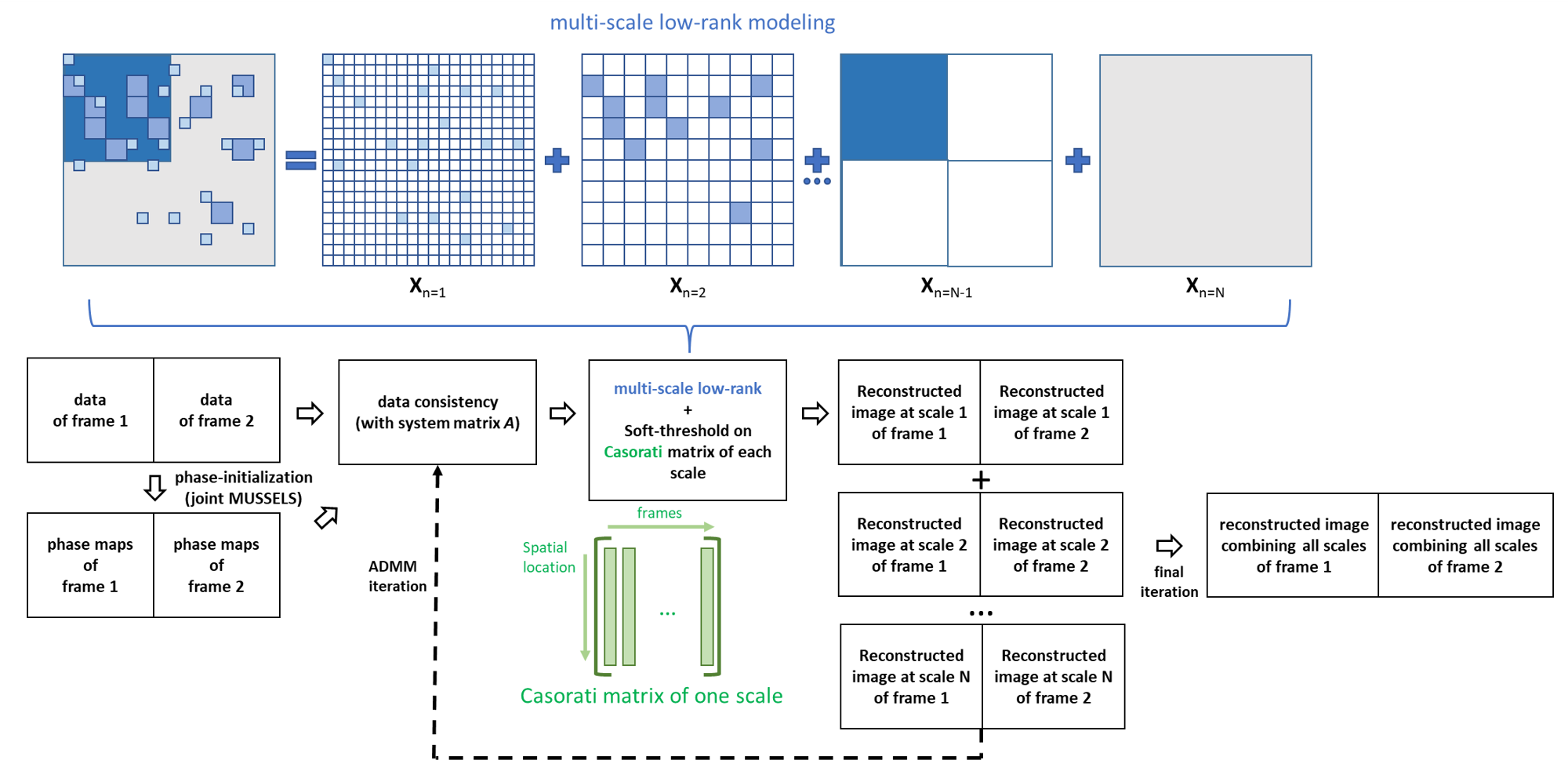
The multi-scale low-rank modelling, typically applied to dynamic imaging, models the image into spatial scales as $$$\mathbf{x}_j=\sum_{n=1}^N\mathbf{x}_{j,n}$$$, where $$$\mathbf{x}_{j, n}$$$ represents one image at timeframe $$$j$$$ at the $$$n$$$-th scale. It integrates global low-rank ($$$n=N$$$), sparse modeling ($$$n=1$$$), and various local low-rank scales ($$$1<n<N$$$)9. For msh-EPI DWI in prostate imaging, factors like motion, susceptibility changes, and eddy currents can cause spatial mismatch between scan partitions. In this study, we treat data from each diffusion direction of each b-value as having a distinct temporal spatial-state (frame). The joint reconstruction across all b-values/diffusion directions ($$$J$$$ frames) is thus formulated as:$$\left\{\hat{\mathbf{x}}_{1,\ldots,J}\right\}=\underset{\mathbf{x}_{1,\ldots,J}}{\operatorname{argmin}}\sum_{j=1}^J\left\|A\left(\sum_{n=1}^N\mathbf{x}_{j,n}\right)-\mathbf{y}\right\|_2^2+\sum_{n=1}^N \sum_{b=1}^{B_n}\lambda_n\left\|R_{b,n}\left\{x_{1,\ldots,J,n}\right\}\right\|_{*^{\prime}}$$For each b-value/diffusion direction image frame $$$\mathbf{x}_{j}$$$, the system matrix $$$A$$$ is a block-diagonal matrix containing each frame-specific system matrix $$$A_{j}$$$ describing multiplication of the k-space sampling mask ($$$M$$$), the Fourier transform ($$$F$$$), the coil-sensitivity maps ($$$S$$$) and the phase map of each shot ($$$P$$$). $$$\mathbf{y}$$$ contains the measured k-space data across all diffusion-directions/b-values, while $$$R_{b,n}$$$ extracts the $$$b$$$-th spatial block at the $$$n$$$-th scale (each scale has $$$B_n$$$ blocks) and combines blocks of all frames to form the Casorati matrix. By minimizing the nuclear norm (as a constraint on the rank of $$$R_{b,n}$$$) at each scale $$$n$$$, the correlation of each scale is enforced, sharing the across-frame redundant information. This guides the reconstruction with compact representations of different low-rank/sparse structures using varying low-rank block sizes. This could especially enhance SNR in high b-value images while keeping frame-specific spatial/contrast information.Prostate DWI experiments were performed on 7 healthy volunteers at a 3T Scanner (Philips) using 16-channel anterior and 12-channel posterior coils. Measurements were taken in low- (1.6×1.6×3mm³, matrix-size:192×188) and medium-resolution (1.3×1.3×3mm³, matrix-size:240×236) with fat-suppressed 3-shot DW EPI. One subject was additionally scanned with high-resolution (1.0×1.0×3mm³, matrix-size:256×240) using 4-shot EPI, and another with ssh-EPI (1.6×1.6×3mm³) for comparison. All multi-shot EPI scans had four b-values (b=0,150,500,1000 s/mm²) with varying numbers of diffusion directions (1,1,2,4 for each b-value), producing =8 frames. Each frame was repeated 3 times (NSA=3) with TE=59/TR=4000ms. ssh-EPI had 3 diffusion directions per b-value (b=0,150,500,1000 s/mm²), NSA=1,1,2,4, and TE=83/TR=4000ms, matching the 5-minute scan time of low-/medium-resolution msh-EPI; the 4-shot scan took 6:15 mins. SPAIR was used for fat-suppression for all datasets.
For reconstruction, 20 iterations of ADMM with automatic regularization-parameter selection was used with $$$N=5$$$ scales (1×1,4×4,16×16,64×64,full-matrix-size). Shot-to-shot phase estimation was performed using a modified IRLS-MUSSELS method (with 8-outer/8-inner iterations), assuming consistent magnitudes but varying phases between images for each b-value (similar to ref.11). The estimated phases were simply used to construct $$$P$$$. With estimated initial phase maps, each of the 8 frames was individually reconstructed using IRLS-MUSSELS7 as the reference method (phase-initialized MUSSELS). Coil sensitivities are estimated from b=0 s/mm2 images using ESPIRiT12. The reconstruction pipeline is shown in Fig.1.
Results
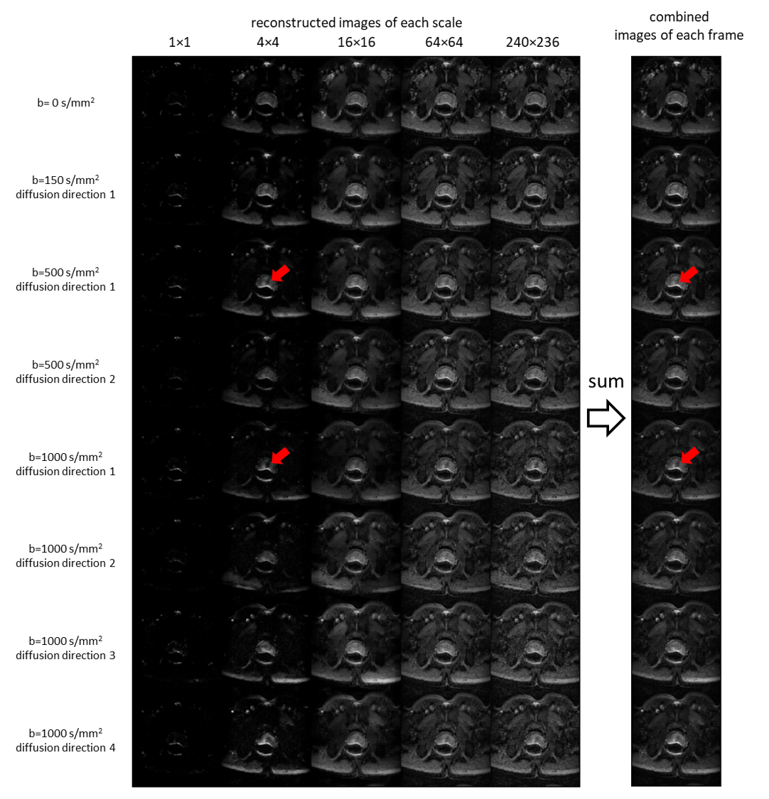
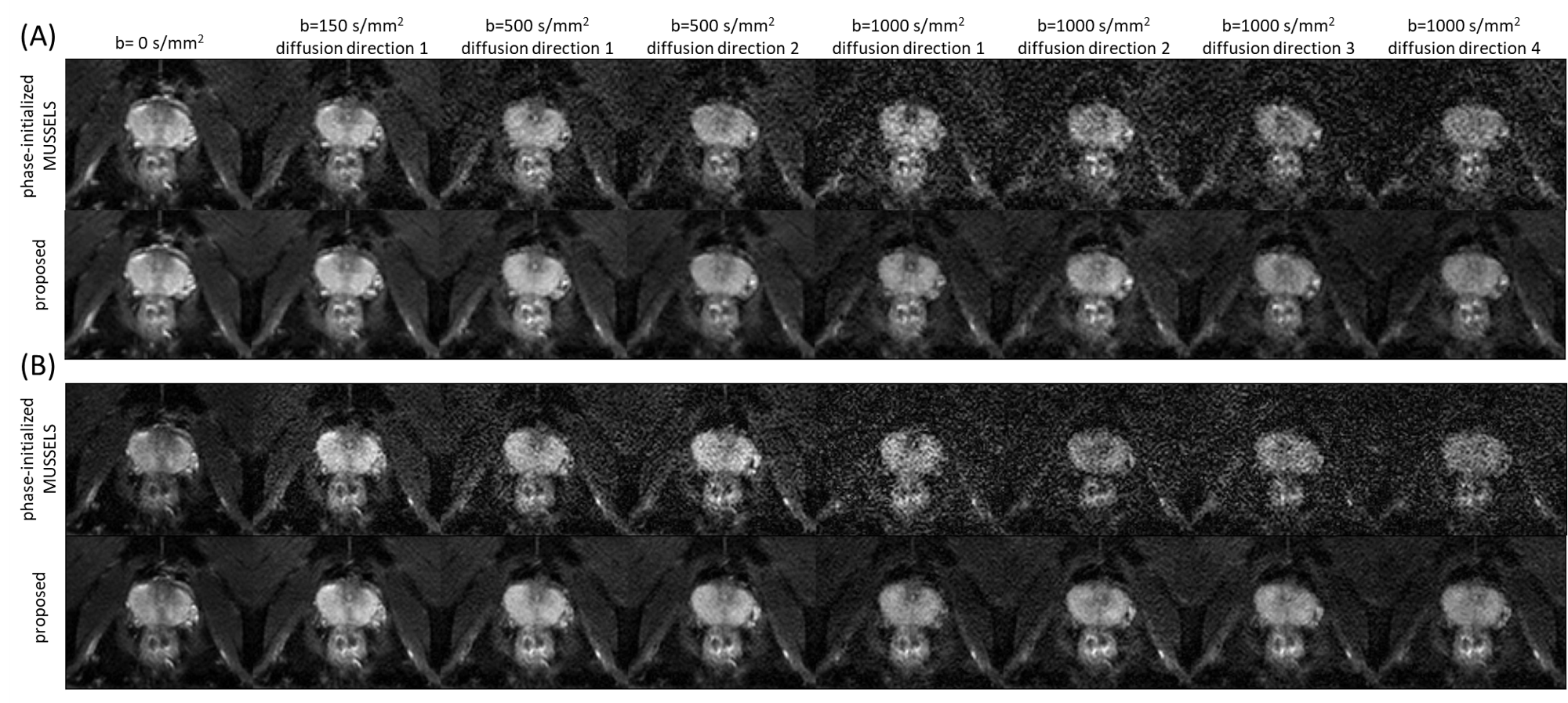
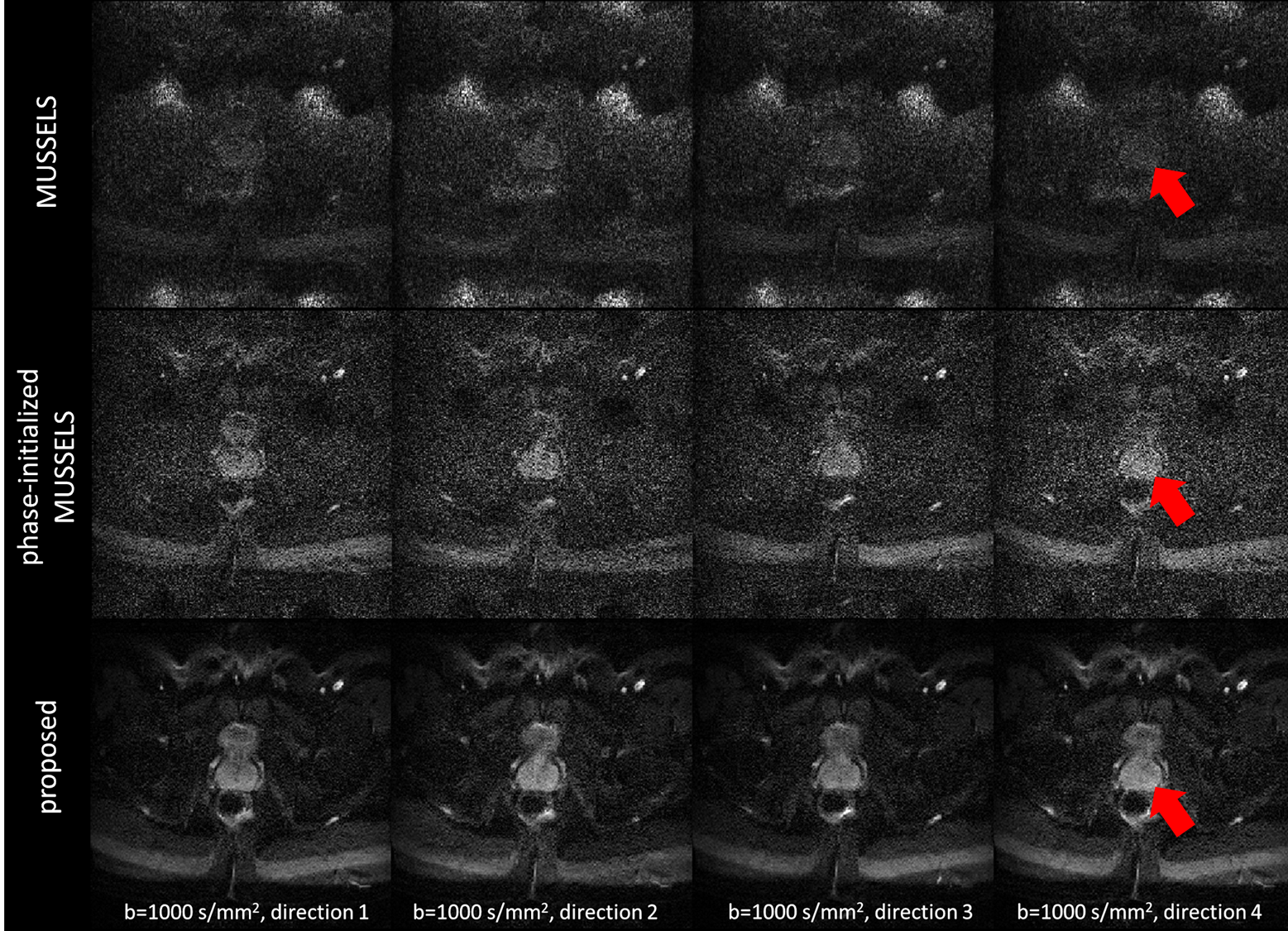
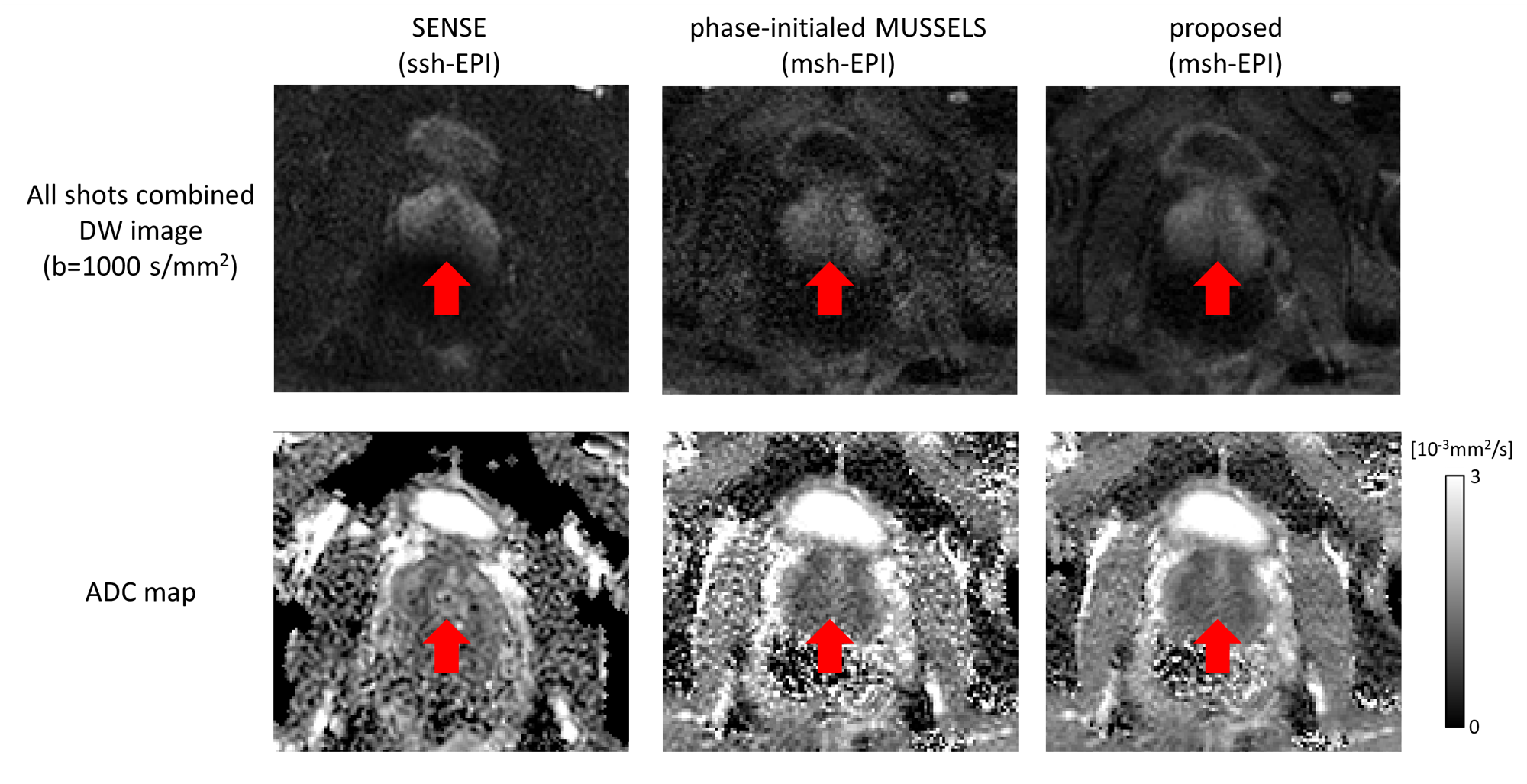
Fig.2 shows one dataset reconstructed with the multi-scale low-rank concept over 8 frames highlighting low-rank information at different scales. Fig.3 shows a subject’s prostate at different resolutions compared to the reference method. Fig.4 shows the high-resolution (1 mm2) b=1000 s/mm² images of four diffusion directions reconstructed with original/phase-initialized MUSSELS and the proposed algorithm, demonstrating the denoising capability of the proposed method. Fig.5 shows a comparison between ssh-EPI and msh-EPI (with two different reconstructions), highlighting the reduced geometric distortion with msh-EPI, and improved denoising especially using multi-scale low-rank for ADC mapping.Discussion and conclusion
In this study, we employed a multi-scale low-rank method for msh-EPI prostate DWI reconstruction across b-values and directions, effectively using across-frame redundant information to guide the reconstruction. The technique yielded high-SNR DW images and ADC maps within a clinically feasible five-minute scan. Although there is minimal diffusion anisotropy in the prostate13, there are noticeable temporal-spatial changes that are captured by the proposed algorithm (see different frames in Fig.2-4). Future applications may include regions with more severe macroscopic motion, such as liver or breast DWI, or high-resolution SNR-starved brain DTI.Acknowledgements
The authors would like to acknowledge NWO-TTW (HTSM-17104).References
1. Maurer MH, Heverhagen JT. Diffusion weighted imaging of the prostate-principles, application, and advances. Transl Androl Urol. 2017;6(3):490-498.
2. Tamada T, Kido A, Ueda Y, et al. Comparison of single-shot EPI and multi-shot EPI in prostate DWI at 3.0 T. Sci Rep. 2022;12(1):1-10.
3. Jeong HK, Gore JC, Anderson AW. High-resolution human diffusion tensor imaging using 2-D navigated multishot SENSE EPI at 7 T. Magn Reson Med. 2013;69(3):793-802.
4. Hu Y, Wang X, Tian Q, et al. Multi-shot diffusion-weighted MRI reconstruction with magnitude-based spatial-angular locally low-rank regularization (SPA-LLR). Magn Reson Med. 2020;83(5):1596-1607.
5. Hu Y, Levine EG, Tian Q, et al. Motion-robust reconstruction of multishot diffusion-weighted images without phase estimation through locally low-rank regularization. Magn Reson Med. 2019;81(2):1181-1190.
6. Mani M, Jacob M, Kelley D, Magnotta V. Multi-shot sensitivity-encoded diffusion data recovery using structured low-rank matrix completion (MUSSELS). Magn Reson Med. 2017;78(2):494-507.
7. Mani M, Aggarwal HK, Magnotta V, Jacob M. Improved MUSSELS reconstruction for high-resolution multi-shot diffusion weighted imaging. Magn Reson Med. 2020;83(6):2253-2263.
8. Ong F, Zhu X, Cheng JY, et al. Extreme MRI: Large-scale volumetric dynamic imaging from continuous non-gated acquisitions. Magn Reson Med. 2020;84(4):1763-1780.
9. Ong F, Lustig M. Beyond Low Rank + Sparse: Multi-scale Low Rank Matrix Decomposition. IEEE J Sel Top Signal Process. 2016;10(4):672.
10. Tang X, Gao J, Yang F, Hu C. Acceleration of Multi-b-value Multi-shot Diffusion-weighted Imaging using Interleaved Keyhole-EPI and Locally Low Rank Reconstruction. In: Proceedings - 2022 15th International Congress on Image and Signal Processing, BioMedical Engineering and Informatics, CISP-BMEI 2022.
11. Dong Y, Koolstra K, Li Z, Riedel M, Osch MJP van, Börnert P. Structured low-rank reconstruction for navigator-free water/fat separated multi-shot diffusion-weighted EPI. Magn Reson Med. Published online September 27, 2023. doi:10.1002/MRM.29848
12. Uecker M, Lai P, Murphy MJ, et al. ESPIRiT - An eigenvalue approach to autocalibrating parallel MRI: Where SENSE meets GRAPPA. Magn Reson Med. 2014;71(3):990-1001.
13. Bourne RM, Bongers A, Chatterjee A, Sved P, Watson G. Diffusion anisotropy in fresh and fixed prostate tissue ex vivo. Magn Reson Med. 2016;76(2).
Figures