2419
Pre-Excitation Gradients for Eddy Current Nulled Convex Optimized Diffusion Encoding (Pre-ENCODE)1Department of Radiology, Stanford University, Stanford, CA, United States, 2Department of Electrical Engineering, Stanford University, Stanford, CA, United States
Synopsis
Keywords: Diffusion Acquisition, Diffusion/other diffusion imaging techniques, eddy currents, image distortion, time-optimal
Motivation: Eddy current-induced image distortions in DWI require either tedious image-based corrections or time inefficient pulse sequence acquisitions that increase the minimum echo time (TEmin) and limit SNR.
Goal(s): Pre-excitation gradients for eddy current-nulled convex optimized diffusion encoding (Pre-ENCODE) was used to mitigate eddy current-induced image distortions without increasing TEmin.
Approach: Simulations, phantoms, and volunteer DWI with monopolar (MONO), eddy current-nulled convex optimized diffusion (ENCODE), and Pre-ENCODE were used to evaluate TEmin, eddy current-induced image distortions, and ADC.
Results: Pre-ENCODE offers shorter TEmin compared to MONO and ENCODE as well as reduced eddy current-induced image distortions and more accurate ADC values compared to MONO.
Impact: Pre-ENCODE reduces eddy current-induced image distortions by incorporating an additional pre-excitation gradient. Pre-ENCODE provides a shorter TEmin, increased SNR, improved image registration, and more accurate ADC quantification compared with conventional DWI encoding techniques.
Introduction
Time-varying magnetic fields from diffusion gradients in Diffusion Weighted Imaging (DWI) generate eddy currents, which create unwanted magnetic fields that contribute an additional spatial encoding field leading to geometric image distortions such as image shearing, scaling, and/or bulk shifting [1].Our previously described eddy current nulled convex optimized diffusion encoding (ENCODE) [4] approach nulls the impact of eddy currents from the diffusion encoding gradients using our Gradient Optimization toolbox [5]. Herein, we modified the ENCODE approach to include an additional pre-excitation gradient (Pre-ENCODE) to enable eddy current-nulled diffusion encoding gradient waveforms that mitigate eddy current-induced image distortions with a shorter TEmin.
Methods
Our GrOpt toolbox was used to design diffusion encoding waveforms that minimize both B0 eddy currents and TEmin for spin echo-based DWI. The eddy current constraint was formulated by modeling eddy current-induced fields, ε(λi,t) from an encoding gradient, G(t), as a resistive-inductive circuit [6,7]:$$\epsilon(\lambda_{i},t)=\vec{S}(t)\circledast e^{-t/\lambda_{i}},$$
where $$$\vec{S}(t)$$$ is the slewrate ($$$\vec{S}(t)=\frac{d\vec{G}(t)}{dt}$$$), $$$\circledast$$$ is the convolution operator, and λi is the ith eddy current time constant. The Pre-ENCODE framework optimized for the constraint ε(λnull,TDiff)=0, where λnull is the target eddy current time constant to be nulled at the end of diffusion encoding, TDiff. This constraint ensured that eddy current-induced fields are zero at TDiff. Unlike our previous ENCODE framework, we modified the optimization to include eddy current fields generated by an additional gradient played before the excitation pulse (Figure 1). This new pre-excitation gradient generates additional eddy current fields, which offset the eddy current fields generated later by the diffusion gradients, but does not contribute to diffusion encoding or add to the TE. The Pre-ENCODE framework used λnull=100ms, which was empirically found [2] to be the optimal value for our 3T system (SIGNA Premier, GE HealthCare).
Pulse sequences were simulated using the protocol parameters outlined in Table 1 for conventional monopolar (MONO) waveforms designed using analytic solutions [8] as well as ENCODE and Pre-ENCODE waveforms designed using GrOpt, all in Python (Figure 1). We compared TEmin values across a range of b-values and in-plane spatial resolutions.
Phantom experiments and in vivo imaging in volunteers (N=6) were performed using the parameters in Table 1 and sequences from Figure 1 to evaluate the impact of eddy current-induced image distortions and ADC accuracy.
MONO and ENCODE waveforms were designed on the scanner using the vendor-provided analytic solution and GrOpt, respectively, while Pre-ENCODE waveforms were designed offline using GrOpt in Python, written to external waveform files, and loaded onto the scanner. All images were reconstructed using the vendor-provided pipeline without additional image registration, post-processing, or distortion correction. ADC maps and eddy current-induced image distortions were measured in MATLAB using the voxel-wise coefficient of variation (CoV) across the diffusion encoding directions in both masked edge (CoVEdge) and global (CoVGlobal) pixels.
Results
The protocol parameters and pulse sequences used for all experiments performed as part of this study are shown in Table 1 and Figure 1, respectively.Pre-ENCODE provided a shorter TEmin than both MONO (71.0±17.7ms vs. 77.6±22.9ms) and ENCODE (71.0±17.7ms vs. 86.2±14.2ms) in all tested cases (Figure 2).
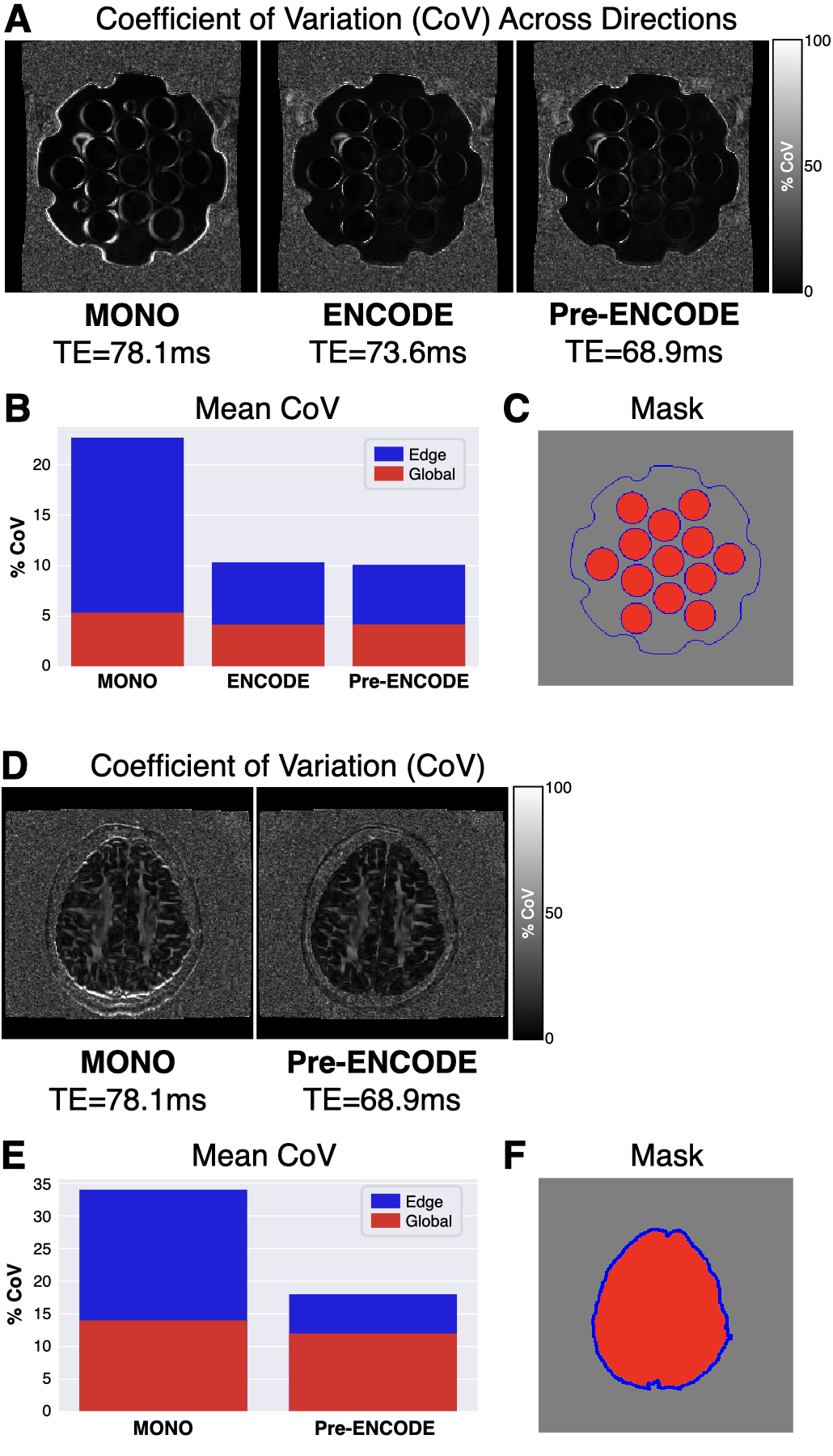
Figure 3 shows the CoV analysis from both the phantom (A-C) and in vivo (D-F) images. The phantom CoVEdge values were highest for MONO (22.7%) near the outer edges (water-air interfaces) and near the edges of each vial (polyvinylpyrrolidone-water interface) and reduced for both ENCODE (10.4%) and Pre-ENCODE (10.1%). Phantom CoVGlobal values were similar across all sequences (MONO=5.4%, ENCODE=4.2%, and Pre-ENCODE=4.2%). In vivo CoVEdge values near the tissue-bone interface were significantly higher (p-value=7e-7) for MONO (34.4±24.7%) compared to Pre-ENCODE (18.4±10.4%) while CoVGlobal values were similar (14.2±9.5% for MONO vs. 13.9±8.3% for Pre-ENCODE, p-value=0.36).
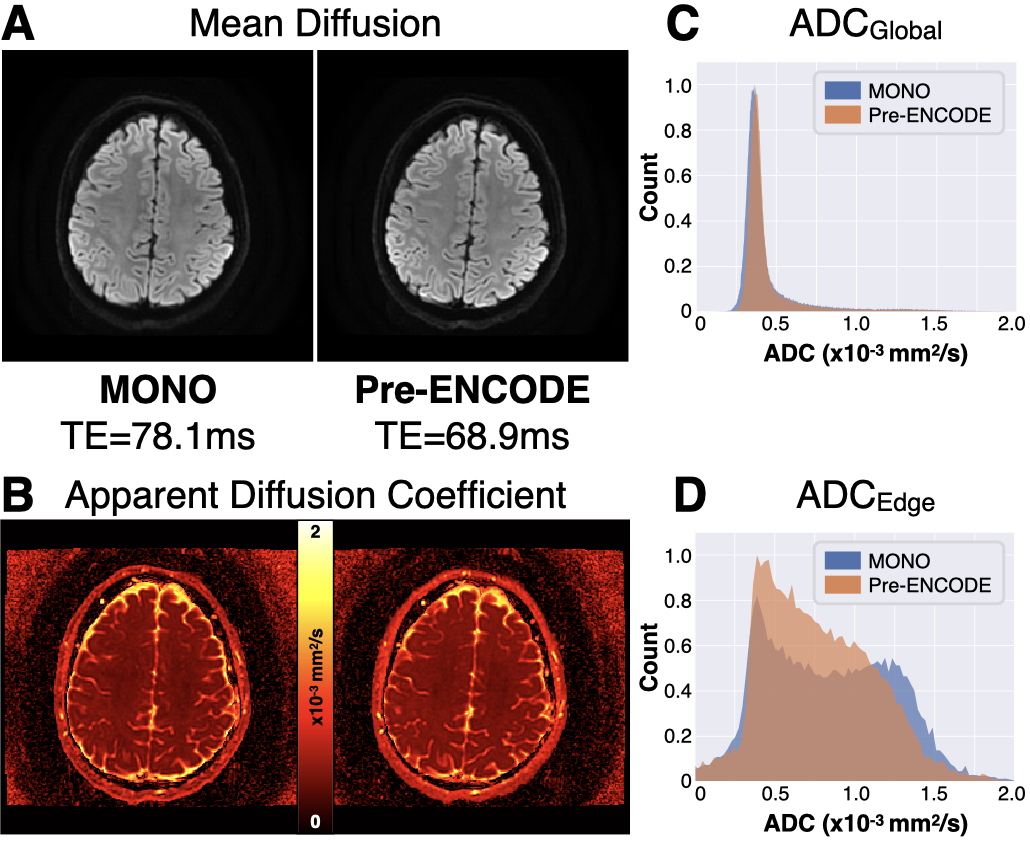
Figure 4 shows representative in vivo images. Qualitatively, MONO and Pre-ENCODE appear similar both in terms of mean diffusivity and ADC, except near the edges of the brain where MONO values are brighter (more yellow), indicating an increase in ADC. Quantitatively, these findings are corroborated by the ADC histograms which show similar median and 95%-CIs for ADCGlobal values (0.37 [0.28,1.45]×10-3mm2/s for MONO vs. 0.38 [0.28,1.45]×10-3mm2/s for Pre-ENCODE, p-value=0.25) and increased ADCEdge values (0.80 [0.17,1.49]×10-3mm2/s for MONO vs. 0.70 [0.18,1.48]×10-3mm2/s for Pre-ENCODE, p-value=0.02). The phantom and in vivo images show artificially increased ADC values in MONO edge pixels, where eddy current-induced image distortions are prevalent, which Pre-ENCODE alleviates.
Conclusions
Pre-ENCODE offers time-optimal mitigation of eddy current-induced image distortions in DWI by incorporating an additional pre-excitation gradient into the ENCODE strategy. Pre-ENCODE affords a shorter TEmin compared to MONO and ENCODE as well as improved image registration and more accurate ADC quantification compared with MONO.Acknowledgements
This work was partially supported by NIH U01-EB025162 and NIH R01-MH116173.References
1. Jezzard P, Barnett AS, Pierpaoli C. Characterization of and correction for eddy current artifacts in echo planar diffusion imaging. Magn Reson Med. 1998;39(5):801–812.
2. Reese TG, Heid O, Weisskoff RM, Wedeen VJ. Reduction of eddy current-induced distortion in diffusion MRI using a twice-refocused spin echo. Magn Reson Med. 2003;49(1):177–182.18.
3. Wider G, Dotsch V, Wuthrich K. Self-compensating pulsed magnetic-field gradients for short recovery times. J Magn Reson.1994;108(2):255–258.
4. Aliotta E, Moulin K, Ennis DB. Eddy current-nulled convex optimized diffusion encoding (EN-CODE) for distortion free diffusion tensor imaging with short echo times. Magn Reson Med. 2018;79(2):663–672.
5. Loecher M, Middione MJ, Ennis DB. A gradient optimization toolbox for general purpose time-optimal MRI gradient waveform design. Magn Reson Med. 2020;84(6):3234–3245.
6. Jezzard P, Barnett AS, Pierpaoli C. Characterization of and correction for eddy current artifacts in echo planar diffusion imaging. Magn Reson Med. 1998;39(5):801–812.
7. Mansfield P. Multi-planar image formation using NMR spin echoes. J Solid State Phys. 1977;10(3):L55.
8. Stejskal EO, Tanner JE. Spin diffusion measurements: spin echoes in the presence of a time-dependent field gradient. J Chem Phys. 1965;42(1):288–292.
Figures

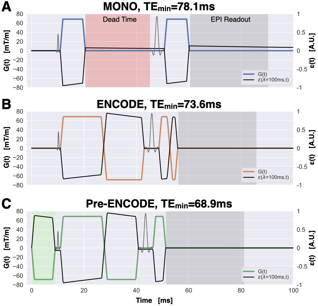
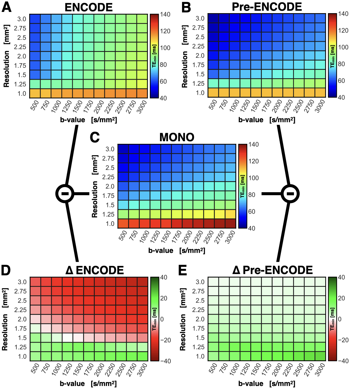
Figure 2. TEmin for a range of b-values (500-3000s/mm2, Δ=250s/mm2) and isotropic in-plane resolutions (1.0-3.0mm2, Δ=0.25mm2) for (C) MONO as well as (A) ENCODE and (B) Pre-ENCODE with λnull=100ms. Differences in TEmin for (D) ΔENCODE=MONO-ENCODE and (E) ΔPre-ENCODE=MONO-Pre-ENCODE. ENCODE provides a shorter TEmin than MONO in 28% of the tested cases whereas Pre-ENCODE offers a shorter TEmin than ENCODE and MONO in 100% of the tested cases.