2417
CATERPillar : a fast and flexible framework framework for generating synthetic white matter numerical phantoms1CHUV, Lausanne, Switzerland, 2EPFL, Lausanne, Switzerland
Synopsis
Keywords: Simulation/Validation, Software Tools, Phantom
Motivation: Building realistic and complex white matter numerical phantoms is needed for accurate diffusion MRI simulations but challenging to achieve.
Goal(s): The creation of white matter (WM) numerical phantoms by mimicking realisitic parallel axonal growth.
Approach: This tool uses overlapping spheres to build realistic axons and parallels their growth to decrease the running time while reliably preventing collisions.
Results: High intracellular volume fraction values can be reached (up to 70%) for tortuous, variable-caliber axons. The parallelism of the growth decreases the run-time.
Impact: Creating numerical phantoms that accurately represent white matter can improve the accuracy of results in diffusion MRI studies using Monte Carlo simulations.
Introduction
Monte-Carlo diffusion simulations are useful for validating tissue microstructure models. By generating synthetic diffusion-weighted magnetic resonance signals (DW-MRI) these simulations allow the mapping between features derived from DW-MRI and microstructure parameters1. They require numerical phantoms that accurately represent a specific tissue2, which is in this work, cerebral white matter(WM). Current methods, like the MEDUSA1 and CACTUS2 algorithms, generate WM numerical substrates by first placing axons and then trying to eliminate overlap. However, real axons grow guided by chemical cues and occupy space naturally3. Emulating this natural growth may have a certain importance for more realistic phantoms. Another method known as CONFIG3 tackles this problem by growing fibers with a set of rules motivated by realistic axonal guidance mechanisms. CONFIG's limitation lies in its need for densely sampled space in the growth network, demanding many nodes for large phantoms, leading to memory-intensive and potentially unfeasible scenarios3. This study introduces CATERPillar (Computational Axonal Threading Engine for Realistic Proliferation), a method simulating natural axonal growth by using overlapping spheres as elementary units as in1. It allows parallel axon growth while preventing collisions and offers user flexibility, enabling control over parameters like density, tortuosity, and beading. Users can even visualize axon growth in real-time.Methods
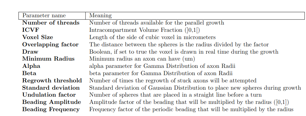
Initialising Axonal Chemoattraction: CATERPillar generates axons with radii drawn from a Gamma distribution, the amount determined by voxel size and density. These axons grow simultaneously in batches, their count defined by user-set processor count. Batches start within a cube voxel on an x-y plane, strategically placed to avoid collisions. An attractor target is set on the opposite side, guiding each axon's growth. This approach mimics natural axon growth, influenced by chemical cues that attract or repel fibers3.Parallel axonal growth: During growth, spheres are added at half the axon's radius using Gaussian-based spherical coordinates to maintain tortuosity. Tortuosity is controlled by the Gaussian standard deviation (Table 1), regulating deviation from the attractor's path. Bead-like structures are shaped with the mean beading amplitude and beading frequency (Table 1). When stuck, the size of the newly added sphere is continuously reduced until it fits or reaches a specified minimum threshold set by the user. Overlaps are detected using a "Sweep and Prune" algorithm4. Colliding axons are pruned, and new ones start elsewhere.
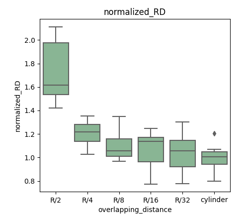
Synthetic dMRI signals: After generating the phantoms, if the user specifies an overlapping factor (Table 1), extra spheres can be inserted between the ones already present in each axon to enhance their overlap. Augmenting the overlap can be advantageous prior to Monte-Carlo DW-MRI simulations because it results in a smoother axonal surface. To assess this effect, we investigate how the spacing between spheres influences the radial diffusivity (RD) derived from Monte-Carlo DW-MRI simulations6. We compare the RD values obtained from phantoms containing straight overlapping spheres (with varying distances between overlaps) with those containing cylinders.
Results and Discussion
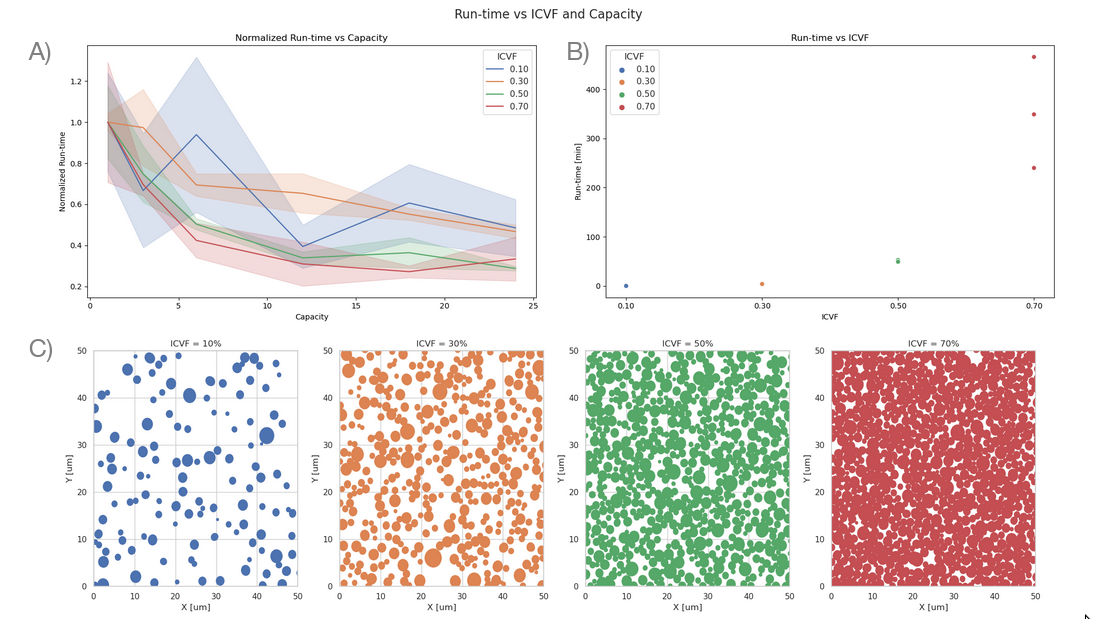
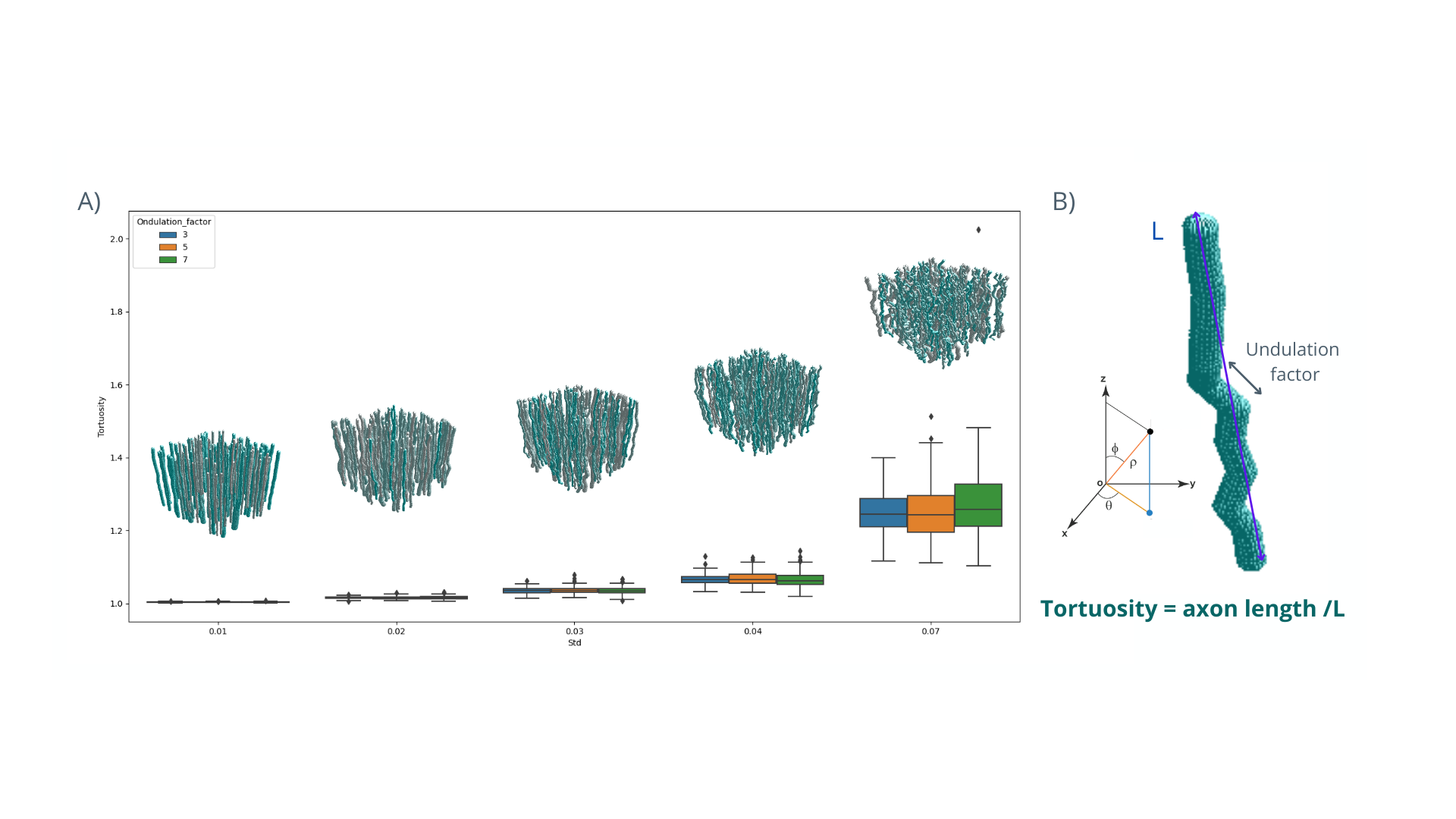
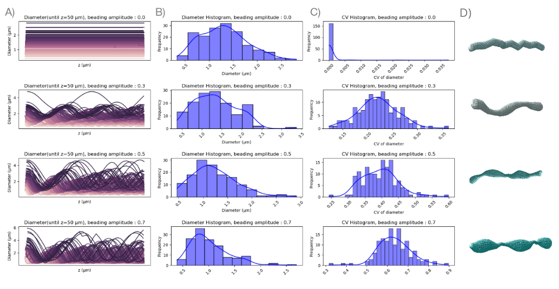
Figure 1 demonstrates the impact of parallel growth threads on duration, revealing a non-linear decrease influenced by growth coding intricacies and Amdahl's law. Densely packed substrates benefit more from higher capacities due to simultaneous axon growth. In Figure 2, the influence of standard deviation and undulation factor parameters on axon tortuosity is depicted. Tortuosity rises with the standard deviation, while the undulation factor has no effect.Figure 3 showcases axonal beading's effect on morphology, displaying the CV concerning axonal beading. Previous electron microscopy data5 suggests that axons have a radius CV of 0.2, resulting in a mean beading amplitude at 0.3 times the radius. In Figure 4, the impact of overlapping sphere distances on RD is explored. RD in axons with closer spheres approaches cylindrical RD values. The optimal balance between accuracy and computation time occurs at R/8 sphere distance. Future studies will delve into optimal overlaps for contrasting tortuous spheres with meshed axons, accounting for varied diffusion times.
Conclusion
We demonstrated the versatility of CATERPillar in generating numerical white matter substrates. Our assessment highlighted the influence of parameters like the number of parallel threads, ICVF, tortuosity and axonal beading on runtime and morphology, and of sphere overlap on DWI signals. Future work will also examine the influence of morphology on the DWI signals.Acknowledgements
This work was supported by ERC Starting Grant ’FIREPATH’, SERI no.MB22.00032References
[1] Kévin Ginsburger, Felix Matuschke, Fabrice Poupon, Jean-François Mangin, Markus Axer, Cyril Poupon, MEDUSA: A GPU-based tool to create realistic phantoms of the brain microstructure using tiny spheres, NeuroImage, Volume 193, 2019, Pages 10-24, ISSN 1053-8119, https://doi.org/10.1016/j.neuroimage.2019.02.055.
[2] Villarreal-Haro, J. L., Gardier, R., Canales-Rodríguez, E. J., Fischi-Gomez, E., Girard, G., Thiran, J. P., and Rafael-Patiño, J. (2023). CACTUS: a computational framework for generating realistic white matter microstructure substrates. Frontiers in neuroinformatics, 17, 1208073. https://doi.org/10.3389/fninf.2023.1208073
[3] Ross Callaghan, Daniel C. Alexander, Marco Palombo, Hui Zhang, ConFiG: Contextual Fibre Growth to generate realistic axonal packing for diffusion MRI simulation, NeuroImage,Volume 220,2020,117107,ISSN 1053-8119,https://doi.org/10.1016/j.neuroimage.2020.117107.
[4] Avril, Quentin and Gouranton, Valérie and Arnaldi, Bruno. (2011). Dynamic Adaptation of Broad Phase Collision Detection Algorithms. ISVRI 2011 - IEEE International Symposium on Virtual Reality Innovations 2011, Proceedings. 41 - 47. 10.1109/ISVRI.2011.5759599.
[5] Lee, HH., Yaros, K., Veraart, J. et al. Along-axon diameter variation and axonal orientation dispersion revealed with 3D electron microscopy: implications for quantifying brain white matter microstructure with histology and diffusion MRI. Brain Struct Funct 224, 1469–1488 (2019). https://doi.org/10.1007/s00429-019-01844-6
[6] Rafael-Patino Jonathan, Romascano David, Ramirez-Manzanares Alonso, Canales-Rodríguez Erick Jorge, Girard Gabriel, Thiran Jean-Philippe TITLE=Robust Monte-Carlo Simulations in Diffusion-MRI: Effect of the Substrate Complexity and Parameter Choice on the Reproducibility of Results, Frontiers in Neuroinformatics, 10.3389/fninf.2020.00008 ISSN=1662-5196
Figures